Mites, caterpillars and moths
Section Editor
Prof. Dr. D. Nashan, Dortmund
Animal mites are only rarely detected in humans.
Given that the skin lesions are nonspecific, it is essential to take a thorough environmental history (pets, occupation, hobbies) and to possibly consult a veterinarian.
Causal treatment of the affected animal is paramount.
In their 2014 publication, Chen and Plewig proposed to classify demodicosis into a primary and a secondary form.
(1) Patients have no history of a preexisting inflammatory skin disease; (2) there is an increased density of Demodex mites (> 5 mites/cm²) in the affected area; and (3) skin lesions heal with acaricidal treatment alone.
Combination treatments are more effective than monotherapies. Relapses are a major problem.
Grouped, severely pruritic papules and papulovesicles, especially in areas of thin skin.
Lesions occur in areas that act as barriers to the migration of the larvae (e.g., tight-fitting clothes).
Affected individuals include those exposed to grain, straw or dried plant produce.
Given that Pyemotes mites are hardly visible to the naked eye, the diagnosis is based on detecting the source of infection including the host insects.
In many Southeast Asian countries, Tsutsugamushi fever is the second most common febrile disease after malaria.
A member of the Rickettsia family, the causative agent is exclusively transmitted by mites.
Worldwide, studies have shown sensitization rates to vary between 20 % and 80 %.
The fact that these allergens have irritant effects and also activate the innate immune system is thought to contribute to augmenting their allergenic potency.
Clinical symptoms of storage mite hypersensitivity are the same as for HDMs.
There is usually little cross-reactivity between storage mites and HDMs as well as among the various types of storage mites.
Hossler has proposed that ’erucism’ be used for any reaction secondary to contact with caterpillars and ’lepidopterism’ for any reaction that results from exposure to butterflies/moths.
Disease symptoms are usually caused by setae.
Irritant-toxic substances may cause cutaneous reactions such as pruritus, edema and erythema, papules and wheals, and in some cases even generalized eczematous eruptions.
Immediate-type hypersensitivity reactions may also be triggered by caterpillars.
There are also toxin-induced reactions related to enzymes with fibrinolytic, proteolytic, hemolytic, or procoagulatory activity.
Severe reactions such as osteoarthritis and hemorrhagic diathesis have also been reported.
The symptoms frequently do not allow for any conclusions to be drawn as to the individual pathomechanism involved. Any such attempt is further complicated by the fact that more than one reaction type may contribute to the clinical presentation.
Clinical manifestations can be classified as cutaneous, mucosal, and systemic reactions.
Widespread/generalized cutaneous reactions include disseminated papules, papulovesicles, or dermatitis as well as urticaria and angioedema.
Mucosal reactions may affect the conjunctivae as well as the oral and bronchial mucosa.
Systemic reactions include nausea, vertigo, or vomiting.
Irrespective of the pathogenesis, a common early feature of all skin lesions caused by oak processionary caterpillars is severe pruritus.
Airborne exposure to setae may result in cough, dyspnea, palpebral edema, and conjunctival symptoms.
After contact with caterpillars, all visible setae should be carefully removed.
Lonomia (L.) obliqua and L. achelous inject toxins through their setae.
Despite pathophysiological differences, contact with both species results in similar clinical symptoms with (sometimes fatal) cutaneous, mucosal, visceral or intracranial hemorrhage.
Various moth species from the genus Calyptra sting larger mammals and feed on their blood.
There are various moths from other genera that feed on the protein-rich tears of other animals.
Especially workers in the silk industry may develop immediate-type hypersensitivity to silk, including allergic asthma, allergic rhinitis or allergic conjunctivitis.
Ingesting silkworm pupae (as practiced quite commonly in Asia) may cause food allergic reactions.
Summary
Besides conditions such as scabies and hypersensitivity to house dust mites, other diseases caused by mites and caterpillars tend to be more uncommon in everyday practice. Nevertheless, there is a broad spectrum of medically relevant disorders associated with these arthropods. Mites may act as parasites that infect or colonize the skin (e.g., scabies, pseudoscabies, demodicosis) or they may pierce the host’s skin and feed on tissue fluid and blood (trombiculosis). In the latter case, they also play a role as vectors transmitting Orientia tsutsugamushi, the pathogen that causes Tsutsugamushi fever. In addition to house dust mites, storage mites, too, are characterized by their allergenic potential.
The terms erucism and lepidopterism are used for the various diseases caused by caterpillars and moths. Both terms are not used consistently. With respect to pathogenesis, various mechanisms have been described, including type I and type IV hypersensitivity as well as irritant and toxin-induced reactions. In Germany, skin reactions following exposure to the hairs of oak processionary caterpillars are particularly common. Extracutaneous manifestations including nausea, vomiting, hemorrhage, arthropathy or even life-threatening reactions have been reported in association with certain exotic species. Some species act as parasites by feeding on blood or tears.
As natural silk can cause immediate and delayed-type hypersensitivity reactions, workers in the silk industry may develop allergic asthma, rhinitis, or conjunctivitis. Consumption of silkworm pupae is associated with the risk of food allergy.
Introduction
The present CME article focuses on those mites, caterpillars and moths in particular whose clinical significance is less well known and might therefore be underestimated. Given the increase in long-distance traveling to exotic places as well as in migration, it is useful to have at least some elementary knowledge about species that occur outside Europe. This article will, however, not cover scabies; please refer to the recent comprehensive review by Sunderkötter et al. 1. House dust mite-related issues will be addressed, albeit briefly.
Diseases caused by mites can be classified into three categories: 1) parasitic disorders, 2) diseases in which mites act as vectors, and 3) mite-induced allergic diseases without parasitism (Table 1). Apart from scabies, parasitic disorders also include pseudoscabies, demodicosis, trombiculosis, and Pyemotes dermatitis (grain itch) 2-5. In case of demodicosis, however, the line between commensalism and parasitism is somewhat blurred 3 (Table 2). Naturally, parasitism is also a prerequisite for mite-borne infectious diseases to occur. The most important disease transmitted in this manner is Tsutsugamushi fever 6. Allergic disorders without parasitism are caused by house dust mites and storage mites 7, 8.
| Parasitic disorders | Scabies Pseudoscabies Demodicosis Trombiculosis Pyemotes dermatitis |
| Mites as vectors | Tsutsugamushi fever Hantaviruses? Ehrlichiosis? Borreliosis? |
| Mites as allergens | House dust mites Storage mites |
| Parasitism | Transient or permanent interaction of two species that is favorable for one (the parasite) but detrimental to the other (the host). |
| – Ectoparasitism | Parasitism with the parasite living on the surface of the host. |
| – Endoparasitism | Parasitism with the parasite living inside the host. |
| Commensalism | Transient or permanent interaction of two species that is favorable for one (the commensal organism) and neither favorable nor detrimental to the other (the host). |
| – Ectocommensalism | Commensalism with the commensal organism living on the surface of the host. |
| – Endocommensalism | Commensalism with the commensal organism living inside the host. |
Diseases caused by caterpillars and moths are characterized by a multitude of different pathomechanisms and clinical manifestations, which rarely show a clear correlation. Likewise, it is frequently difficult to match specific pathomechanism or symptoms with a particular caterpillar species 9, 10. We will therefore present pathomechanisms and clinical presentations in separate chapters. In Europe, the most important species in terms of their medical significance are the oak processionary caterpillar and the brown-tail caterpillar. Caterpillars from tropical regions may cause severe and sometimes life-threatening reactions, commonly referred to as lonomism or dendrolimiasis 10, 11. Some parasitic moths have developed the ability to feed on blood or tears 12. Finally, we will cover allergic reactions to silkworm-derived products 13, 14.
Scabies-like disorders transmitted by animals – “pseudoscabies”
Various mite species cause mange in animals. Many of them can be transmitted to humans and induce transient scabies-like eruptions. These include a variety of Sarcoptes mites found in dogs, foxes, ferrets, pigs, cattle, goats, chamois, sheep, horses, camels and tapirs. Transmission of Notoedres cati from cats, Cheyletiella species from dogs, cats and rabbits, Trixacarus caviae from guinea pigs, Dermanyssus gallinae from birds and Ornithonyssus bacoti from rodents have also been reported. Even reptiles may be a source of transmission (snake mite, Ophionyssus natricis). Given the high degree of host specificity, animal mites infest humans only transiently and do not dig burrows. Animal mites are only rarely detected in humans. Pseudoscabies is characterized by pruritic papules and papulovesicles (2–6 mm in diameter). Lesions are not found in areas typically affected by scabies but rather in those areas that come in direct contact with the host animal. These usually include the neck, arms and abdomen. Given that the skin lesions are nonspecific, it is essential to take a thorough environmental history (pets, occupation, hobbies) and to possibly consult a veterinarian. The poultry red mite Dermanyssus gallinae deserves special mention. It feeds on blood and can apparently cover relatively long distances, so transient infestation does not require direct contact with birds or contaminated objects. Causal treatment of the affected animal is paramount. Whenever Dermanyssus is involved, it is recommended to remove the nest, clean the surroundings with a vacuum cleaner, seal the vacuum cleaner bag tightly and dispose of it outside the house, and lastly, to disinfest the affected area using pyrethroids 15. Human patients are treated symptomatically with anti-inflammatory and antipruritic agents 2.
Follicular mites: Demodex folliculorum and Demodex brevis
Demodex mites are permanent ectoparasites that reside in hair follicles in humans and other mammals. The two species relevant to humans are Demodex folliculorum and Demodex brevis. They are primarily found in the infundibula of regular hair follicles on the face and scalp as well as in sebaceous and meibomian glands, where they feed on sebum and epithelial debris. Given their anatomical limitations (e.g., lack of an anus), they are unable to excrete digesta. While this results in a shortened life span, it also prevents the host from developing an inflammatory response to fecal matter, as is observed in scabies. Infestation rates in humans (measured by microscopic analysis of removed eyelashes) increase with age; with approximately 11 % of children under the age of ten affected and nearly 100 % of 90-year-olds. The prevalence among 46–65-year-olds has been reported to be 36–38 % 16. There is conflicting data in terms of gender differences. Greasy skin as well as a warm and humid climate seem to facilitate colonization. The exact pathogenetic role of Demodex mites is controversial. In humans, Demodex infestation is usually asymptomatic and thus tends to constitute a form of commensalism. As a consequence, clinical symptoms primarily seem to be the result of Demodex overgrowth 17. In their 2014 publication, Chen and Plewig proposed a pathogenetic classification of demodicosis into a primary and a secondary form. Further subclassification is subsequently guided by the clinical presentation. Accordingly, primary demodicosis is a distinct disease entity defined by the following three criteria: (1) Patients have no history of a preexisting inflammatory skin disease; (2) there is an increased density of Demodex mites (> 5 mites/cm²) in the affected area; and (3) skin lesions heal with acaricidal treatment alone. Based on disease severity, the condition is divided into three clinical variants. Secondary demodicosis, on the other hand, is characterized by increased density of Demodex mites in association with preexisting skin or systemic diseases such as rosacea, seborrheic dermatitis, perioral dermatitis, leukemia, HIV infection, malignancies or chronic renal disease (Table 3) 3. For rosacea in particular, it has been suggested that Demodex mites play an important pathogenetic role 18. Some authors even question whether there should be a distinction between rosacea and demodicosis at all 19. The increased density of Demodex mites in rosacea 20 and the fact that the acaricidal agent ivermectin has proven effective suggest a possible pathogenetic role; however, it should be borne in mind that ivermectin has also been shown to have anti-inflammatory and antibacterial effects 21, 22. The frequent occurrence of asymptomatic Demodex infestation further complicates assessment of their exact pathogenetic role.
| Primary demodicosis | Spinulate demodicosis | |
| Papulopustular demodicosis | ||
| Nodulocystic demodicosis | ||
| Secondary demodicosis | Associated with inflammatory dermatoses |
|
| Associated with tumors |
|
|
| Associated with systemic diseases |
|
|
| Iatrogenic |
|
Treatment options include topical ivermectin, metronidazole, and tetracyclines. However, the latter two agents have as yet not been proven to have specific acaricidal effects 3. Tea tree oil may be used to reduce palpebral Demodex infestation in particular. The substance has been demonstrated to have acaricidal effects in vitro, which are attributed to the ingredient terpinen-4-ol 23, 24. Combination treatments are more effective than monotherapies. Relapses are a major problem 15. The exact mechanisms of recolonization have not been elucidated. One possible reason may be insufficient reduction in the number of mites 25. Chen et al. have suggested the possibility that mites may develop drug resistance 3.
Trombiculosis (chigger bites): Trombiculidae
The causative agents of trombiculosis (chigger bites), trombiculid mites (primarily Neotrombicula autumnalis) lay their eggs on grass or soil. After hatching a few days later, the tiny six-legged larvae are almost invisible to the human eye and thus frequently go unnoticed. Following direct contact, they parasitize and subsequently feed on their host. These bites primarily occur in late summer/early fall and present as grouped, severely pruritic papules and papulovesicles, especially in areas of thin skin (ankles, popliteal fossae, interdigital spaces, penis and scrotum). Trombiculosis lesions are not infrequently confused with scabies. However, they typically occur in areas that act as barriers to the migration of the larvae (e.g., tight-fitting clothes).
A special variant, summer penile syndrome is characterized by significant swelling of the penis caused by a localized immediate-type hypersensitivity reaction to mite larvae 4. Given their tiny size (< 0.5 mm), the larvae are difficult to see with the naked eye, and dermoscopy has proven useful in their detection 26. There have been varying reports as to the time larvae remain on their host, ranging from a few hours 27 to ten days 26.
Treatment is symptomatic and includes topical antipruritic agents. The effect of topical corticosteroids is delayed and may be improved by using occlusive dressings and additional application of ice packs. Diethyltoluamide (DEET) is the most commonly used agent for prevention. Its efficacy can be increased by permethrin-treated clothing 4.
Pyemotes dermatitis (straw itch)
Pyemotes dermatitis is typically caused by mites of the genus Pyemotes (straw itch mites). They live on grain kernels and stems where female mites usually feed on the larvae and pupae of various insect species. As a result, they develop the eponymous round belly (Pyemotes ventricosus) in which their offspring grows. Young mites are born fully mature in the summer, and females are impregnated shortly thereafter. Humans too may be parasitized by these mites; consequently, stings typically occur in the summer. Affected individuals include those (occupationally) exposed to grain, straw or dried plant produce, provided these are infested with storage pests that are hosts for the mites. While epidemics of up to 2,000 affected patients have been reported in the past, such events are rare today, given the widespread use of pesticides.
Clinically, patients present with severely pruritic, erythematous wheals with a central blister. Commonly affected sites are the exposed areas of the arms and trunk; the disease is self-limiting. Systemic symptoms such as fever, chills, tachycardia, headache, diarrhea, nausea, vomiting, arthralgia or asthma may also occur. The exact pathogenesis of these symptoms is unclear. Pyemotes infestation is transient, as humans are accidental hosts. Given that Pyemotes mites are hardly visible to the naked eye, the diagnosis is based on detecting the source of infection including the host insects. Skin lesions can be treated with topical corticosteroids and permethrin. In addition, the hosts may be killed by spraying infested plant produce with insecticides 5, 28.
Mites as vectors: Leptotrombidium deliense (Tsutsugamushi fever)
Related trombiculid mites may also act as disease vectors. In this context, the mite-borne bacterium Orientia tsutsugamushi, the causative agent of tsutsugamushi fever (scrub typhus), should be mentioned. Transmitted by Leptotrombidium species, the disease typically occurs in the Asia-Pacific region, covering a triangle formed by Afghanistan, China, Japan, and Australia (tsutsugamushi triangle). Tsutsugamushi fever is a zoonosis associated with symptoms such as fever, headache, myalgia, eschar formation at the bite site, lymphadenopathy and skin eruptions. In many Southeast Asian countries, tsutsugamushi fever is the second most common febrile disease after malaria. Complications include myocarditis, pneumonia, meningoencephalitis, acute renal failure, gastrointestinal bleeding and acute respiratory distress syndrome (ARDS). Tetracyclines and chloramphenicol are the treatment of choice. A member of the Rickettsia family, the causative agent is exclusively transmitted by mites. Leptotrombidium deliense larvae are the main vectors, thus rendering the infection cycle unusual and complex. The larvae bite only once during their lifetime and feed on tissue or lymph fluid. Thus, they are infected by small animals (usually rodents) that are carriers of Orientia tsutsugamushi. In the adult mite, the obligately intracellular bacterium is subsequently transmitted to the next generation of larvae by vertical (transovarial/testicular) transmission. This next generation may then infect rodents (or humans). The mites are more active in the morning and evening and less so during bright daylight. In addition, they prefer a warm and humid climate. Their number and the incidence of Tsutsugamushi fever therefore vary by region and season 6. Mites are also suspected of transmitting other diseases such as hantavirus, ehrlichiosis, or borreliosis 4.
House dust mites: Dermatophagoides species, Blomia tropicalis, Euroglyphus maynei
House dust mites (HDMs) are found worldwide. The most common species (in descending order) are Dermatophagoides pteronyssinus, Dermatophagoides farinae, Euroglyphus maynei, and Blomia tropicalis. Relative humidity is a critical factor for their survival. Given that global climate change is expected to increase humidity levels, it is safe to assume that there will be a continued uptick in the overall prevalence of HDMs and that the proportion of thermophilic species such as Blomia tropicalis will likely increase as well 29. Worldwide, studies have shown sensitization rates to vary between 20 % and 80 % in patients with allergic asthma or allergic rhinitis. The prevalence of hypersensitivity to HDMs is estimated at around 1–2 %. Moderate amounts of HDM allergens in dust appear to carry the highest risk of sensitization. The main allergens are digestive proteases, which are excreted along with the mites’ feces. The fact that these allergens have irritant effects and also activate the innate immune system is thought to contribute to augmenting their allergenic potency. It is thought that the development of HDM hypersensitivity is facilitated by genetic predisposition. Associated food allergies against crustaceans and mollusks are likely mediated through tropomyosins (Der p 10) as well as amylases and lipid-binding proteins (Der p 4, 5 and 7) 7, 29.
Storage mites: Acarus siro, Glycyphagus domesticus, Lepidoglyphus destructor, Tyrophagus putrescentiae
Storage mites are another potential source of domestic allergens. Under optimal living conditions (temperature: 25°C; relative humidity: 90 %), they have been shown to reproduce up to 30 times per year. Thus, storage mites procreate much more rapidly than HDMs. They feed on grain, flour, plant matter, and molds. The most relevant species are Acarus siro (flour mite), Glycyphagus domesticus (furniture mite), Lepidoglyphus destructor (hay mite) and Tyrophagus putrescentiae (mold mite). Clinical symptoms of storage mite hypersensitivity are the same as for HDMs (allergic rhinitis, allergic conjunctivitis, and allergic asthma). As a consequence, storage mite hypersensitivity should be suspected in patients who present with the aforementioned symptoms but show no sensitization to HDMs. In some parts of Asia, storage mites are the most common source of inhalation allergens overall. The diagnostic workup includes prick testing and/or measurement of serum IgE levels.
There is usually low cross-reactivity between storage mites and HDMs as well as among the various types of storage mites (exception: between Glycyphagus domesticus and Lepidoglyphus destructor) 8. The relevant allergens have been characterized on a molecular level, and some of them are available for diagnostic tests 8, 30. There have been reports of allergic reactions to food contaminated with storage mite allergens.
The two most important treatment options for storage mite hypersensitivity are allergen avoidance and specific immunotherapy. Given that storage mites are considered rare allergens, extracts for immunotherapy are only available in the form of extemporaneous formulations. Their quality is therefore not monitored by the Paul Ehrlich Institute in Germany 8.
Caterpillars and moths – general remarks
Moths and butterflies (Lepidoptera) comprise one of the largest orders of insects (more than 165,000 species). Prior to their becoming true moths/butterflies, they undergo complete metamorphosis, which includes four stages 31. Moths and butterflies represent the adult stage. They lay eggs from which larvae (caterpillars) hatch. Caterpillars (growth phase) are unable to fly as they have no wings. After several rounds of molting, they enter the resting phase (pupa), from which the butterfly or moth (imago stage) subsequently emerges 9.
There are two different terms used for disorders caused by exposure to Lepidoptera: erucism (deriving from the Latin eruca, caterpillar) and lepidopterism. Hossler 9 has proposed that ’erucism’ be used for any reaction secondary to contact with caterpillars and ’lepidopterism’ for any reaction that results from exposure to butterflies/moths. However, there is no clear correlation between either term and specific clinical manifestations 9, 10.
Small hairs (urticating setae) found at various sites on the bodies of caterpillars and adult moths are primarily responsible for causing disease symptoms. There have also been reports of symptoms caused by setae found on cocoons, webs or clutches (of eggs), as well as airborne and mechanical transmission 32. In addition, caterpillar secretions or products (e.g. silk) may also trigger disease manifestations 13.
Pathogenetic mechanisms
There are roughly six pathogenetic mechanisms of diseases caused by contact with caterpillars or moths: (1) irritant-toxic reactions, (2) non-allergic immediate-type reactions, (3) type IV hypersensitivity reactions, (4) type I hypersensitivity reactions, (5) toxin-induced reactions, (6) sting reactions (caused by mouthparts)
Irritant-toxic substances may cause cutaneous reactions such as pruritus, edema and erythema, papules and wheals, and in some cases even generalized eczematous eruptions. Asthmatic symptoms or conjunctivitis may ensue. The aforementioned symptoms are usually caused by irritant substances in the caterpillars’ setae, secretions or stingers 11, 33, 34. The setae of some species contain acetylcholine or formic acid, which may likewise cause irritant-toxic reactions. Other compounds that have been detected include histamine, proteins with vasodegenerative and fibrinolytic effects, as well as enzymes such as phospholipase and phosphoesterase. The line between irritant-toxic and non-allergic immediate-type reactions is somewhat blurred 9. Thaumetopoein, for example, is a protein that causes direct mast cell degranulation 9.
Type IV hypersensitivity reactions present as localized or even generalized eczematous eruptions that may develop up to 48 hours after exposure. The types of cutaneous reactions vary depending on the postulated mechanism 9.
Immediate-type hypersensitivity reactions may also be triggered by caterpillars. Processionary caterpillars (genus Thaumetopoea) in particular are known to cause symptoms such as urticaria, asthma, conjunctivitis, and angioedema. It has been shown that this may be a “true” allergic event not triggered by mere mechanical irritation or direct cytokine release 9, 10. In a study, 58.8 % of patients had a positive prick test to pine processionary caterpillars, and in 72 % of those individuals, serological tests showed specific IgE antibodies 35.
Patients with a positive prick test significantly more commonly developed generalized skin lesions and edema. Moreover, their symptoms occurred more rapidly and persisted for a longer period of time 9, 22. In addition, specific IgE antibodies directed against setae extract was found in the majority of individuals with a history of exposure, especially in those who had reported asthma, angioedema, or urticaria following contact with these caterpillars 9. The structures of Tha p1, which is identical to the above-mentioned thaumetopoein, and Tha p2 have been characterized on a molecular level 36.
There are also toxin-induced reactions related to enzymes with fibrinolytic, proteolytic, hemolytic, or procoagulatory activity. Toxin-induced reactions may cause headache, nausea, vomiting, abdominal pain, tachycardia, hypotension, and seizures. Severe reactions such as osteoarthritis and hemorrhagic diathesis have also been reported. While the pathophysiology of osteoarthritis occurring after contact with the genus Dendrolimus (dendrolimiasis) has not yet been fully elucidated, granulomatous inflammation is a characteristic finding. There have been some cases in which setae were detected in the immediate vicinity of the joints. Apart from toxin-induced reactions, other relevant pathogenetic factors that have been proposed include IgE-mediated hypersensitivity, autoimmune reactions, and secondary infection 9, 10, 11. Hemorrhagic diathesis typically occurs after exposure to the genus Lonomia. The pathomechanisms involved will be discussed in a separate chapter (see below).
The toxins (venom) are produced by hypodermal gland cells, circulate in the hemolymph (the body fluid of a caterpillar), and may subsequently be stored in setae or stingers. While some caterpillars carry venom in their entire body, others (phanerotoxic species) only store it in their setae or stingers; cryptotoxic species carry venom only in their hemolymph 11, 31.
Moths of the genus Calyptra are medically relevant in a completely different manner. They possess specially modified mouthparts with which they sting mammals to feed on their blood. The sting itself is painful, but there have been no reports of any particular subsequent cutaneous reactions (neither irritant nor allergic) 37.
The symptoms frequently do not allow for any conclusions to be drawn as to the individual pathomechanism involved. Any such attempt is further complicated by the fact that more than one reaction type may contribute to the clinical presentation.
Clinical manifestations
Clinical manifestations can be classified as cutaneous, mucosal, and systemic reactions. Cutaneous reactions may be either localized or widespread/generalized. Localized reactions include wheals, papules and papulovesicles, as well as dermatitis. They usually develop at the site of envenomation and are associated with pruritus; however, severely painful sting reactions have also been reported. In some cases, localized reactions present with a characteristic pattern, such as the grid-like pattern of hemorrhagic papules associated with burning pain and edema caused by Megalopyge opercularis 10.
Widespread/generalized cutaneous reactions include disseminated papules, papulovesicles, or dermatitis as well as urticaria and angioedema. Urticaria and angioedema are frequently caused by Thaumetopoea (see following chapter) 9, 10, 11. Unlike localized reactions, generalized/widespread reactions are usually not caused through direct contact but by airborne exposure.
Mucosal reactions may affect the conjunctivae as well as the oral and bronchial mucosa. Ocular exposure to setae, either by mechanical or airborne transmission, may lead to conjunctivitis with palpebral edema. The most severe form of these ophthalmic symptoms, which is associated with a number of Lepidoptera species, is referred to as ophthalmia nodosa. The condition is characterized by chemosis, foreign body sensation, iritis, uveitis and granulomatous lesions. Cough, dyspnea, and asthma are signs of bronchial involvement 10, 38.
Systemic reactions include nausea, vertigo, or vomiting. Besides the characteristic grid-like pattern, the aforementioned species Megalopyge opercularis (east coast of the USA down to Central America; summer months until October) may cause lymphadenopathy, headaches, abdominal pain, muscle spasms, vertigo, faintness, dyspnea, dysphagia and seizures in about one-third of affected patients 10. Severe and sometimes fatal hemorrhage has been reported for the genus Lonomia, which will be described in more detail below. Primarily found in China, caterpillars and cocoons of the pine-tree lappet (genus Dendrolimus) may cause not only self-limiting cutaneous lesions, such as papular or urticarial eruptions, but also polyarthritis symptoms and even chronic osteoarthritis in about two-thirds of affected individuals. The condition is referred to as dendrolimiasis.
Medically relevant species in Europe – processionary caterpillars and brown-tail caterpillars
Classic caterpillar dermatitis usually presents as irritant-toxic or, in some cases, allergic dermatitis. It is caused by setae-induced mechanical irritation or release of histamine, thaumetopoein, and other kinins 39. In Europe, the species primarily responsible for causing skin lesions are processionary caterpillars (genus Thaumetopoea) and brown-tail caterpillars (Euproctis chrysorrhoea).
The peak season for oak and pine processionary caterpillars is between May and July. Pine processionary caterpillars are frequently found in Great Britain and Southern Europe, whereas oak processionary caterpillars tend to occur in Southern and Central Europe. In Germany, too, there have been a number of epidemic-like outbreaks associated with cutaneous reactions and allergic symptoms caused by the latter species.
Oak processionary caterpillars go through six larval stages. Setae only appear during the third stage and subsequently increase in number until the last stage 32. Processionary caterpillars are predominantly associated with immediate-type reactions 10.
Irrespective of the pathogenesis, a common early feature of all skin lesions caused by oak processionary caterpillars is severe pruritus. Roughly an hour after exposure, affected individuals may develop burning erythematous wheals or toxic-irritant dermatitis, which usually resolve within one day. Papular or nodular lesions (similar to those seen after insect stings) have also been reported and may persist for several weeks. Likewise, there have been reports of inflammatory papular reactions that occur several hours after exposure to the caterpillar and that may persist for longer periods of time 9, 10, 39. Setae of processionary caterpillars may remain in the skin for up to three weeks and trigger localized inflammatory reactions 33, 34. Airborne exposure to setae may result in cough, dyspnea, palpebral edema, and conjunctival symptoms 38.
Symptoms can vary greatly depending on the degree of sensitization 10. Given that setae of oak processionary caterpillars may remain active for more than ten years, they may continue to cause symptoms even long after the caterpillars themselves have disappeared. Dry and warm weather is beneficial for caterpillar proliferation; windy conditions facilitate the dissemination of setae. The caterpillars prefer single trees and trees at the edges of forests 34, 38, 40.
After contact with caterpillars, all visible setae should be carefully removed (e.g., with adhesive tape or tissue) to avoid any additional release of reactive substances due to abrasion. Clothes should be washed at a minimum temperature of 60°C. Taking a shower (including hair washing) is also recommended. Ocular involvement is associated with unilateral hyperemia, conjunctival and palpebral edema as well as photophobia and severe lacrimation. In the following days, these symptoms deteriorate, and yellowish conjunctival nodules that contain setae subsequently develop. The eyes should be rinsed as soon as possible, and an ophthalmologist consulted. In the presence of intracorneal setae, patients must be informed about the risk of ocular penetration and the necessity for close follow-up over a period of at least six months 33, 38.
Asthmatic symptoms should be treated with bronchodilators. Cutaneous reactions are treated topically with antihistamines and medium to high-potency corticosteroids. Severe cases may necessitate systemic corticosteroid treatment 38.
Professional pest control measures are required, e.g., by the forestry office or fire brigade. Apart from insecticides, such measures may also include biological agents (e.g., products that contain Bacillus thuringiensis var. kurstaki) 39, 41. The German Umweltbundesamt (Federal Environmental Agency) provides a graded containment plan, which takes into account the level of infestation and the risk for the general population 42.
Brown-tail moths and their caterpillars are found all over Europe, including Great Britain and the Canary Islands, as well as in North Africa. The brown caterpillars are about 2–3 cm long, have white hairs and distinctive red spots on the back. They feed on blackthorn and hawthorn bushes as well as on most fruit trees and ornamental trees 11. Individuals exposed to these caterpillars may develop dermatitis, urticarial papules and wheals, vesicles, pustules and bruises. Other symptoms may include conjunctivitis, rhinitis, bronchitis, and, in some cases, severe hypersensitivity reactions 10.
Toxin-induced reactions to caterpillars and moths/butterflies – lonomism
Two South American species, Lonomia (L.) obliqua and L. achelous, inject toxins through their setae. Apart from localized reactions, these toxins may cause severe or even fatal coagulation disorders. L. achelous caterpillars possess lonomins, proteins that interfere with the coagulation cascade by direct fibrinolysis through activation of prothrombin and degradation of coagulation factors VIII and X. L. obliqua caterpillars, on the other hand, carry two procoagulatory toxins: losac (Lonomia obliqua Stuart-factor activator), a factor X activator, and lopap (Lonomia obliqua prothrombin activator protease), a prothrombin activator. Both lead to prolonged coagulation and secondary fibrinolysis 10, 11.
Despite their pathophysiological differences, contact with both species results in similar clinical symptoms with (sometimes fatal) cutaneous, mucosal, visceral or intracranial hemorrhage 9, 10. While thromboses (in some cases, associated with disseminated intravascular coagulation) have also been reported, the clinical picture is dominated by hemorrhage 43. Acute renal failure is another possible complication, likely due to a combination of direct nephrotoxicity and fibrin deposits 11.
Parasitic moths
Various moth species from the genus Calyptra sting larger mammals and feed on their blood. These moths are endemic to Thailand, Laos, and Malaysia. Stings usually occur at night, and the feeding process takes an average of twelve minutes. In rare cases, the insects will also lick blood and secretions from existing wounds. To date, only male moths have been observed to sting 12. Humans too have been stung, under experimental conditions. The stings are described as exceedingly painful. Possible transmission of infectious diseases has been proposed but has not yet been confirmed 37.
There are various moths from other genera that feed on the protein-rich tears of other animals (lachryphagy) 12.
Hypersensitivity reactions to silkworm products
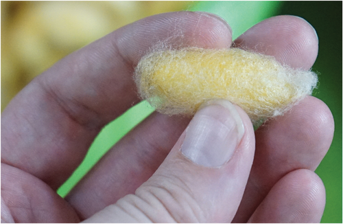
Natural silk is manufactured from the cocoons produced by silkworms (Figure 1). While there are various silkworm species, the most important is the caterpillar of the silk moth, Bombyx mori, which is bred for commercial silk production 13. Silk is not only used for textiles such as clothing and furniture covers but also for bed fillings or in cosmetic products (e.g., hair care) 9, 44. Silk threads consist of a double strand of the protein fibroin, linked by the glycoprotein sericin 13.

Especially workers in the silk industry may develop immediate-type hypersensitivity to silk, including allergic asthma, allergic rhinitis or allergic conjunctivitis 13. This is attributed to the silk fibers themselves, but also to caterpillar scales and excretions 14. Hypersensitivity reactions have likewise been reported following non-occupational exposure. Here, pillows filled with silk remnants may cause nocturnal asthma attacks 13, 45. Cases of contact urticaria caused by silk have also been described, with wet silk being particularly problematic. A possible reason for this might be the fact that sericin, the allergenic protein, is water soluble. To date, there has only been one report of an allergic reaction to fibroin 46. In rare cases, sericin may also cause allergic contact dermatitis 47.
Ingesting silkworm pupae (as practiced quite commonly in Asia) may cause food allergic reactions. In this context, various allergens have been characterized on a molecular level, e.g., Bomb m1, an arginine kinase with cross-reactivity to cockroaches, as well as a tropomyosin with cross-reactivity to cockroaches and HDMs 14.
Acknowledgements
Open access funding enabled and organized by Projekt DEAL. [Correction added on 11 November 2022, after first online publication: Projekt DEAL funding statement has been added.]
Conflict of interest
None.


