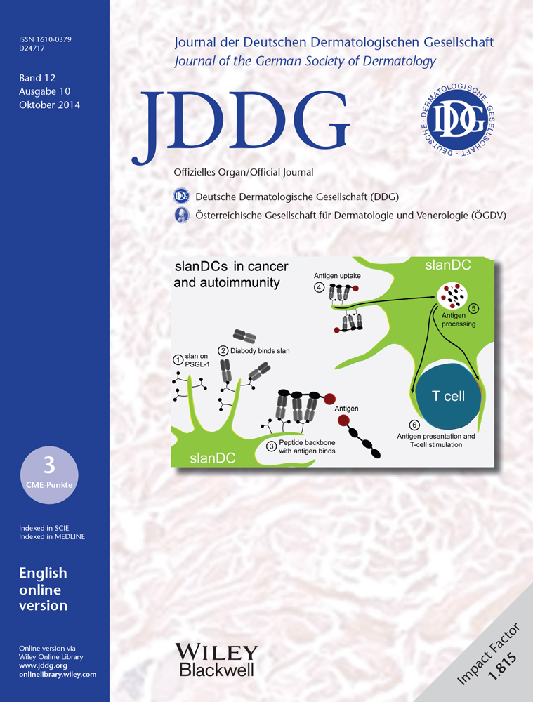
The role of human 6-sulfo LacNAc dendritic cells (slanDCs) in autoimmunity and tumor diseases
Summary
Dendritic cells play a central role in the regulation of immunological reactivity. The existence of functionally specialized populations of dendritic cells in the skin is a consequence of qualitatively different attacks on our organism. slanDCs are human inflammatory dendritic cells that are characterized by the specific expression of the carbohydrate 6-sulfo LacNAc (slan). After phenotypic maturation, slanDCs are capable of producing very high amounts of proinflammatory mediators such as IL-12, TNF-α, IL-1β and IL-23. Recent data describe a potential role of slanDCs in a number of different diseases like psoriasis, lupus erythematosus, and tumors, thus opening up new areas of research on their respective pathogenesis. Furthermore, a slanDC-specific targeting system has been developed as a basis for direct therapeutic manipulation. Future challenges of slanDC research include deepening our understanding of the significance of slanDCs in the regulation of adaptive and innate immune responses, as well as translating this knowledge into therapeutic options.
Introduction
In recent years, our knowledge of dendritic cells (DCs) in humans has expanded rapidly. Thereby, the system of DCs in the skin has proved to be a complex and dynamic component of the cutaneous immune system. While it was previously assumed that DCs in the skin were solely represented by Langerhans cells of the epidermis, studies have since shown that the dermis also contains a dense system of different DCs and related cells, as well as macrophages, some of which are difficult to distinguish phenotypically. Complicating this further, recent mouse studies have shown that the Langerhans cell network in the epidermis is established independently of hematopoietic stem cells and is derived from fetal precursor cells, which renew themselves locally through proliferation 1, 2. Thus, Langerhans cells are not conventional DCs (i.e., derived from hematopoietic stem cells in the bone marrow), at least ontogenetically, even if they share functional characteristics with DCs 3, 4.
There are two sub-types of conventional DCs in human blood which are derived from hematopoietic stem cells in the bone marrow: CD1c+ DCs (mDC1) and CD141+ DCs (mDC2), both of which can also be found in the skin 5. Besides these, the blood contains plasmacytoid dendritic cells (pDC), which are rarely found in healthy skin but may be recruited during inflammation 6. The skin additionally contains phenotypically heterogeneous CD14+ cells, which are probably derived from monocytes. Under inflammatory conditions these cells also display a highly proinflammatory phenotype 7. While Langerhans cells are largely limited to the epidermis, DCs, including slanDCs, are mainly located in the upper dermis (0–20 μm below the dermoepidermal junction zone). In addition, the deeper dermis harbours macrophages (40–60 μm below the dermoepidermal junction zone), the majority of which seem to be associated with vessels 8.
slanDCs are human inflammatory DCs which can be identified and characterized by their specific expression of the carbohydrate modification 6-sulfo N-acetyllactosamine (6-sulfo LacNAc, slan) 9-11. Slan is a terminal, O-glycosidic bound, sulfated disaccharide on PSGL-1, an important adhesion molecule on immune cells 10. Unlike slanDCs, CD1c+ DCs and pDCs express cutaneous lymphocyte antigen (CLA), which results in a specific binding of selectins. slanDCs in blood exhibit an immature phenotype. However, after isolation they mature spontaneously into professional antigen-presenting cells with the ability to stimulate naïve T cells. As they mature, slanDCs develop a phenotype of inflammatory DCs, which is characterized by upregulation of typical maturation markers (CD40, CD80, CD83, CD86, HLA-DR) and a marked stimulation by pathogen-associated molecular patterns (PAMPs) 10, 12. As a result, stimulation of slanDCs by various TLR ligands leads to production of large quantities of pro-inflammatory cytokines such as IL-12, TNF-α, IL-6, IL-β, and IL-23 11, 12. In co-cultures, this cytokine profile causes programming of Th1 and Th17 T cells, sub-types of effector T cells which are relevant to various autoimmune diseases such as psoriasis, rheumatoid arthritis, and lupus erythematosus.
In the following, we discuss recent data which highlight a potential role of slanDCs in chronic inflammatory diseases and autoimmune diseases of the skin. We then turn to a few recently discovered aspects of DC biology.
Psoriasis
Compared to other autoimmune skin disorders, psoriasis has been relatively well researched. The causes of disease are complex – and stem from environmental and genetic factors, the importance of which can vary from person to person – but at least the general cellular mechanisms in the development of psoriatic plaques are comparatively well understood. The characteristic lesions of psoriasis seem to be mediated by interactions between DCs, T cells, and keratinocytes. In early lesions, the production of IFN-α by pDCs, as well as IL-12 and IL-23 by mDCs, is important. In chronic lesions, the production of IL-12 and IL-23 by mDCs and inflammatory DCs, as well as the activity of Th1, Th17, and Th22 T cells play an important role 13. Thereby, inflammatory DCs were originally defined by their expression of the DC marker CD11c and the absence of the markers for CD1c+ DCs and CD141+ DCs. Among the closely studied inflammatory DC populations are TNF- and iNOS-producing DCs (TIP-DCs) 14, which partly overlap with slanDCs in their characteristic expression of TNF-α and iNOS.
slanDCs are found in small amounts in healthy skin 15. Yet, abundant slanDCs are found in the lesional skin of psoriasis patients, where they are mainly located in the papillary dermis, a typical site for DCs 11, 12. slanDCs in psoriatic plaques exhibit an activated phenotype and are largely positive for IL-23, TNF-α, and iNOS 12. The phenotype of slanDCs suggests a contribution in sustaining the chronic inflammation. In vitro experiments with isolated slanDCs have confirmed their ability to produce pro-inflammatory cytokines (IL-1β, IL-23, IL-12, and IL-6), with the potential for programming Th1 and Th17 T cells. slanDC/T cell co-cultures have also shown that T cells induced by slanDCs may be characterized as Th17/Th1 T cells, which demonstrate active production of IL-17, IL-22, TNF-α, and IFN-γ. Thus, both the cytokine profile of slanDCs, as well as that of slanDC-stimulated effector T cells, point to a pathogenetic relevance of slanDCs in sustaining chronic plaques (Figure 1). Treatment of psoriasis with etanercept, a TNF-α antagonist, can strongly reduce the number of slanDCs in healing lesions 16. The few slanDCs that remain in the skin continue to express TNF-α and IL-23 and may be responsible for a rapid relapse after the drug is discontinued 17.

The importance of slanDCs in the initiation of inflammatory lesions cannot yet be evaluated. Yet it is notable that, in the presence of the antimicrobial peptide LL37 (cathelicidin) and RNA, slanDCs are effectively stimulated to produce TNF-α. Complexes made up of LL37 and nucleic acids have been shown to be associated with the development of psoriatic plaques 18, 19. It is therefore possible that slanDCs contribute to the development as well as the maintenance of the plaques. Given that along with myeloid DCs, pDCs in particular appear to be important in the development of psoriatic plaques, it would be interesting to study the effect of factors related to pDCs on slanDCs. Thereby, the ability of pDCs to produce large amounts of type I interferons has to be especially considered (Figure 1).
Several studies have shown that slanDCs interact with neutrophilic granulocytes and natural killer (NK) cells and that this considerably enhances their pro-inflammatory function 20-22. Both contact-dependent (CD18/ICAM-1) and cytokine-mediated (IL-12, IFN-γ) signals are involved in this positive feedback loop. In vivo studies have found co-localization of slanDCs, neutrophilic granulocytes, and NK cells in the inflammatory infiltrate in psoriasis and Crohn's disease. Given that neutrophilic granulocytes may play a so far under-appreciated auto-inflammatory role in the early phase of psoriasis 23, future studies should also take into account the interactions of these cells in psoriasis.
Lupus erythematosus
Lupus erythematosus (LE) refers to a group of cutaneous and systemic autoimmune diseases which are associated with the formation of autoantibodies against various nuclear antigens. Autoantigens in LE may be DNA itself, nuclear proteins, or RNA-containing ribonucleoproteins. A typical feature of LE is the deposition of autoimmune complexes in tissues and the vascular bed. These immune complexes stimulate Fc receptor-bearing immune cells and contribute to the disease pathogenesis. pDCs play an important role in the development of, mediated by their production of IFN-α after detection of nucleic acids or nucleic acid-containing immune complexes 24. A potential role of Th1 and Th17 T cells has also been described, which points to the activity of myeloid DCs 25.
An increased frequency of slanDCs has been detected in various sub-types of LE (chronic discoid LE, subacute cutaneous LE, systemic LE). Thereby, slanDCs are not only scattered throughout the dermis, but are often associated with lymph follicle-like structures, within which they co-localize with T cells, but not B cells. As a sign of their activation, slanDCs were shown to express TNF-α in these lesions 26.
slanDCs have at least two phenotypical features which could have a functional significance in the pathogenesis of LE. Unlike other DCs, slanDCs express two receptors for IgG immune complexes (CD16/FcγRIII and CD32/FcγRII), and thus have a high capacity for binding immune complexes. Immune complexes that are bound by slanDCs are internalized, processed, and antigenic peptides are presented on MHC molecules which provides stimulation of antigen-specific T cells 27. In addition, slanDCs, unlike CD1c+ DCs and pDCs, exhibit co-expression of TLR7 and TLR8, and thus express two endosomal pattern-recognition receptors for single-stranded RNA. Both receptors are functional, and the stimulation of both receptors leads to effective production of TNF-α and IL-12 26. In line with this, serum taken from LE patients, which contains nucleic acid-containing immune complexes as stimulatory components 28, 29, can induce TNF-α production by slanDCs, while serum from healthy subjects cannot 26.
In LE, as in psoriasis, a presence of an elevated concentration of the cationic, antimicrobial peptide LL37 (cathelicidin) has been described. LL37 can bind nucleic acids and thereby stabilizing them. In higher concentrations, LL37 may promote the uptake of nucleic acids by the body's own cells. It is interesting to note here that slanDCs, compared to CD1c+ DCs, produce much higher amounts of TNF-α after stimulation with RNA and LL37 12.
Tumors
DCs are also found in the cellular milieu of tumors. However, their proper function seems to be interfered with by the suppressive tumor microenvironment. This suppression is believed to partially explain the absence of an effective tumor-specific immune response. A better understanding of tumor-associated DCs is therefore important for discovering novel ways of breaking the tumor-induced immunological tolerance 30.
In a recently published paper, slanDCs were connected with a new form of tumor immunosurveillance or tumor immunoediting in carcinoma 31. The study examined various primary tumors as well as metastatic, draining lymph nodes. Primary tumors were only rarely associated with slanDCs, while metastatic, draining lymph nodes demonstrated recruitment of slanDCs along the metastatic tumor tissue. An analysis of metastatic and non-metastatic sentinel lymph nodes of breast cancer patients has shown that slanDCs are only recruited after the tumor cells have arrived in the lymph node. In metastatic lymph nodes, other DCs seem to maintain their specific intranodal location despite an increased cell number. Yet slanDCs were recruited very close to the tumor cells, and were therefore located at the intersection between tumor cells and T lymphocytes. slanDCs had contact with apoptotic tumor cells, and thus were optimally located to present potential tumor antigens to T cells. In vitro studies have shown that slanDCs can induce tumor-specific cytotoxic T cells 9. However, inside the lymph nodes, the T cells were mostly positive for GATA-2 and thus were probably Th2-polarized, which was interpreted as an escape mechanism of the tumor. If one considers that slanDCs taken from the blood of carcinoma patients are unlike other DCs fully functional, then slanDCs emerge as a novel cell type for targeted tumor therapies. Even independent of their ability to present antigen, various studies have shown immune reactivity of slanDCs against tumor cells: effective antibody-dependent cell-mediated cytotoxicity (ADCC) 32, direct cytotoxicity against tumor cells 33, and a marked stimulation of tumor-targeted NK-cell mediated cytotoxicity 22, 34. Interestingly, a co-localization of slanDCs with NK cells and neutrophilic granulocytes has been observed in metastatic lymph nodes.
Therapeutic applications
There are currently no approved targeted therapies for human DCs. An experimental modular targeting system for slanDCs, consisting of two components of recombinant proteins, has been developed by M. Bachmann (Dresden, Germany) (Figure 2). One of the components is a bispecific diabody (scBsDb) with a specificity for the slan epitope, and another specificity for the second component of the system, a peptide backbone. This peptide backbone has several binding sites for the diabody, and thus multivalent binding leads to increased avidity of the interaction. Along with the binding sites for the diabody, the peptide can bear antigen. If both components of the targeting system and slanDCs are present, slanDCs are specifically loaded with the antigen. To test the functioning of this system a peptide backbone with a tetanus toxin peptide was applied. slanDCs were pre-incubated with the components of the targeting system, washed, and then cultivated with autologous T cells. In the co-culture, the targeting of the slanDCs with a relatively low concentration of antigens thus led to efficient reactivation of tetanus toxin-specific memory T cells 35.

The potential of such targeting systems is clear: they would be suitable for novel vaccination strategies against infectious disease and for experimental tumor therapies. Another possibility would be the coupling of chemotherapeutics or other drugs to the peptide backbone, which would achieve the specific manipulation of slanDCs.
Conclusions
In the conditions investigated so far slanDCs manifest as highly proinflammatory subset of inflammatory DCs. This proinflammatory character and additional functional properties suggest a pathogenetic relevance of slanDCs for diseases like psoriasis and lupus erythematosus. However, it is highly probable that the recruitment of slanDCs as inflammatory DCs is not limited to the diseases described here. Other examples of the presence of slanDCs are inflammatory tonsils 31, 36, the ileum of patients with Crohn's disease 36, the inflammatory synovia of patients with rheumatoid arthritis 11, and intestinal graft-versus-host disease 37.
Along with the role of slanDCs in autoimmune diseases, the field of research on slanDC biology has been growing steadily. Newer findings include the potential role of slanDCs in immunosurveillance or immuno editing in metastatic carcinomas 31. slanDCs also demonstrate a specific reaction pattern in certain viral diseases, as has been described for HIV 38. Finally, through the co-expression of two immune complex receptors, slanDCs appear to exhibit a specialization for the detection and processing of immune complexes 27. With the advancement of techniques for the specific targeting of slanDCs 35, the future will eventually see a translation of the knowledge into slanDC-specific therapies.
Financing
This work was supported by the Collaborative Research Center (Sonderforschungsbereich) 938 (T.D. and K.S.).
Conflict of interest
None.


