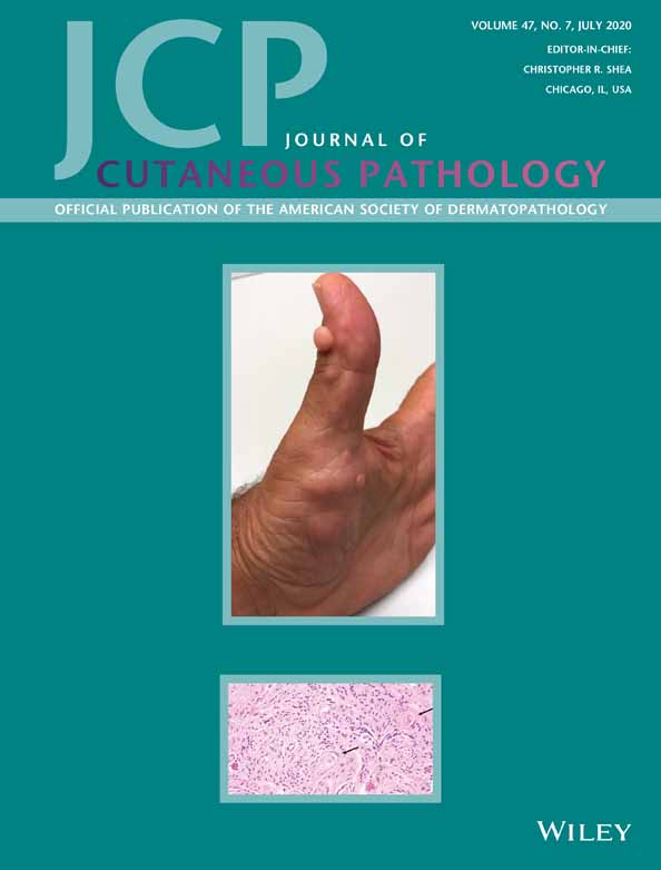
Scleromyxedema histopathologically mimicking hypercellular fibrous papules (angiofibomas): Case report of an unusual histopathological presentation
Abstract
Scleromyxedema (SMX) is an inflammatory condition of unknown etiology strongly associated with monoclonal gammopathy. Classical histopathology of SMX is characterized with the triad of diffuse mucin deposits, increased amount of collagen, and presence of stellate fibroblasts. Herein, we report an unusual histopathological variant of SMX in a 41-year-old female with lesions of the nose histopathologically mimicking cellular angiofibromas. The dome-shaped papules were characterized by increased collagen bundles and fascicles of spindle cells. Widened vessels were seen at the periphery of the proliferation. Cells expressed CD68. Factor XIIIa was expressed only by dendritic cells. The mucin was highlighted with colloidal iron. In sum, we draw attention to this unusual variant of SMX, which should be suspected in a setting of multiple “angiofibromas/fibrous papules” on the face with presence of mucin.
CONFLICT OF INTEREST
None.


