PLGA+HA/βTCP scaffolds embedding simvastatin induced mineral deposition by mesenchymal stem cells
15576 POSTER DISPLAY BASIC RESEARCH
Background
The search for bone substitutes that might promote osteoconduction, osteoinduction, and osteogenesis, has fomented research on tissue engineering. Studies based on the association of new approaches, including scaffolds, biological inducers, and mesenchymal stem cells, have shown promising results.
Aim/Hypothesis
The aim of this study was to evaluate the deposition of mineralization nodules in the extracellular matrix induced by simvastatin (SIM) embedded in scaffolds of poly(lactic-co-glycolic) acid (PLGA) and biphasic ceramic composed of hydroxyapatite (HA) and β-tricalcium phosphate (βTCP).
Materials and Methods
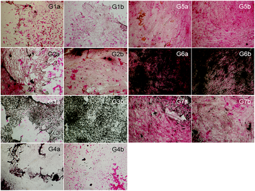
Samples were obtained by the solvent evaporation technique and leaching of sucrose particles. Biphasic ceramic particles (70% HA and 30% βTCP) were added to the PLGA in 1:1 ratio. Samples with simvastatin received 1% (w:w) of this medication. Samples covered with 10% collagen (COL) were included in the study to improve wettability of scaffolds. Samples were sterilized by ethylene oxide. Four experimental groups: G1) PLGA+HA/βTCP, G2) PLGA+HA/βTCP+COL, G3) PLGA+HA/βTCP+SIM, G4) PLGA+HA/βTCP+COL+SIM, with stem cells from human exfoliated deciduous teeth (SHED) in non-osteogenic medium, and three control groups: G5) SHED in non-osteogenic medium; G6) SHED in osteogenic medium; and G7) MC3T3-E1 in osteogenic medium, were determined. Von Kossa staining was performed after 21 days of cell culture to assess the capacity of scaffolds to induce mineral deposition by SHED. After staining, samples were taken to optical microscope and two images of different fields were captured.
Results
G6 visibly presented the highest amount of mineralization nodules followed by G3 and G7. G5 presented a high density of eosin-colored cells, but just a few points of mineralization. G1, G2, and G4 showed a variable distribution of both eosin-colored cells and mineralization nodules. These results are presented in Figure 1.
Conclusion and Clinical Implications
PLGA+HA/βTCP+SIM scaffolds were the most beneficial among experimental groups regarding deposition of mineralization nodules in the extracellular matrix by SHED. This signalizes that SIM embedded in scaffolds of PLGA+HA/βTCP might be a promising biomaterial for tissue engineering, comprehending osteoconduction, osteoinduction, and osteogenesis. Further analyses are suggested to confirm the osteoinductive potential of these scaffolds to be used as bone substitutes for clinical applications.


