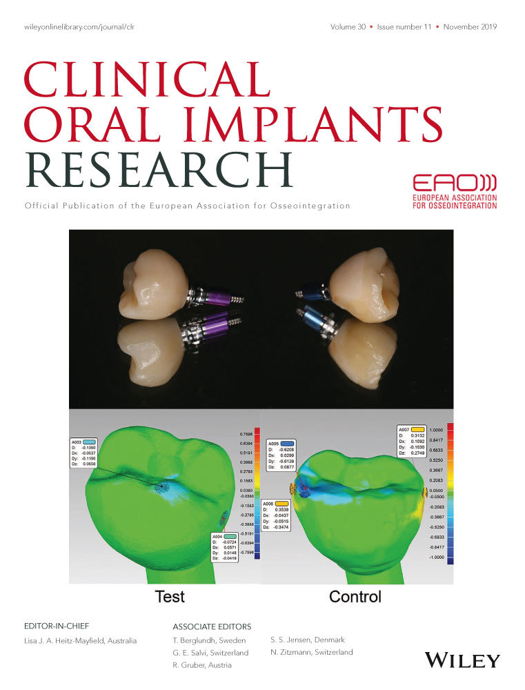
Quantitative clinical adjustment analysis of posterior single implant crown in a chairside digital workflow: A randomized controlled trial
Yifan Zhang
Department of Oral Implantology, Peking University School and Hospital of Stomatology & National Clinical Research Center for Oral Disease & National Engineering Laboratory for Digital and Material Technology of Stomatology & Beijing Key Laboratory of Digital Stomatology, Beijing, China
Search for more papers by this authorJiehua Tian
Department of Oral Implantology, Peking University School and Hospital of Stomatology & National Clinical Research Center for Oral Disease & National Engineering Laboratory for Digital and Material Technology of Stomatology & Beijing Key Laboratory of Digital Stomatology, Beijing, China
Search for more papers by this authorDonghao Wei
Department of Oral Implantology, Peking University School and Hospital of Stomatology & National Clinical Research Center for Oral Disease & National Engineering Laboratory for Digital and Material Technology of Stomatology & Beijing Key Laboratory of Digital Stomatology, Beijing, China
Search for more papers by this authorCorresponding Author
Ping Di
Department of Oral Implantology, Peking University School and Hospital of Stomatology & National Clinical Research Center for Oral Disease & National Engineering Laboratory for Digital and Material Technology of Stomatology & Beijing Key Laboratory of Digital Stomatology, Beijing, China
Correspondence
Ping Di and Ye Lin, Department of Oral Implantology, Peking University School and Hospital of Stomatology & National Clinical Research Center for Oral Disease & National Engineering Laboratory for Digital and Material Technology of Stomatology & Beijing Key Laboratory of Digital Stomatology, 22 Zhongguancun South Avenue, Haidian District, Beijing 10081, China.
Emails: [email protected] (PD); [email protected] (YL)
Search for more papers by this authorCorresponding Author
Ye Lin
Department of Oral Implantology, Peking University School and Hospital of Stomatology & National Clinical Research Center for Oral Disease & National Engineering Laboratory for Digital and Material Technology of Stomatology & Beijing Key Laboratory of Digital Stomatology, Beijing, China
Correspondence
Ping Di and Ye Lin, Department of Oral Implantology, Peking University School and Hospital of Stomatology & National Clinical Research Center for Oral Disease & National Engineering Laboratory for Digital and Material Technology of Stomatology & Beijing Key Laboratory of Digital Stomatology, 22 Zhongguancun South Avenue, Haidian District, Beijing 10081, China.
Emails: [email protected] (PD); [email protected] (YL)
Search for more papers by this authorYifan Zhang
Department of Oral Implantology, Peking University School and Hospital of Stomatology & National Clinical Research Center for Oral Disease & National Engineering Laboratory for Digital and Material Technology of Stomatology & Beijing Key Laboratory of Digital Stomatology, Beijing, China
Search for more papers by this authorJiehua Tian
Department of Oral Implantology, Peking University School and Hospital of Stomatology & National Clinical Research Center for Oral Disease & National Engineering Laboratory for Digital and Material Technology of Stomatology & Beijing Key Laboratory of Digital Stomatology, Beijing, China
Search for more papers by this authorDonghao Wei
Department of Oral Implantology, Peking University School and Hospital of Stomatology & National Clinical Research Center for Oral Disease & National Engineering Laboratory for Digital and Material Technology of Stomatology & Beijing Key Laboratory of Digital Stomatology, Beijing, China
Search for more papers by this authorCorresponding Author
Ping Di
Department of Oral Implantology, Peking University School and Hospital of Stomatology & National Clinical Research Center for Oral Disease & National Engineering Laboratory for Digital and Material Technology of Stomatology & Beijing Key Laboratory of Digital Stomatology, Beijing, China
Correspondence
Ping Di and Ye Lin, Department of Oral Implantology, Peking University School and Hospital of Stomatology & National Clinical Research Center for Oral Disease & National Engineering Laboratory for Digital and Material Technology of Stomatology & Beijing Key Laboratory of Digital Stomatology, 22 Zhongguancun South Avenue, Haidian District, Beijing 10081, China.
Emails: [email protected] (PD); [email protected] (YL)
Search for more papers by this authorCorresponding Author
Ye Lin
Department of Oral Implantology, Peking University School and Hospital of Stomatology & National Clinical Research Center for Oral Disease & National Engineering Laboratory for Digital and Material Technology of Stomatology & Beijing Key Laboratory of Digital Stomatology, Beijing, China
Correspondence
Ping Di and Ye Lin, Department of Oral Implantology, Peking University School and Hospital of Stomatology & National Clinical Research Center for Oral Disease & National Engineering Laboratory for Digital and Material Technology of Stomatology & Beijing Key Laboratory of Digital Stomatology, 22 Zhongguancun South Avenue, Haidian District, Beijing 10081, China.
Emails: [email protected] (PD); [email protected] (YL)
Search for more papers by this authorAbstract
Objectives
To compare the three-dimensional changes in quantity and morphology following clinical adjustment of a posterior single implant crown between chairside digital workflow (test) and hybrid digital workflow (control).
Materials and Methods
A total of 33 participants were included for single-tooth replacement with screw-retained crowns in posterior sites of either the maxillary or mandible. A total of 17 participants were carried to a chairside digital workflow, receiving monolithic lithium disilicate (LS2)-crowns (test), while the remaining 16 participants were fitted with CAD/CAM-fabricated zirconia superstructures and hand-layered ceramic veneering crowns (control). As each crown underwent intraoral scanning (3Shape TRIOS Color, 3Shape), 3D digital models were rendered. These scans were taken both before and after try-in. Clinical adjustment dimensional changes were measured by superimposing the optical scans of models within a reverse software (Geomagic Control 2014). Adjustment counts and amounts (from vertical dimension) between two workflows were assessed and compared. Time consumption was recorded for efficiency analysis.
Results
All patients were successfully treated in both groups. The median maximum vertical adjustment (taking both occlusal and interproximal surfaces into consideration) was 237 μm ± 112 in the test group and 485 μm ± 195 in the control group (p < .0001), respectively. The median adjustment count was 2.00 ± 1.09 in test group and 3.00 ± 1.05 in control group (p = .001), respectively. The total active working time/ total time for two workflows was 92.3/113.7 min for the test group and 146.3/676.3 min for the control group, respectively.
Conclusion
The test group showed fewer adjustments and apparent precision on the occlusal surface compared with the control group with only a fifth of the consumption of a hybrid workflow.
CONFLICT OF INTEREST
The authors declare no potential conflict of interests with respect to the authorship and/or publication of this article.
REFERENCES
- Al-Bakri, I. A., Hussey, D., & Al-Omari, W. M. (2007). The dimensional accuracy of four impression techniques with the use of addition silicone impression materials. The Journal of Clinical Dentistry, 18(2), 29–33.
- Deferm, J. T., Schreurs, R., Baan, F., Bruggink, R., Merkx, M. A. W., Xi, T., … Maal, T. J. J. (2017). Validation of 3D documentation of palatal soft tissue shape, color, and irregularity with intraoral scanning. Clinical Oral Investigations, 22(3), 1303–1309. https://doi.org/10.1007/s00784-017-2198-8
- Di Fiore, A., Vigolo, P., Graiff, L., & Stellini, E. (2018). Digital vs Conventional workflow for screw-retained single-implant crowns: A comparison of key considerations. The International Journal of Prosthodontics, 31(6), 577–579. https://doi.org/10.11607/ijp.5938
- Ender, A., Attin, T., & Mehl, A. (2016). In vivo precision of conventional and digital methods of obtaining complete-arch dental impressions. Journal of Prosthetic Dentistry, 115(3), 313–320. https://doi.org/10.1016/j.prosdent.2015.09.011
- Ender, A., & Mehl, A. (2013). Accuracy of complete-arch dental impressions: A new method of measuring trueness and precision. Journal of Prosthetic Dentistry, 109(2), 121–128. https://doi.org/10.1016/S0022-3913(13)60028-1
- Ender, A., & Mehl, A. (2014). Accuracy in dental medicine, a new way to measure trueness and precision. Journal of Visualized Experiments, (86). e51374. https://doi.org/10.3791/51374
- Ender, A., Zimmermann, M., Attin, T., & Mehl, A. (2016). In vivo precision of conventional and digital methods for obtaining quadrant dental impressions. Clinical Oral Investigations, 20(7), 1495–1504. https://doi.org/10.1007/s00784-015-1641-y
- Gan, N., Xiong, Y., & Jiao, T. (2016). Accuracy of intraoral digital impressions for whole upper jaws, including full dentitions and palatal soft tissues. PLoS ONE, 11(7), e0158800. https://doi.org/10.1371/journal.pone.0158800
- Guth, J. F., Keul, C., Stimmelmayr, M., Beuer, F., & Edelhoff, D. (2013). Accuracy of digital models obtained by direct and indirect data capturing. Clinical Oral Investigations, 17(4), 1201–1208. https://doi.org/10.1007/s00784-012-0795-0
- Joda, T., & Bragger, U. (2014). Complete digital workflow for the production of implant-supported single-unit monolithic crowns. Clinical Oral Implants Research, 25(11), 1304–1306. https://doi.org/10.1111/clr.12270
- Joda, T., & Bragger, U. (2015a). Digital vs. conventional implant prosthetic workflows: A cost/time analysis. Clinical Oral Implants Research, 26(12), 1430–1435. https://doi.org/10.1111/clr.12476
- Joda, T., & Bragger, U. (2015b). Time-efficiency analysis comparing digital and conventional workflows for implant crowns: A prospective clinical crossover trial. International Journal of Oral and Maxillofacial Implants, 30(5), 1047–1053. https://doi.org/10.11607/jomi.3963
- Joda, T., & Bragger, U. (2016a). Patient-centered outcomes comparing digital and conventional implant impression procedures: A randomized crossover trial. Clinical Oral Implants Research, 27(12), e185–e189. https://doi.org/10.1111/clr.12600
- Joda, T., & Bragger, U. (2016b). Time-efficiency analysis of the treatment with monolithic implant crowns in a digital workflow: A randomized controlled trial. Clinical Oral Implants Research, 27(11), 1401–1406. https://doi.org/10.1111/clr.12753
- Joda, T., Ferrari, M., & Bragger, U. (2017a). Monolithic implant-supported lithium disilicate (LS2) crowns in a complete digital workflow: A prospective clinical trial with a 2-year follow-up. Clinical Implant Dentistry and Related Research, 19(3), 505–511. https://doi.org/10.1111/cid.12472
- Joda, T., Ferrari, M., & Bragger, U. (2017b). A prospective clinical cohort study analyzing single-unit implant crowns after three years of loading: Introduction of a novel functional implant prosthodontic score (FIPS). Clinical Oral Implants Research, 28(10), 1291–1295. https://doi.org/10.1111/clr.12955
- Joda, T., Ferrari, M., Bragger, U., & Zitzmann, N. U. (2018). Patient reported outcome measures (PROMs) of posterior single-implant crowns using digital workflows: A randomized controlled trial with a three-year follow-up. Clinical Oral Implants Research, 29(9), 954–961. https://doi.org/10.1111/clr.13360
- Joda, T., Katsoulis, J., & Bragger, U. (2016). Clinical fitting and adjustment time for implant-supported crowns comparing digital and conventional workflows. Clinical Implant Dentistry and Related Research, 18(5), 946–954. https://doi.org/10.1111/cid.12377
- Jung, R. E., Zembic, A., Pjetursson, B. E., Zwahlen, M., & Thoma, D. S. (2012). Systematic review of the survival rate and the incidence of biological, technical, and aesthetic complications of single crowns on implants reported in longitudinal studies with a mean follow-up of 5 years. Clinical Oral Implants Research, 23(Suppl 6), 2–21. https://doi.org/10.1111/j.1600-0501.2012.02547.x
- Kapos, T., & Evans, C. (2014). CAD/CAM technology for implant abutments, crowns, and superstructures. International Journal of Oral and Maxillofacial Implants, 29(Suppl), 117–136. https://doi.org/10.11607/jomi.2014suppl.g2.3
- Kurbad, A. (2016). Final restoration of implants with a hybrid ceramic superstructure. International Journal of Computerized Dentistry, 19(3), 257–279.
- Lee, C. Y., Wong, N., Ganz, S. D., Mursic, J., & Suzuki, J. B. (2015). Use of an intraoral laser scanner during the prosthetic phase of implant dentistry: A pilot study. Journal of Oral Implantology, 41(4), e126–e132. https://doi.org/10.1563/AAID-JOI-D-13-00132
- Lee, S. J., Betensky, R. A., Gianneschi, G. E., & Gallucci, G. O. (2015). Accuracy of digital versus conventional implant impressions. Clinical Oral Implants Research, 26(6), 715–719. https://doi.org/10.1111/clr.12375
- Mangano, C., Mangano, F., Piattelli, A., Iezzi, G., Mangano, A., & La Colla, L. (2010). Prospective clinical evaluation of 307 single-tooth Morse taper-connection implants: A multicenter study. International Journal of Oral and Maxillofacial Implants, 25(2), 394–400.
- Mangano, F., & Veronesi, G. (2018). Digital versus analog procedures for the prosthetic restoration of single implants: A randomized controlled trial with 1 year of follow-up. BioMed Research International, 2018, 5325032. https://doi.org/10.1155/2018/5325032
- Mehl, A., Ender, A., Mormann, W., & Attin, T. (2009). Accuracy testing of a new intraoral 3D camera. International Journal of Computerized Dentistry, 12(1), 11–28.
- Mehl, A., Koch, R., Zaruba, M., & Ender, A. (2013). 3D monitoring and quality control using intraoral optical camera systems. International Journal of Computerized Dentistry, 16(1), 23–36.
- Muhlemann, S., Kraus, R. D., Hammerle, C. H. F., & Thoma, D. S. (2018). Is the use of digital technologies for the fabrication of implant-supported reconstructions more efficient and/or more effective than conventional techniques: A systematic review. Clinical Oral Implants Research, 29(Suppl 18), 184–195. https://doi.org/10.1111/clr.13300
- Schepke, U., Meijer, H. J., Kerdijk, W., & Cune, M. S. (2015). Digital versus Analog complete-arch impressions for single-unit premolar implant crowns: Operating time and patient preference. Journal of Prosthetic Dentistry, 114(3), 403–406 e401. https://doi.org/10.1016/j.prosdent.2015.04.003
- Spies, B. C., Pieralli, S., Vach, K., & Kohal, R. J. (2017). CAD/CAM-fabricated ceramic implant-supported single crowns made from lithium disilicate: Final results of a 5-year prospective cohort study. Clinical Implant Dentistry and Related Research, 19(5), 876–883. https://doi.org/10.1111/cid.12508
- van der Zande, M. M., Gorter, R. C., & Wismeijer, D. (2013). Dental practitioners and a digital future: An initial exploration of barriers and incentives to adopting digital technologies. British Dental Journal, 215(11), E21. https://doi.org/10.1038/sj.bdj.2013.1146
- Windisch, S. I., Jung, R. E., Sailer, I., Studer, S. P., Ender, A., & Hammerle, C. H. (2007). A new optical method to evaluate three-dimensional volume changes of alveolar contours: A methodological in vitro study. Clinical Oral Implants Research, 18(5), 545–551. https://doi.org/10.1111/j.1600-0501.2007.01382.x
- Wismeijer, D., Bragger, U., Evans, C., Kapos, T., Kelly, R., Millen, C., … Taylor, T. (2014). Consensus statements and recommended clinical procedures regarding restorative materials and techniques for implant dentistry. International Journal of Oral and Maxillofacial Implants, 29(Suppl), 137–140. https://doi.org/10.11607/jomi.2013.g2
- Wismeijer, D., Mans, R., van Genuchten, M., & Reijers, H. A. (2014). Patients' preferences when comparing analogue implant impressions using a polyether impression material versus digital impressions (Intraoral Scan) of dental implants. Clinical Oral Implants Research, 25(10), 1113–1118. https://doi.org/10.1111/clr.12234
- Zaruba, M., & Mehl, A. (2017). Chairside systems: A current review. International Journal of Computerized Dentistry, 20(2), 123–149.


