Novel and efficient method for culturing patient-derived gastric cancer stem cells
Abstract
Experimental techniques for patient-derived cancer stem-cell organoids/spheroids can be powerful diagnostic tools for personalized chemotherapy. However, establishing their cultures from gastric cancer remains challenging due to low culture efficiency and cumbersome methods. To propagate gastric cancer cells as highly proliferative stem-cell spheroids in vitro, we initially used a similar method to that for colorectal cancer stem cells, which, unfortunately, resulted in a low success rate (25%, 18 of 71 cases). We scrutinized the protocol and found that the unsuccessful cases were largely caused by the paucity of cancer stem cells in the sampled tissues as well as insufficient culture media. To overcome these obstacles, we extensively revised our sample collection protocol and culture conditions. We then investigated the following second cohort and, consequently, achieved a significantly higher success rate (88%, 29 of 33 cases). One of the key improvements included new sampling procedures for tumor tissues from wider and deeper areas of gastric cancer specimens, which allowed securing cancer stem cells more reproducibly. Additionally, we embedded tumor epithelial pieces separately in both Matrigel and collagen type-I as their preference to the extracellular matrix was different depending on the tumors. We also added a low concentration of Wnt ligands to the culture, which helped the growth of occasional Wnt-responsive gastric cancer stem-cell spheroids without allowing proliferation of the normal gastric epithelial stem cells. This newly improved spheroid culture method may facilitate further studies, including personalized drug-sensitivity tests prior to drug therapy.
Abbreviations
-
- CI
-
- confidence interval
-
- CM
-
- conditioned medium
-
- CRC
-
- colorectal cancer
-
- EGF
-
- epidermal growth factor
-
- GC
-
- gastric cancer
-
- GEI
-
- growth effect index
-
- NGE-SC
-
- normal gastric epithelial stem cell
-
- PD
-
- patient-derived
-
- RNA-seq
-
- RNA sequencing
-
- SC
-
- stem cell
-
- WHO
-
- World Health Organization
1 INTRODUCTION
Gastric cancer (GC) is the fifth most common cancer in the world and fourth leading cause of cancer death even with significant improvements in surgical techniques and chemotherapy.1, 2 Histopathologically, GC comprises intestinal and diffuse types according to Lauren's classification,3 which are further subdivided according to the World Health Organization (WHO) classification.4 Recently, The Cancer Genome Atlas5 and Asian Cancer Research Group6 proposed molecular classifications based on the gene expression profiles. However, these classifications are of limited help in determining the most efficacious treatments, necessitating a personalized strategy. Currently, a few diagnostic markers are available to select suitable GC patients for treatment with therapeutic antibodies, such as those against HER27 and PD-1/PD-L1.8, 9 Since only a small proportion of patients can benefit from each therapy, more diagnostic tools are needed to stratify patients for current and upcoming therapies so that specific GC subpopulations can be effectively targeted.
Among possibly promising strategies for personalized cancer treatments, a more direct approach is to test the drug sensitivity of patient-derived (PD) cancer stem cells (SCs) in vitro and/or in mouse xenografts. Recently, testing PD cancer stem-cell organoids have become feasible as a clinically relevant tool for investigating personalized therapeutics,10, 11 as exemplified by those derived from colorectal cancer (CRC).12 When it comes to GC, however, the success rates for establishing GC-SC lines are substantially lower than those for CRC-SC, with cumbersome culture methods owing to various supplementary factors and selection drugs needed for specific subtypes of GC.13-20
Recently, we have reported an efficient method for culturing PD–CRC-SCs21 based on the method for normal intestinal epithelial stem cells.22-24 These cells embedded in Matrigel form nearly spherical structures, termed spheroids, that are comprised of nearly all mitotic stem/progenitor cells, in contrast to intestinal organoids with the budding structures that comprise mixed populations of mitotic and post-mitotic cells.25 In the present study, we have modified this conventional culture method for propagating PD–GC-SC spheroids so that we can apply it for personalized clinical diagnosis and treatment.
2 MATERIALS AND METHODS
2.1 Human samples
Tumor samples were collected from GC patients who underwent primary resections at the Kyoto University Hospital (KUHP, Kyoto, Japan) and Medical Research Institute Kitano Hospital (Osaka, Japan) from January 2016 to November 2022. Their diagnosis was confirmed through histopathological examinations by board-certified diagnostic pathologists.
2.2 L-WRN conditioned medium
The L-WRN cells expressing mouse Wnt3a, R-spondin 3, and Noggin were obtained from Dr. Thaddeus S. Stappenbeck (Cleveland Clinic). Conditioned medium (CM) from L-WRN cells was prepared according to a previous protocol.22 Quality control testing of L-WRN CM was conducted according to the validation procedures and guidelines reported previously.26 A commercial L-WRN CM was purchased from Sigma-Aldrich.
2.3 Spheroid culture of human gastric cancer and normal gastric epithelial cells
Immediately after surgical resection, the excised stomach by operation was opened longitudinally, wrapped in gauze moistened with saline to prevent drying, and kept at room temperature. Sample specimens were collected within 1 h after the resection operation. From each stomach, one to four tumor pieces (100–1000 mm3 each) and one to two pieces of normal mucosa (500–2000 mm3) were collected in separate 15-mL conical tubes containing 5–10 mL ice-cold washing medium (Table S1). Sample tubes were kept on ice during transportation to the laboratory, and the isolation of epithelial cells and preparation of stem cell culture were performed within 6 h after sample collection (i.e., 7 h after the resection operation) according to a step-by-step protocol.22 Specifically, the specimen pieces were minced in a 60-mm Petri dish, digested with 1–3 mL collagenase solution (Table S1) at 37°C for 40–60 min and dissociated by pipetting. Epithelial cell clusters were filtered through a 100-μm cell strainer (Corning), collected in a 1.5-mL tube, and resuspended in Matrigel (Corning) or collagen type-I matrix (Cellmatrix, Nitta Gelatin). The cell-matrix mixture was placed at the center of each well of the 12-well cell-culture plate (30 μL/well; TPP). After polymerization of matrix materials at 37°C, GC and normal gastric epithelial (NGE) cells were cultured with the cancer medium and eL-WRN medium (epidermal growth factor [EGF]-containing 50% L-WRN CM), respectively (Table S1). The medium was changed every other day. To passage, we collected Matrigel-embedded spheroids and treated them with 2.5 g/L trypsin solution (Nacalai Tesque) at 37°C for 2–5 min. Collagen type-I–embedded spheroids were treated with collagenase solution at 37°C for 30 min, followed by trypsinization. Spheroids were dissociated into small cell aggregates by pipetting, and they were resuspended in Matrigel or collagen type-I. Dilution (based on the volume of matrix materials) was adjusted to one to six times depending on the growth rate and spheroid density. It should be noted that too much trypsinization and pipetting caused poor cell survival when spheroids grew poorly in early passages. The spheroid culture was considered successful when spheroids were expanded to 12 wells of a 12-well cell-culture plate.
2.4 Growth monitoring in spheroid culture using a cell imager
To monitor cell growth, we resuspended trypsinized spheroids in Matrigel or collagen type-I at a density of approximately 150 cell aggregates/μL. Subsequently, 3 μL cell-matrix mixture was distributed in each well of the 96-well cell-culture plate (TPP). After polymerization of matrix materials, cells were cultured in 100 μL of media. High-resolution cell images were obtained using a cell imager (Cell3iMager duos, SCREEN) every 3–4 days (Figure S1A). The area of each spheroid in each well was outlined using image processing software (Figure S1B). The volume of each spheroid was estimated using the following formula: spheroid volume (μm3) = 4/3 × {[spheroid area (μm2)]3/π}1/2. The cell growth rate for each well was estimated as the proportion of total spheroid volume to that on initial measurement, and the growth effect index (GEI) was defined as the relative growth rate of an experimental group to that of its control group. At least three independent experiments were performed for each analysis.
2.5 Mutational analysis
The exonic regions of 409 cancer-related genes in GC-SC spheroids were sequenced using the Ion AmpliSeq Comprehensive Cancer Panel (Thermo Fisher), and the sequence alignment to the reference genome (hg19) and variant calling were performed at Macrogen Japan. We omitted the analyses of the primary tumors because we and others had shown homogeneity of driver-gene mutations in cancer and their stability during ex vivo culture.14, 27, 28 Detection of cancer-specific mutations was performed as we described previously with modifications.27 Specifically, polymorphic alleles were removed from the called variants using the VCFtools program (V.0.1.13)29 by referring to the GEM Japan Whole Genome Aggregation (GEM-J WGA) panel (https://togovar.biosciencedbc.jp/doc/datasets/gem_j_wga) or the profiles of NGE-SC spheroids from the same patients (when available). The selected variants were annotated using the ANNOVAR program,30 and polymorphic alleles were removed again by referring to the Human Genetic Variation Database.31, 32 Subsequently, they were filtered to select nonsynonymous, frameshift, and splicing mutations with more than 20% frequency. Variant calls that appeared in more than two lines were eliminated as false-positive except for those identified in the COSMIC database. Other erroneous mutations were eliminated by surveying their coverage tracks on the Integrative Genomics Viewer software (V.2.12.3, Broad Institute).
2.6 Mutation detection from RNA sequencing (RNA-seq) data
To save time and cost, we took advantage of our transcriptome analysis data that we completed in most GC-SC spheroid lines. Namely, mutations in cancer-related genes were determined by deducing from the sequences of the RNA-seq data. Spheroid RNA samples were purified using the NucleoSpin RNA II kit (Takara Bio), and RNA-seq analysis was performed at Macrogen Japan. The sequence alignment to the reference genome (hg19) and variant calling were performed using the Subio Platform software (V.1.24.5853, Subio). Cancer-specific mutations in the exonic regions of expressed genes were detected with the same workflow as for the cancer panel.
Additional Materials and Methods can be found in Appendix S1.
3 RESULTS
3.1 Improvement of patient-derived gastric cancer stem-cell spheroid culture efficiency using a revised protocol
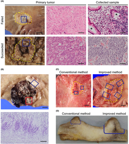
To culture GC-SC spheroids, we conducted two sets of experiments in which we collected tumor samples from 71 patients of the first cohort, followed by those from 33 patients of the second. To the first cohort samples, we applied our conventional method originally developed for CRC-SC spheroids (Table 1). Namely, we cultured tumor epithelial cells in a serum-containing cancer medium (Table S1) to propagate GC-SC spheroids.21 In contrast, NGE-SC spheroids were also established from normal mucosa of the same patients using the eL-WRN medium (Table S1) containing mouse Wnt3a, R-spondin 3, and Noggin.21, 22 The success rate for establishing GC-SC spheroids was 25% (18 of 71 cases; 95% CI, 15%–35%), whereas that for NGE-SC spheroids was 94% (67 of 71 cases; 95% CI, 89%–100%; Table 1; Table S2). To improve the low success rate, we revised our protocol in the following three points and tested its feasibility with fresh GC samples of the second patient cohort (Table 1). First and foremost, we scrutinized the sample collection maneuver from cancer tissues. One of the major reasons for our earlier failure in GC-SC spheroid establishment by our conventional method was likely the paucity of cancer stem cells in the sampled tumor pieces as estimated histopathologically in a retrospective manner (47% with 95% CI, 30%–64%; in 16 of the 34 failed cases; Figure 1A). Another minor cause was fungal contamination (9% with 95% CI, 2%–17%; in five of the 53 failed cases), particularly, of those samples from necrotic lesions that tended to accumulate fungi and/or hyphae (Figure 1B). Therefore, we collected more tumor pieces from wider and deeper areas, avoiding necrotic lesions to harvest cancer stem cells more reproducibly (Figure 1C,D). Importantly, the revised protocol reviewed by board-certified diagnostic pathologists of the collaborating hospitals did not affect pathological and molecular pathological assessment. Second, we embedded tumor epithelial pieces of each patient in both Matrigel and collagen type-I separately. This was because the different extracellular matrix (ECM) was preferred in some minority cases. Third, we added 5% L-WRN CM (containing Wnt ligands) to the cancer medium to help propagate Wnt-responsive GC-SCs, as the extent of dependence of GC-SC organoids on Wnt ligands has been variable.13, 33 Owing to these changes, we achieved a significantly higher success rate (88% with 95% CI, 77%–99%; 29 of 33 cases) as compared to that (25% with 95% CI, 15%–35%; 18 of 71 cases) with the first patient cohort (Table 1; Table S3). We failed in four of 33 cases because of heavy contamination with yeasts (two cases) or poor cell growth in early passages (two cases). Notably, five of 29 lines (17%) were established only when embedded in collagen type-I with a statistically significant difference (p = 0.008, Fisher's exact test), whereas three lines (10%) were only in Matrigel (Figure 2A). Regarding Wnt dependency, five GC-SC lines required L-WRN CM to maintain spheroid lines (Figure 2A). Our revised method also improved the culture efficiency in terms of the time needed for spheroid culture establishment, as the median time of the second cohort (21 days) was significantly shorter than that of the first cohort (33.5 days; Figure 2B).
| Our conventional method | Our improved method | Nanki et al.13 | Yan et al.14 | |
|---|---|---|---|---|
| Sampling method | ||||
| Site | Inside the tumor boundary | Both sides of the tumor boundary | NS | NS |
| Number of tissue pieces | 1–2 | 3–4 | NS | NS |
| Area (mm2)/Depth (mm) | 50–150/2–3 | 100–200/3–5 | NS | NS |
| Matrix material | Matrigel | Matrigel and collagen-I, separately | Matrigel | Matrigel |
| Medium composition | ||||
| Growth factor | EGF, FGF2, FBS | EGF, FGF2, FBS | EGF, FGF10 | EGF, FGF10, FBS (as CM) |
| Stem cell niche factor | – | L-WRN CM | Afamin-Wnt3a CM, RSPO1, Noggin | Wnt3a CM, RSPO1 CM, Noggin CM |
| Inhibitor | SB431542, Y27632 | SB431542, Y27632 | A83-01 | A83-01, Y27632 |
| Other supplements | B27, NECA | B27, NECA | B27, Gastrin, NAC | B27, Gastrin, NAC |
| Selection procedure for cancer cell enrichment | No selection | No selection | +Nutlin-3, –A83-01/+TGF-β, –EGF/–FGF10, or single-cell dissociation | Manual picking or +Nutlin-3 |
| Success rate | 25% (18/71) (95% CI, 15%–35%) | 88% (29/33) (95% CI, 77%–99%) | 75% (44/59) | >50% |
- Note: Two representative methods reported previously are also shown as references.
- Abbreviations: −, no or withdrawal from the culture medium; +, addition to the culture medium; CI, confidence interval; CM, conditioned medium; EGF, epidermal growth factor; FBS, fetal bovine serum; FGF, fibroblast growth factor; NAC, N-Acetyl-l-cysteine; NECA, 5’-N-ethylcarboxamine adenosine; NS, not specified; RSPO1, R-spondin 1; TGF-β, transforming growth factor beta.
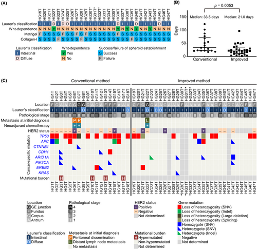
Typically, GC cells formed spherical aggregates in either Matrigel or collagen type-I (Figure S2A), and they were highly proliferative in the cancer medium (Figure S2B). Their structures and expression of markers such as CDX2 and MUC2 recapitulated those in the epithelial components of their primary cancer tissues (Figure S2C). Consistent with a previous study,33 culturing a Wnt-dependent spheroid line (HG22T) in the Wnt-free cancer medium accumulated signet-ring cell-like cells that were prominent in the primary tumor (Figure S2D). To assess the tumor-initiating activity in vivo, we injected GC-SC spheroids subcutaneously into immunodeficient mice, as we reported previously.34 Three of the five GC-SC spheroid lines formed subcutaneous tumors in nude or NSG mice, and their epithelial structures were similar to those of the primary tumors (Figure S3A,B), indicating that most of our GC-SC spheroid lines contained abundant tumor-initiating cells. Genetic alterations of TP53 and APC were detected frequently in the first patient cohort (13 and five lines, respectively, of 18), whereas they were less frequent in the second cohort (10 and three lines, respectively, of 25), suggesting that the improved culture condition helped propagate niche factor-sensitive GC-SCs that did not carry these key driver mutations (Figure 2C; Tables S4 and S5). Based on the estimated amounts of mutational burden, we identified four hypermutated GC-SC spheroid lines in the first patient cohort (22%; four of 18 lines; Figure 2C; Figure S4A), which was confirmed for lack of mismatch repair proteins by immunohistochemistry (Figure S4B,C; Table S6).
Collectively, these results demonstrated that our revised method for GC-SC spheroids was more efficient than our previous one.
3.2 Collagen type-I stimulates the growth of some slow-growing gastric cancer stem-cell spheroids
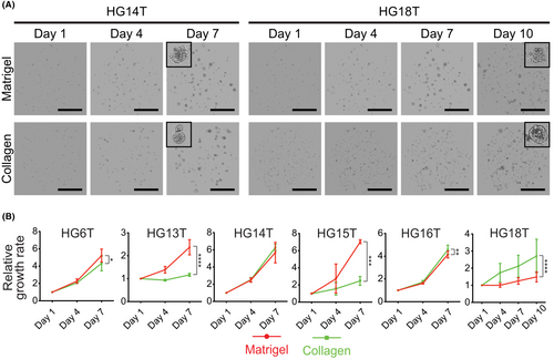
A diffuse-type GC-SC spheroid line (HG18T) embedded in Matrigel grew very slowly in vitro compared with other lines in the first patient cohort. Diffuse-type GC cells often invade the stromal layer of gastric mucosa,4 suggesting that these cells have a higher affinity to collagen (e.g., collagen type-I) than Matrigel extracellular scaffold rich in laminin-1.35 Therefore, we cultured HG18T and other spheroid lines separately in Matrigel and collagen type-I. Notably, HG18T spheroids preferentially proliferated in collagen type-I, whereas HG13T and HG15T in Matrigel. Other lines, HG6T, HG14T, and HG16T, showed little differences in growth between the two matrix materials without affecting the maintenance of spheroid lines because they more than quadrupled their cell volume in 6 days in either Matrigel or collagen type-I (Figure 3A,B). Thus, we decided to try both Matrigel and collagen type-I simultaneously but separately for primary culture of PD–GC-SCs, and empirically determine the matrix best suited for each GC-SC spheroid line.
3.3 Exogenous Wnt ligands stimulate the growth of some slow-growing gastric cancer stem-cell spheroids
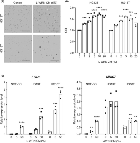
Previous studies have shown that a subset of GC organoids is dependent on exogenous Wnt ligands such as Wnt and/or R-spondin for growth.13, 14 However, Wnt ligands cause predominant growth of NGE-SCs in primary culture, which necessitates another selection procedure to enrich GC-SCs.13, 14, 33 To resolve this problem, we hypothesized that a low concentration of L-WRN CM that contained Wnt ligands could stimulate the growth of Wnt-responsive GC-SC spheroids without affecting NGE-SCs. Before determining such a concentration of L-WRN CM, we titrated its activity to ensure the reproducibility of culture conditions. We determined mRNA expression levels of MKI67 (proliferation marker) and LGR5 (stem cell marker) in normal colonic epithelial SC spheroids cultured with eL-WRN media containing serially diluted L-WRN CM according to the previous guidelines for quality control testing.26 As a result, we found that low concentrations of L-WRN CM (1%–10%) from two different sources (in-house and commercial media) stimulated MKI67 mRNA expression in a dose-dependent manner but failed to maintain LGR5 mRNA levels (Figure S5). Next, we conducted serial dilutions of in-house L-WRN CM with the cancer medium in the range of 0%–20% to titrate its effects on the growth of HG13T and HG18T, which showed the lowest growth rates among our GC-SC lines that we have established so far (Figure 3B). In both spheroid lines, 5%–10% of L-WRN CM supported the proliferation of GC-SC spheroids, whereas 5% CM of NGE-SC spheroids did not (Figure 4A,B; Figure S6A–C). Interestingly, 5% L-WRN CM stimulated the expression of the stem cell marker LGR5 in both HG13T and HG18T but not in NGE-SCs (Figure 4C). In contrast, L-WRN CM had smaller effects on the expression of the proliferation marker MKI67 in GC-SC lines than those in NGE-SCs (Figure 4C). These results suggested that supplementation with a low concentration (e.g., at 5%) of L-WRN CM should support self-renewal of Wnt-responsive GC-SCs without allowing that of NGE-SCs.
4 DISCUSSION
In this study, we propagated PD–GC-SCs using our spheroid culture method modified from that originally developed for PD–CRC-SCs.21 Although non-serum culture media are commonly used for organoid culture,36 the present method takes advantage of the serum-containing media that allow cost-efficient propagation of pure populations of normal epithelial stem cells as undifferentiated spheroids.22, 24 We previously applied this strategy to culture PD–CRC-SCs, and established more than 160 such spheroid lines at a high efficiency (up to approximately 90%).21 Although the establishment of PD–GC-SC lines was more challenging than CRC-SC lines with the first patient cohort (25% success rate), we finally achieved a higher success rate (88%) by improving our previous culture protocol specifically for GC-SCs (Table 1).
Importantly, we experienced difficulty in localizing the GC-SCs by macroscopic observation of patient samples (Figure 1A) as well as more frequent contamination of fungi, likely Candida species (7%; in seven of 104 cases),37, 38 than in CRC (3%; in four of 148 cases). Therefore, we decided to sample tumor tissue pieces from a wider and deeper area, avoiding necrotic lesions as antifungal drugs appeared ineffective (Figure 1C,D).38 We then re-evaluated culture conditions and newly employed collagen type-I matrix, which for the first time, shed light on the importance of ECM preference in the primary culture. Further studies are needed to determine the molecular features underlying the ECM preferences by GC-SC lines.
We also overcame the previously addressed limitations of GC organoid culture, including the high cost of niche factors and concomitant propagation of NGE-SCs,18, 39-41 by simply adding a low concentration of L-WRN CM, a cost-efficient source of stably active Wnt ligands (Figure S5).26 These modifications should help propagate distinct populations of GC-SCs that exhibit different dependencies on the niche factors without the need for negative selection to eliminate NGE-SCs.
In conclusion, we developed a simple and efficient method to propagate PD–GC-SC spheroids by improving our conventional sample collection protocol and culture conditions. Recent studies have shown that the drug sensitivity test on PD-CRC organoids can predict patient outcomes with 100% sensitivity,42, 43 even if some intra-tumor heterogeneity is lost in the spheroid/organoid line.44 Our PD–GC-SC spheroids can be utilized to investigate new molecular targeted therapies and their companion diagnostics for patient selection,45, 46 as we recently identified a subset of PD–CRC-SC spheroid lines that responded to fibroblast growth factor receptor inhibitors.47, 48 Additionally, the genomic and expression profiles of GC-SC spheroids will help determine novel molecular subtypes and diagnostic gene signatures. Thus, our improved method may open a new horizon for personalized GC diagnosis and treatment.
AUTHOR CONTRIBUTIONS
Conception and design, T. Morimoto (TMo), MMT, and H. Miyoshi (HMi); Development of methodology, TMo, YT, T. Miura (TMi), and HMi; Investigation, TMo, T. Yamamoto, FK, HA, H. Maekawa (HMa), T. Yamaura, and HMi; Analysis and interpretation of data, TMo, YT, TMi, and HMi; Administrative and material support, HMa, KK, YS, YY, HT, and KO; Manuscript writing, TMo, MMT, and HMi.
ACKNOWLEDGMENTS
The authors thank members of the Department of Surgery at KUHP and Medical Research Institute Kitano Hospital for help collecting surgical specimens; Hiromi Kikuchi for technical assistance; and the Medical Research Support Center, Graduate School of Medicine, Kyoto University for the use of the facility. We also thank Dr. Thaddeus S. Stappenbeck for providing L-WRN cells. We are grateful to Dr. Masanobu Oshima for comments on the manuscript.
FUNDING INFORMATION
This work was supported by Grants-in-Aid for Scientific Research (JP18H02639 and JP22K07187 to H.Miyoshi and JP21K06948 to FK) from the Japan Society for the Promotion of Science; research funds from Kyo Diagnostics K.K. and SCREEN Holdings Co., Ltd. (to H.Miyoshi and KO); the Program for Creating Start-ups from Advanced Research and Technology (ST261001TT) from the Japan Science and Technology Agency (to MMT); the Practical Research for Innovative Cancer Control (ck0106195h) from the Japan Agency for Medical Research and Development (to MMT); the Kyoto University Venture Incubation from the Kyoto University Office of Society-Academia Collaboration for Innovation (to MMT); and the Dynamic Project for Colon Cancer Personalized Therapy from the Institute for Advancement of Clinical and Translational Science, KUHP (to MMT).
CONFLICT OF INTEREST STATEMENT
H. Miyoshi and KO received research funds from Kyo Diagnostics K.K. and SCREEN Holdings. MMT owns stock in Kyo Diagnostics K.K. YT and H.Maekawa belong to the Department of Personalized Cancer Medicine at the Graduate School of Medicine, Kyoto University, which is supported by Kyo Diagnostics K. K., AFI, and SCREEN Holdings. T.Miura is an employee of SCREEN Holdings. The other authors have no conflicts of interest to declare.
ETHICS STATEMENTS
Approval of the research protocol by an Institutional Reviewer Board: The study protocol was approved by Kyoto University Graduate School and Faculty of Medicine, Ethics Committee (No. R0915 and R0857) as well as that of Medical Research Institute Kitano Hospital (extension of the Kyoto University study as a collaboration).
Informed Consent: Written informed consent was obtained from all patients.
Registry and the Registration No. of the study/trial: N/A.
Animal Studies: All animal experiments were conducted according to the protocol approved by the Institutional Animal Care and Use Committee of Kyoto University Graduate School of Medicine (Nos 14546, 15091, 16047, 16654, 17086, 18080, and 19601).


