A 56-year-old woman with back pain and lower limbs weakness
CLINICAL HISTORY
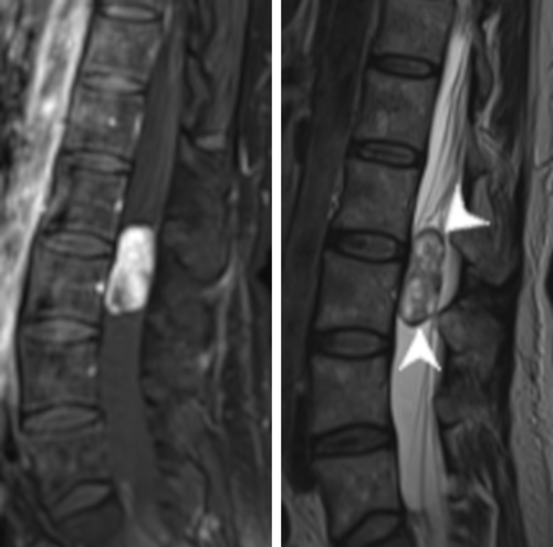
A 56-year-old woman previously healthy presented with a one-year history of low back pain. More recently, she also complained of radicular pain in both lower limbs, with nocturnal resurgences. Clinical examination did not find any motor, sensory or sphincter control disorders. MRI of the lumbar spine revealed a well-delineated, intradural extramedullary tumor, with strong enhancement on postcontrast T1-weighted images (Figure 1, left). T2-weighted images showed serpentine flow voids inside the tumor, and a “cap sign” (i.e., hypointense borders) traducing previous hemorrhage with hemosiderin deposits (Figure 1, right).

The patient underwent a L2-L3-L4 laminectomy. The dural sac opening revealed a well-circumscribed, purplish tumor surrounded by the cauda equina spinal nerve roots. The filum terminale was shifted posteriorly but not invaded. Strong adhesion of the tumor capsule to the right L4 spinal nerve root was observed. Total removal was obtained in a piecemeal fashion using the ultrasonic aspirator and sharp dissection. Clinical examination after surgery was normal.
1 FINDINGS
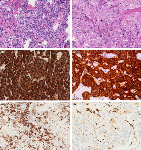
At low magnification, the tumor architecture was made of nests or lobules. The tumor was composed of areas of small tumor cells with high nuclear-cytoplasmic ratio and fine granular chromatin (Box 1). In other areas numerous ganglion cells, representing more than 20% of tumor cells, were present (Figure 2B1,B2). The vessels were thin, forming a delicate vascular network. No more than one mitosis for 10 high power field was found and there was no necrosis. Tumor cells were stained by antibodies directed to chromogranin A, synaptophysin, INSM1 as well as cytokeratin AE1/AE3 (Figure 2B3–B6). S100 protein was limited to a small number of sustentacular cells. Ki67-proliferation index was less than 5%. What is your diagnosis?
BOX 1. Slide scan
Access the scan at https://isn-slidearchive.org/?col=ISN&fol=Archive&file=BPA-21-05-102-2nd.svs
[Correction added on 20 February 2023, after first online publication: Link to slide scan has been updated.]

2 FINAL DIAGNOSIS
Cauda equina gangliocytic paraganglioma, WHO grade I.
A whole genome methylation profile was obtained and matched to the “paraganglioma, spinal non-CpG island methylator phenotype” group with a calibrated score of 0.99 (Brain Methylation classifier, v11b4).
3 DISCUSSION
Gangliocytic paraganglioma is a term usually used for paraganglioma (PGL) with nests of paraganglia cells with “zellballen” organization admixed with population of large mature ganglion cells [1]. Gangliocytic paraganglioma is a rare histologic subtype of paraganglioma usually found besides the central nervous system, especially in the duodenum. Spinal localization is rare, typically involving the cauda equina [2].
The prognosis of paragangliomas is usually good, especially if the tumor is entirely resected and recurrences are local. A recent next generation sequencing study has described genomic alteration such as hypoxia metabolic pathway correlated to poor outcome or metastasis evolution [3]. Due to the rarity of cauda equina paraganglioma and of this scarce morphological gangliocytic variant, it is not known whether the amount of gangliocytic cells has any prognostic significance.
As for genetic characteristics, Schweizer et al. [2] have shown that cauda equina paraganglioma (CEP) presents a distinct methylation-profile, distant from those of other paragangliomas (from head and neck, pheochromocytoma or extra-adrenal localization), showing that CEPs are independent tumor than standard PGL, and may arise from a different cell of origin.
Thus, our case illustrates that CEPs can commonly show gangliocytic differentiation, but also intense pan-cytokeratin labeling, which is unusual in paragangliomas from other sites [2]. These immunostaining particularities must be known, especially as PGLs express neuroendocrine markers that can mislead for—very rarely—a neuroendocrine tumor metastasis [4].
ACKNOWLEDGMENT
The authors thank the French neuropathological network for brain tumors (RENOCLIP-LOC).
CONFLICT OF INTEREST
None.
AUTHOR CONTRIBUTIONS
D.L. and F.F. conceived of the presented idea. D.L. wrote the manuscript with input from all authors. C.B. and F.V. contributed their expertise for the description of the radiological images as well as the clinical and surgical details. C.G. helped supervise the project and approved final diagnosis. All authors approved the final version of the manuscript.
Open Research
DATA AVAILABILITY STATEMENT
Data available under justified request to corresponding authors.


