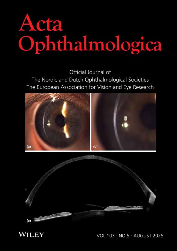
Changes in choroidal thickness and blood flow in myopic children with 0.01% atropine or orthokeratology and atropine combination therapy
Corresponding Author
Saiko Matsumura
Department of Ophthalmology, Toho University Faculty of Medicine, Tokyo, Japan
Correspondence
Saiko Matsumura, Department of Ophthalmology, Toho University Faculty of Medicine, 6-11-1 Omorinishi, Ota-ku, Tokyo 143-8540, Japan.
Email: [email protected]
Search for more papers by this authorTakashi Itokawa
Department of Ophthalmology, Toho University Faculty of Medicine, Tokyo, Japan
Search for more papers by this authorMomoko Kawakami
Department of Ophthalmology, Toho University Faculty of Medicine, Tokyo, Japan
Search for more papers by this authorTadashi Matsumoto
Department of Ophthalmology, Toho University Faculty of Medicine, Tokyo, Japan
Search for more papers by this authorHitoshi Ishikawa
Department of Orthoptics and Visual Science, School of Allied Health Sciences, Kitasato University, Kanagawa, Japan
Search for more papers by this authorYuichi Hori
Department of Ophthalmology, Toho University Faculty of Medicine, Tokyo, Japan
Search for more papers by this authorCorresponding Author
Saiko Matsumura
Department of Ophthalmology, Toho University Faculty of Medicine, Tokyo, Japan
Correspondence
Saiko Matsumura, Department of Ophthalmology, Toho University Faculty of Medicine, 6-11-1 Omorinishi, Ota-ku, Tokyo 143-8540, Japan.
Email: [email protected]
Search for more papers by this authorTakashi Itokawa
Department of Ophthalmology, Toho University Faculty of Medicine, Tokyo, Japan
Search for more papers by this authorMomoko Kawakami
Department of Ophthalmology, Toho University Faculty of Medicine, Tokyo, Japan
Search for more papers by this authorTadashi Matsumoto
Department of Ophthalmology, Toho University Faculty of Medicine, Tokyo, Japan
Search for more papers by this authorHitoshi Ishikawa
Department of Orthoptics and Visual Science, School of Allied Health Sciences, Kitasato University, Kanagawa, Japan
Search for more papers by this authorYuichi Hori
Department of Ophthalmology, Toho University Faculty of Medicine, Tokyo, Japan
Search for more papers by this authorAbstract
Purpose
To evaluate changes in choroidal thickness (CT), choroidal blood flow and axial length (AL) after therapy with either 0.01% atropine eye drops (AT) or the combination of orthokeratology and 0.01% AT (OKA) among Japanese myopic children.
Methods
We analysed changes in CT, choroidal blood flow and AL among myopic children who received either 0.01% AT or the OKA therapy at Toho University Omori Hospital from January 2021 through December 2022 (n = 38 eyes, 8.26 ± 2.13 years old in the 0.01% AT group and n = 44 eyes, 8.36 ± 1.54 years old in the OKA group). Comprehensive ophthalmologic examinations were performed at baseline, 3, 6 and 12 months.
Results
Subfoveal CT increased in the OKA group more than that in the AT group at 3, 6 and 12 months. Choroidal blood flow increase was greater in the OKA group compared to that in the AT group at 6 and 12 months. AL increase was less in the OKA group than that in the AT group at 6 and 12 months. AL changes were negatively correlated with choroidal blood flow and CT changes at both time points. In multivariate analysis, age, male, CT change and choroidal blood flow were independently associated with AL changes at 12 months.
Conclusion
The increase in CT was more pronounced in the combination therapy group compared to the AT group. The increase in CT and choroidal blood flow was associated with a reduced AL progression.
Supporting Information
| Filename | Description |
|---|---|
| aos17537-sup-0001-Supinfo1.docxWord 2007 document , 17.1 KB |
Data S1. |
| aos17537-sup-0002-Supinfo2.docxWord 2007 document , 38.7 KB |
Data S2. |
Please note: The publisher is not responsible for the content or functionality of any supporting information supplied by the authors. Any queries (other than missing content) should be directed to the corresponding author for the article.
REFERENCES
- Aizawa, N., Yokoyama, Y., Chiba, N., Omodaka, K., Yasuda, M., Otomo, T. et al. (2011) Reproducibility of retinal circulation measurements obtained using laser speckle flowgraphy-NAVI in patients with glaucoma. Clinical Ophthalmology, 5, 1171–1176.
- Cevher, S., Ucer, M.B. & Sahin, T. (2022) Choroidal thickness in emmetropia: an enhanced depth imaging spectral-domain optical coherence tomography-based study. Beyoglu Eye Journal, 7, 115–120.
- Chakraborty, R., Read, S.A. & Collins, M.J. (2011) Diurnal variations in axial length, choroidal thickness, intraocular pressure, and ocular biometrics. Investigative Ophthalmology & Visual Science, 52, 5121–5129.
- Chen, Z., Niu, L., Xue, F., Qu, X., Zhou, Z., Zhou, X. et al. (2012) Impact of pupil diameter on axial growth in orthokeratology. Optometry and Vision Science, 89, 1636–1640.
- Chen, Z., Xue, F., Zhou, J., Qu, X. & Zhou, X. (2016) Effects of orthokeratology on choroidal thickness and axial length. Optometry and Vision Science, 93, 1064–1071.
- Chia, A., Li, W., Tan, D. & Luu, C.D. (2013) Full-field electroretinogram findings in children in the atropine treatment for myopia (ATOM2) study. Documenta Ophthalmologica, 126, 177–186.
- Chiang, S.T., Turnbull, P.R.K. & Phillips, J.R. (2020) Additive effect of atropine eye drops and short-term retinal defocus on choroidal thickness in children with myopia. Scientific Reports, 10, 18310.
- Enaida, H., Okamoto, K., Fujii, H. & Ishibashi, T. (2010) LSFG findings of proliferative diabetic retinopathy after intravitreal injection of bevacizumab. Ophthalmic Surgery, Lasers & Imaging, 41, e1–e3.
10.3928/15428877-20101124-11 Google Scholar
- Hao, Q. & Zhao, Q. (2021) Changes in subfoveal choroidal thickness in myopic children with 0.01% atropine, orthokeratology, or their combination. International Ophthalmology, 41, 2963–2971.
- Hieda, O., Hiraoka, T., Fujikado, T., Ishiko, S., Hasebe, S., Torii, H. et al. (2021) Efficacy and safety of 0.01% atropine for prevention of childhood myopia in a 2-year randomized placebo-controlled study. Japanese Journal of Ophthalmology, 65, 315–325.
- Itokawa, T., Matsumoto, T., Matsumura, S., Kawakami, M. & Hori, Y. (2023) Ocular blood flow evaluation by laser speckle flowgraphy in pediatric patients with anisometropia. Frontiers in Public Health, 11, 1093686.
- Kinoshita, N., Konno, Y., Hamada, N., Kanda, Y., Shimmura-Tomita, M., Kaburaki, T. et al. (2020) Efficacy of combined orthokeratology and 0.01% atropine solution for slowing axial elongation in children with myopia: a 2-year randomised trial. Scientific Reports, 10, 12750.
- Kobayashi, T., Shiba, T., Nishiwaki, Y., Kinoshita, A., Matsumoto, T. & Hori, Y. (2019) Influence of age and gender on the pulse waveform in optic nerve head circulation in healthy men and women. Scientific Reports, 9, 17895.
- Li, Z., Hu, Y., Cui, D., Long, W., He, M. & Yang, X. (2019) Change in subfoveal choroidal thickness secondary to orthokeratology and its cessation: a predictor for the change in axial length. Acta Ophthalmologica, 97, e454–e459.
- Maeda, K., Ishikawa, F. & Ohguro, H. (2009) Ocular blood flow levels and visual prognosis in a patient with nonischemic type central retinal vein occlusion. Clinical Ophthalmology, 3, 489–491.
- Matsumura, H. & Hirai, H. (1999) Prevalence of myopia and refractive changes in students from 3 to 17 years of age. Survey of Ophthalmology, 44(Suppl 1), S109–S115.
- Matsumura, S., Chen, C.Y. & Saw, S.M. (2020) Global epidemiology of myopia. In: M. Ang & T. Wong (Eds.) Updates on myopia. Singapore: Springer, pp. 27–51.
10.1007/978-981-13-8491-2_2 Google Scholar
- Ostrin, L.A., Harb, E., Nickla, D.L., Read, S.A., Alonso-Caneiro, D., Schroedl, F. et al. (2023) IMI–the dynamic choroid: new insights, challenges, and potential significance for human myopia. Investigative Ophthalmology & Visual Science, 64, 4.
- Pereira-da-Mota, A.F., Costa, J., Amorim-de-Sousa, A., González-Méijome, J.M. & Queirós, A. (2020) The impact of overnight orthokeratology on accommodative response in myopic subjects. Journal of Clinical Medicine, 17, 3687.
10.3390/jcm9113687 Google Scholar
- Reiner, A., Fitzgerald, M.E.C., Del Mar, N. & Li, C. (2018) Neural control of choroidal blood flow. Progress in Retinal and Eye Research, 64, 96–130.
- Repka, M.X., Weise, K.K., Chandler, D.L., Wu, R., Melia, B.M., Manny, R.E. et al. (2023) Low-dose 0.01% atropine eye drops vs placebo for myopia control: a randomized clinical trial. JAMA Ophthalmology, 141, 756–765.
- Saw, S.M., Matsumura, S. & Hoang, Q.V. (2019) Prevention and management of myopia and myopic pathology. Investigative Ophthalmology & Visual Science, 60, 488–499.
- Shiga, Y., Omodaka, K., Kunikata, H., Ryu, M., Yokoyama, Y., Tsuda, S. et al. (2013) Waveform analysis of ocular blood flow and the early detection of normal tension glaucoma. Investigative Ophthalmology & Visual Science, 54, 7699–7706.
- Spaide, R.F., Koizumi, H. & Pozzoni, M.C. (2008) Enhanced depth imaging spectral-domain optical coherence tomography. American Journal of Ophthalmology, 146, 496–500.
- Sugiyama, T., Araie, M., Riva, C.E., Schmetterer, L. & Orgul, S. (2010) Use of laser speckle flowgraphy in ocular blood flow research. Acta Ophthalmologica, 88, 723–729.
- Tan, C.S., Ouyang, Y., Ruiz, H. & Sadda, S.R. (2012) Diurnal variation of choroidal thickness in normal, healthy subjects measured by spectral domain optical coherence tomography. Investigative Ophthalmology & Visual Science, 53, 261–266.
- Tan, Q., Ng, A.L., Cheng, G.P., Woo, V.C. & Cho, P. (2023) Combined 0.01% atropine with orthokeratology in childhood myopia control (AOK) study: a 2-year randomized clinical trial. Contact Lens and Anterior Eye, 46, 101723.
- Upadhyay, A. & Beuerman, R.W. (2020) Biological mechanisms of atropine control of myopia. Eye & Contact Lens, 46, 129–135.
- Xiong, R., Zhu, Z., Jiang, Y., Wang, W., Zhang, J., Chen, Y. et al. (2023) Longitudinal changes and predictive value of choroidal thickness for myopia control after repeated low-level red-light therapy. Ophthalmology, 130, 286–296.
- Xu, H., Ye, L., Peng, Y., Yu, T., Li, S., Weng, S. et al. (2023) Potential choroidal mechanisms underlying atropine's antimyopic and rebound effects: a mediation analysis in a randomized clinical trial. Investigative Ophthalmology & Visual Science, 64, 13.
- Yam, J.C., Jiang, Y., Lee, J., Li, S., Zhang, Y., Sun, W. et al. (2022) The association of choroidal thickening by atropine with treatment effects for myopia: two-year clinical trial of the low-concentration atropine for myopia progression (LAMP) study. American Journal of Ophthalmology, 237, 130–138.
- Yam, J.C., Jiang, Y., Tang, S.M., Law, A.K.P., Chan, J.J., Wong, E. et al. (2019) Low-concentration atropine for myopia progression (LAMP) study: a randomized, double-blinded, placebo-controlled trial of 0.05%, 0.025%, and 0.01% atropine eye drops in myopia control. Ophthalmology, 126, 113–124.
- Ye, L., Shi, Y., Yin, Y., Li, S., He, J., Zhu, J. et al. (2020) Effects of atropine treatment on choroidal thickness in myopic children. Investigative Ophthalmology & Visual Science, 61, 15.
- Yotsukura, E., Torii, H., Inokuchi, M., Tokumura, M., Uchino, M., Nakamura, K. et al. (2019) Current prevalence of myopia and association of myopia with environmental factors among schoolchildren in Japan. JAMA Ophthalmology, 137, 1233–1239.
- Zhao, W., Li, Z., Hu, Y., Jiang, J., Long, W., Cui, D. et al. (2021) Short-term effects of atropine combined with orthokeratology (ACO) on choroidal thickness. Contact Lens and Anterior Eye, 44, 101348.


