Carl Friedrich Richard Foerster (1825–1902) – the inventor of perimeter and photometer
Abstract
Carl Friedrich Richard Foerster (1825–1902) was a German who was born in the Polish city Leszno. He studied medicine at the Medical Faculty of Breslau (now Wroclaw, Poland) University, and later in Heidelberg and Berlin. From 1855, he worked in Breslau, where he established in 1857 the first ophthalmology clinic. Later, he became a professor in ophthalmology, the first director of the Department of Ophthalmology at the University of Breslau, and even the rector of this University. Forster did many pioneering works on visual fields, invented a photometer and the first perimeter, known for many years as the Foerster perimeter. Moreover, he studied night blindness, visual field changes due to different pathologies, and many eye diseases, including glaucoma, cataract, retinal and choroidal diseases.
Biography
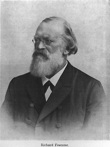
Carl Friedrich Richard Foerster (Fig. 1) was born in Poland on the 15 November 1825 in Leszno (called Lissa during the period of German occupation in 19th century). After graduation from the Gymnasium in Leszno in 1845, Foerster entered the Medical Faculty of Breslau (now Wroclaw, Poland) University (Baer 1902). From 1847 to 1849, he studied medicine at the universities of Heidelberg and Berlin. In 1849, he received his doctorate at Berlin University. Subsequently, he visited the most famous educational institutions in Wien (Vienna), Paris and Prague. In 1855, he took a position as a primary physician (in the surgical and syphilitic ward) at the Allerheiligen hospital in Breslau (Baer 1902). His habilitation was in 1857 and two years later he established the first ophthalmology clinic there. He became associate professor in 1863, and full professor in ophthalmology in 1873 (Hirschberg 1918). He became the first director of the Department of Ophthalmology at the University of Breslau in the years 1876 to 1896. Moreover, he was elected rector of the University in 1884 and was appointed a member of the House of Lords in 1894. He retired at the age of 70 and carried about management of his inherited farm (Dominium) in Bronikowo on the border of Silesia and Wielkopolska (Greater Poland) (Kaufmann 1911).
Dr Foerster's Works on Visual Fields and Perimetry
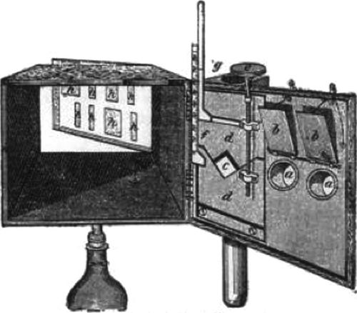
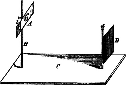
His work included different studies, but he is best remembered for his studies on visual fields and perimetry. His habilitation thesis on night blindness and the photometer opened new paths for diagnostic methods (Foerster 1857c). Foerster's photometer (Fig. 2) was a dark box with black lines on a white paper (one or two centimetres wide and five centimetres long), which was illuminated by a wax candle. By gradual enlargement of the illuminated surface, light intensity was measured. He examined the light intensity in different diseases of the choroid and retina and noted that the light perception was a cardinal sign of diseases affecting the posterior retina (Foerster 1871). As a result of this experience, Foerster took to calling retinitis syphilitica – Chorioiditis syphilitica (Foerster 1874).
In 1857, he published with Hermann Aubert his first work about quantitative visual field measurements (Aubert & Foerster 1857). However, until von Graefe's publication about visual fields in amblyopia, visual field testing was mostly qualitative (Johnson et al. 2011). They conducted several experiments. One of them (Fig. 3) was based on a concept that the duration of electric sparks, which illuminate an object, is so short that the eye cannot move during it. Therefore, the objects are seen through the stationary retina. The purpose of this experiment was to evaluate how far away from the visual axis that objects can be recognized. The experiment was performed in a darkened room. Altogether there were four sheets of paper at ten different distances, and each observer had 10–20 attempts; therefore, a total of 500 attempts were performed. Using this experiment, both a ‘spatial angle’ (visual angle of the room, at which the numbers could be recognized) and the visual angle for the biggest diameter of numbers (the angle of numbers) could be calculated.
The following conclusions were drawn from this experiment:
- If the angle of numbers is equal, the spatial angle is smaller for big and far afield numbers than for small and near numbers (Table 1).
Table 1. The spatial angle is smaller for big and far afield numbers than for small and near numbers with the equal angle of numbers. Source: Aubert & Foerster 1857 . Angle of numbers Spatial angle Distance (metre) 1°3′ 11°52′ 0.7 1 14°28′ 0.4 1°29′ 10°58′ 1 1°29′ 16°36′ 0.5 1°33′ 9°20′ 0.96 1°36′ 20°26′ 0.25 1°51′ 14°36′ 0.8 1°52′ 20°38′ 0.4 2°7′ 12°40′ 0.7 2 27°14′ 0.2 3°42′ 27°2′ 0.4 3°43′ 32°34′ 0.2 6°51′ 40°36′ 0.21 7°27′ 60°40′ 0.1 - The area of retina, which recognizes shapes of a certain size, is not a circle, but an ellipse with the largest diameter horizontally.
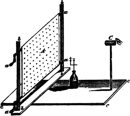
The aim of the second experiment (Fig. 4) was to find out the distance from the retinal centre, in which two points can be recognized from each other. This experiment was performed with eight different sheets of papers (four different sizes of points, each in two different distances) (Table 2). Each position was repeated eight times.
| No. of sheet of paper | Angel of diameter of points | Angel of distant of points | Left eye (1) | Right eye (1) | Left eye (2) | Right eye (2) | Mean | Angel of distant of points divided through angel of distant points from the fixation point |
|---|---|---|---|---|---|---|---|---|
| I | 0°56′ | 0°56′ | 8° | 9° | 10° | 9° | 9° | 9.6 |
| II | 2°28′ | 18° | 18° | 17° | 16° | 17.25° | 5 | |
| I | 0°43′ | 1°52′ | 14° | 14° | 16° | 14° | 14.5° | 7.7 |
| II | 4°7′ | 17° | 17° | 20° | 20° | 18.5° | 4.5 | |
| I | 1° | 2°35′ | 12° | 13° | 18° | 17° | 15° | 5.9 |
| II | 5°43′ | 23° | 22° | 24° | 23° | 23° | 4 | |
| I | 1°4′ | 2°44′ | 16° | 16° | 16° | 16° | 16° | 5.8 |
| II | 5°49′ | 22° | 23° | 21° | 22° | 22° | 3.8 |
The results of the second experiment were as follows:
- The ability to recognize two points from each other depends on distance from the eye axis and on retinal meridian.
- The ability to recognize two points from each other is decreasing if they are farther from the eye axis.
- The size of points does not seem to have an impact on their recognition.
In comparison with the first experiment, there were following changes:
The lighting was continuous.
- An observer had to recognize points instead of letters and numbers.
- The distance between an observer and a sheet of paper was always equal (0.2 metre).
- The measurement was more accurate due to gradual movement of the sheet of paper.
- The measurement was taken in eight different retinal meridians.
- The inability to recognize two points from each other was not related to optical properties of the eye. This conclusion was confirmed with an additional experiment on an extirpated rabbit eye, which was placed behind a screen with two holes. The light fell through these holes. If the eye was located 0.2 metre away from the screen, an image of two holes could be recognized. If the eye was situated 0.7 metre away from the screen, the image could not be recognized.
The following conclusions were drawn from these two experiments:
- The characteristics of spatial orientation decrease from the retinal centre into the periphery, and are different in the both eyes.
- The characteristics of spatial orientation are different for various retinal meridians. A decrease of characteristics of spatial orientation is faster vertically than horizontally.
- The blind spot is ‘a real defect’.
- These experiments confirmed the Weber's assumptions that cones and not rods are responsible for the spatial orientation as they are located in the macula lutea and seldom are in the periphery.
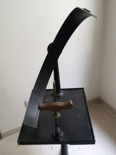
In another series of experiments, Foerster studied the limits of the visual field (Foerster 1862a). A rotary arc with radius of 11.5 inches was used. The rotary arc was composed of 2″ wide semicircular strip from sheet brass revolving around a central axis. Each degree of the arc signified a parallel circle. The back nodal point of the examined eye, the centre of the pupil and the fovea centralis laid along a straight line, which corresponded to the centre of the arc. Due to the division of visual field into meridians and parallel circles, each point of the retina was determinable. During the examination, a reader should take a white square (1x1 cm) to be a 2-dimensional object. The border of visual field was marked in place where an object had disappeared.
The fixation point was not the centre of rotation of the arc, but a spot located 15° inwards. Hence, the centre of rotation corresponded to the blind spot. He observed that the inner and outer borders of the visual field were different: the inner (nasal) visual field was 55–60°, and the outer (temporal) visual field was 85–90°. The inner visual field did not hardly achieve the bridge of the nose; therefore, it was not limited thereby; a retinal area along the ora serrata was not sensitive to light; the vertical meridian divided the retina in two different halves; the fovea centralis was located eccentric.
In the previous works of Foerster (Aubert & Foerster 1857; Foerster 1862a), gradual development of his perimeter can be observed. In 1867, he introduced his perimeter (Fig. 5) to the International Congress of Ophthalmology in Paris and described it in an article in 1869 (Foerster 1869a). There were two different methods of measuring the visual field: (1) using a plane surface and (2) a hollow bowl. The bowl had obvious advantages. If the lateral visual field was measured out to 75°, an object was moved along an arcuate line to the point g; whereas, if the visual field was plotted on the wall, or on a flat screen, the test object would be moved to the point h, which was four times further away than object g (Fig. 6). Thus, the object h would be four times smaller on the retina than the object at point g. The examination was performed in ten different meridians, and two objects were necessary, one for the fixation and another one for measuring the visual field. Both objects were mounted on a mobile track, which was moved using a wheel (Foerster 1869a).

In another study, Foerster investigated the problem of visual field in anaesthesia of the retina (Foerster 1877a). He performed 120 visual field tests in 7 patients. He found that fatigue had a great impact on the visual field because there were great variations of visual field defects not only on different days but also in different hours. Therefore, he noted that ‘the longer the examination, the smaller the visual field’. He also observed that a visual field defect was more meaningful when it was measured using an object moving centrifugal (away from fixation), rather than centripetal (towards fixation), and that glass correction for hyperopia produced prismatic displacement of the stimulus and influenced the size of visual field.
In addition, he presented a chart with a fixation point (earlier the blind spot) in the centre of vision for graphical presentation of visual field (Hirschberg 1918). This chart included meridians, which were numbered 360° with 0° at the top of the vertical meridian, and parallel circles with equal intervals between them (Foerster 1883).
Other Ophthalmic Works
Foerster described a case report patient with a double-sided homonymous hemianopsia due to thrombosis in the cortex of the brain. He observed that due to another blood supply of the point of sharpest vision, the vertical boundary line of the visual field defect was shifted 2–4° in the direction of the blind hemifield and so that it did not cross the fixation point. He offered the suggestion that perhaps the cortical representation of the macula may have had an alternate blood supply. He reported a colour vision defect, but no atrophy of the optic nerve was seen. (Foerster 1889, 1890).
Foerster also characterized the optic disc in glaucoma (Foerster 1857a) and reported that the optic disc has an excavation instead of the prominence. He also reported the kinking of vessels as indicative of the location of the cup boundary.
He studied the development of senile cataract and observed that sweeping the cornea using a squint hook after iridectomy tended to accelerate the maturation of early cataract due to degradation of cortical fibres (Foerster 1857b, 1884). He also described metamorphopsias as a symptom of retinal and choroidal diseases, including retinitis circumscripta and chorioiditis areolaris (Foerster 1862b), fungal infection of the inferior lacrimal duct (Foerster 1869b), amaurosis partialis fugax (Foerster 1869c), accommodation in aphakia (Foerster 1872), traumatic lens luxation in the anterior chamber (Foerster 1887) and pseudo-Egyptian ophthalmia (Foerster 1888).
He published a book about the spread of cholera as a result of contaminated water in public fountains based on his own experiences and observations in Silesia and Posen, thus supporting the 1854 work of Snow in London. (Foerster 1873). In addition, he wrote a book's chapter about relationships between systemic diseases and the eye based on 100 000 references! (Foerster 1877b).
He also addressed the pathogenesis of myopia (Foerster 1885). He believed that the progression of myopia might be caused by spectacle overcorrection. This led him to argue for improvement of the eye hygiene, including full correction of spectacles, a working distance of 40 cm, better lighting etc. (Foerster 1886).
In short, Foerster was a vigorous clinician who worked hard to improve eye care. He loved to encounter a new clinical mystery that challenged his knowledge. He tried hard to understand it so that the next time he saw such a problem he would know what to do!


