Alcohol withdrawal syndrome: mechanisms, manifestations, and management
Abstract
The alcohol withdrawal syndrome is a well-known condition occurring after intentional or unintentional abrupt cessation of heavy/constant drinking in patients suffering from alcohol use disorders (AUDs). AUDs are common in neurological departments with patients admitted for coma, epileptic seizures, dementia, polyneuropathy, and gait disturbances. Nonetheless, diagnosis and treatment are often delayed until dramatic symptoms occur. The purpose of this review is to increase the awareness of the early clinical manifestations of AWS and the appropriate identification and management of this important condition in a neurological setting.
1 Introduction - Medical Burden of Alcohol Abuse
An estimated 76.3 million people worldwide have alcohol use disorders (AUDs), and these account for 1.8 million deaths each year.1 It is estimated that up to 42% of patients admitted to general hospitals, and one-third of patients admitted to hospital intensive care units (ICU) have AUD.2 Alcohol withdrawal syndrome (AWS) is a well-known condition occurring after intentional or unintentional abrupt cessation of heavy/constant drinking, and it occurs in about 8% of hospitalized AUD inpatients.3 Severe AWS more than doubles the length of stay and frequently requires treatment at the ICU. A complicated AWS includes epileptic seizures and/or delirium tremens (DT), the occurrence of which may be as high as 15% in AUD patients.4, 5 Delirious patients show high rates of comorbidities, and their mortality rate is comparable to patients having severe malignant diseases. However, with early detection and appropriate treatment, the expected mortality is in the range of 1% or less.6
AUDs are common in patients referred to neurological departments, admitted for coma, epileptic seizures, dementia, polyneuropathy, and gait disturbances. Nonetheless, diagnosis and treatment are often delayed until dramatic symptoms occur. The purpose of this review is to increase the awareness of the early clinical manifestations of AWS and the appropriate identification and management of this important condition in a neurological setting.
2 Pathophysiology
Ethanol is a central nervous system depressant that produces euphoria and behavioral excitation at low blood concentrations due to increased glutamate binding to N-methyl-D-aspartate (NMDA) receptors; at higher concentrations, it leads to acute intoxication by potentiation of the gamma-aminobutyric acid (GABA) effects,7 particularly in receptors with delta subunits.7, 8 The local distribution of these subunits explains why the cerebellum, cortical areas, thalamic relay circuitry, and brainstem are the main networks that mediate the intoxicating effects of alcohol.9 Prolonged alcohol use leads to the development of tolerance and physical dependence, which may result from compensatory functional changes by downregulation of GABA receptors and increased expression of NMDA receptors with production of more glutamate to maintain central nervous system (CNS) transmitter homeostasis.7
Abrupt cessation of chronic alcohol consumption unmasks these changes with a glutamate-mediated CNS excitation resulting in autonomic overactivity and neuropsychiatric complications such as delirium and seizures.10 The latter are usually of generalized tonic–clonic type and are mediated largely in the brainstem by abrogation of the tonic inhibitory effect of the GABAergic delta subunits.8 Therefore, the trigger zone of these seizures is distinct from that believed to be responsible for seizures in the context of epilepsy, and this may explain why epileptiform activity is rarely observed in the EEG after alcohol withdrawal seizures.8 As upregulation of NMDA receptors as well as reduced GABA-A receptor inhibition largely explain the clinical symptoms, the therapeutic approach to AWS mainly targets these mechanisms. Dopamine is another neurotransmitter involved in alcohol withdrawal states. During alcohol use, increase in dopamine positively influences the reward system thereby maintaining abuse. In withdrawal, increase in dopamine levels contributes to the clinical manifestations of autonomic hyperarousal and hallucinations.10, 11 Moreover, polymorphisms in the dopamine receptor 2 gene seem to influence not only AUD but also the clinical manifestation of alcohol withdrawal symptoms.12 In combination with increased glutamate and norepinephrine, it may also cause the elongation of the QT interval in people who have active epilepsy; this can increase the risk of sudden unexpected death in epilepsy (SUDEP).9 Another excitotoxic compound that is increased in AUD is homocysteine. During active drinking, there is an increase in homocysteine through stimulation of the NMDA receptors. In withdrawal, excitotoxicity is induced by further raise in homocysteine via rebound activation of glutamatergic neurotransmission.7
3 Clinical spectrum
AWS represents a group of symptoms that usually arise 1–3 d after the last drink. Sometimes, the symptoms are already present when the alcohol blood level is above 0 (0.5‰ or even more).3
- A clear evidence of cessation or reduction in heavy and prolonged alcohol use.
- The symptoms of withdrawal are not accounted for by a medical or another mental or behavioral disorder.
Physical examination and investigations should be directed toward detecting common signs and symptoms of AWS that are listed in Table 1.6, 10, 14-18
| Autonomic symptoms | Motor symptoms | Awareness symptoms | Psychiatric symptoms |
|---|---|---|---|
| Tachycardia | Hand tremor | Insomnia | Illusions |
| Tachypnea | Tremulousness of body | Agitation | Delusions |
| Dilated pupils | Seizures | Irritability | Hallucinations |
| Elevated blood pressure | Ataxia | Delirium | Paranoid ideas |
| Elevated body temperature | Gait disturbances | Disorientation | Anxiety |
| Diaphoresis | Hyper-reflexia | Affective instability | |
| Nausea/vomiting | Dysarthria | Combativeness | |
| Diarrhea | Disinhibition |
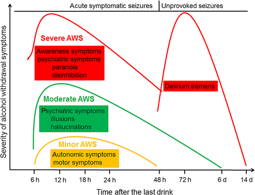
The alcohol withdrawal syndrome is a dynamic and complex process. For this reason, there have been many attempts to classify symptoms of AWS either by severity or time of onset to facilitate prediction and outcome. In early stages, symptoms usually are restricted to autonomic presentations, tremor, hyperactivity, insomnia, and headache. In minor withdrawal, patients always have intact orientation and are fully conscious. Symptoms start around 6 h after cessation or decrease in intake and last up to 4–48 h (early withdrawal).6, 10 Hallucinations of visual, tactile or auditory qualities, and illusions while conscious are symptoms of moderate withdrawal. They can last up to 6 d. The appearance of acute symptomatic seizures may emerge 6–48 h after the last drink.19 Delirium tremens (DT, onset 48–72 h after cessation of drinking) represents characteristics of severe withdrawal that may last for up to 2 weeks (late withdrawal).6, 10, 15, 18 The chronological development of the various symptoms is illustrated in Fig. 1.

The alcohol withdrawal seizure is a symptom occurring primarily during the early phase of withdrawal and is characterized by reduction in the seizure threshold. More than 90% of acute symptomatic seizures emerge within 48 h of cessation of prolonged drinking.20, 21 Seizures frequently occur in the absence of other signs of the AWS. More than half of the individuals present with repeated seizures, and in up to 5%, they may progress to status epilepticus.17 More than 50% of withdrawal seizures are associated with concurrent risk factors such as prior epilepsy, structural brain lesions, or use of other drugs.17, 20 It is remarkable that the development of acute symptomatic seizures during an alcohol withdrawal episode is associated with a fourfold increase in the mortality rate that is due to complications of severe AUD rather than a direct effect of seizures.17, 22 The appearance of a withdrawal seizure represents a strong risk factor for progression into a severe withdrawal state with following development of DT in up to 30% of cases.21 Unprovoked seizures occurring later than 48 h after the last drink suggest other causes such as head trauma or combined drug withdrawal effects.19, 23
Delirium is a clinical syndrome of acute onset characterized by a global confusional state, perceptual abnormalities, and somatic symptoms of vegetative or central nervous presentation.6 Hallucinosis represents a unique form of withdrawal-related psychosis which can begin even while the person is continuing to use alcohol or after cessation of drinking. The sensorium is clear in the beginning, but it often evolves into the syndrome of DT, a specific type of delirium typically associated with psychomotor agitation (hyperactive delirium) which emerges during the late withdrawal phase.14, 18 Delirium can also manifest as a hypoactive state with decreased arousal and psychomotor activity, which is associated with a worse prognosis, delayed diagnosis and treatment as well as later complications.6 In cases of hypoactive delirium, comorbid or other medical illnesses must be ruled out. This is especially important in patients who have not had a previous history of DT. Differential diagnoses for severe alcohol withdrawal are listed in Table 2.10, 15
| Differential diagnosis | Comment |
|---|---|
| Hyponatremia | Due to poor oral intake, dehydration, and uremia; frequently presenting as hypoactive delirium |
| Hepatic encephalopathy | Jaundice, hematemesis, melena, icterus, flapping tremor, ascites, sleep–wake reversal |
| Pneumonia | Fever, cough, low arterial blood oxygen saturation, delirium before cessation of alcohol use |
| Encephalitis/Meningitis | Fever, meningeal signs, and focal neurological deficits; MRI/CSF abnormalities |
| Head injury | Being found unconscious, ear or nose bleeding, pinpoint pupils, focal neurological deficits |
| Thyrotoxicosis | History of thyroid illness; thyromegaly, exophthalmos, lagophthalmos |
| Lithium intoxication | History of psychiatric illness, drug overuse, diarrhea, fever, use of NSAID or diuretics |
| Atropine/Tricyclic intoxication | Fever, hot dry skin, dilated pupils |
| Psychosis | Hallucinations/delusions of long-standing duration, absence of clouding of sensorium |
| Antidepressant intoxication | Use of SSRI; diarrhea, myoclonus, jitteriness, seizures, altered sensorium |
| Subacute encephalopathy with seizures in AUD | Several days after alcohol cessation; complex/simple partial seizures with reversible motor deficits; in EEG focal slowing, periodic lateralized discharges; MRI with reversible T2w flair hyperintensities |
In summary, physical examination and investigations should be directed toward detecting signs of intoxication, seizures, hallucinations, and delirium tremens as well as Wernicke's encephalopathy (one or more symptoms of ataxia, amnesia, and ophthalmoplegia). Apart from neuropsychiatric symptoms, physical injury or medical problems including aspiration pneumonia, dehydration, and electrolyte imbalance should be taken into account.16
4 Biomarkers
In several studies, possible predictors for the development of a severe AWS have been investigated. Medical history and laboratory biomarkers are the two most important methods for the identification of patients at high risk. It appears that the most robust predictor for an incident occurrence of DT or seizures is a history of a similar event.3, 10, 24, 25 Clinical findings such as elevated heart rate, systolic blood pressure, and temperature are all easily verifiable in the initial patient assessment, although their predictive value to identify patients with AWS who are more likely to develop DT is not high.3, 10, 26 In a patient with impaired consciousness, laboratory markers represent helpful tools to confirm the suspected clinical diagnosis of an AUD.
4.1 Markers useful in the emergency setting
The quantitative, measurable detection of drinking is important for the successful treatment of AUD. Therefore, the importance of direct and indirect alcohol markers to evaluate consumption in the acute clinical setting is increasingly recognized. A summary of relevant markers in the emergency setting is given in Table 3. The detection of ethanol itself in different specimens is still a common diagnostic tool to prove alcohol consumption. Alcohol ingestion can be measured using a breath test. Although ethanol is rapidly eliminated from the circulation, the time for detection by breath analysis is dependent on the amount of intake as ethanol depletes according to a linear reduction at about 0,15‰/1 h. Alcohol use can alternatively be detected by direct measurement of ethanol in blood or urine.27 The time course of the ethanol concentration in the blood after the ingestion of an alcoholic beverage is controlled by its pharmacokinetics that represents an interplay between the kinetics of absorption, distribution, and elimination and is thus important in determining the pharmacodynamic responses to alcohol. There is a large degree of variability in alcohol metabolism as a result of both genetic and environmental factors.
| Biomarker | Specimen | Access to laboratory results | Detection over a period of | Specificity/sensitivity | Comments | Ref. |
|---|---|---|---|---|---|---|
| Ethanol | BreathBloodUrine | <6 h | 5–24 hdepletion 0,15‰/1 h | ~ 90%/~ 95% | Conversion factor breath alcohol:blood alcohol 1:2100 within 2–5 h after the last drink | 27, 32 |
| Hypokalemia | Blood | <6 h | Days to weeks | ~ 47%/~ 90% | Serum levels <2,5 mmol/L indicate severe AUD | 20, 30 |
| Thrombocytopenia | Blood | <6 h | 7–12 d | ~ 69%/~ 75% | High NPV, low PPV; rebound thrombocytosis after cessation of alcohol abuse | 24, 25, 29, 33 |
| Mean corpuscular volume | Blood | <6 h | 4 mo | ~ 80%/~ 60% | Dose-dependent increase | 34 |
| γ-glutamyltransferase | Blood | <6 h | 2–8 wk | ~ 80%/~ 65% | Severe AUD with liver damage | 24 |
| Ratio AST/ALT >2 | Blood | <6 h | AST 18 hALT 36 h | ~ 50%/~ 80% | Severe AUD, marker of liver damage | 24, 35, 36 |
Apart from ethanol itself, indirect markers of AUD are widely available and mostly part of routine laboratory testing. Severe AWS involves changes in electrolytes, especially potassium that is due to increased catecholamine activity with activation of the sodium–potassium ATPase pump and elevated vasopressin.25 Hypokalemia is not specific for alcohol consumption but is frequently reported to be associated with DT or seizures.10, 25, 28, 29 The same applies to thrombocytopenia (with high negative predictive value)10, 24, 25, 29, 30 that additionally is predictive of an incident occurrence of DT and seizures.25 More indirect markers, such as AST, ALT, γGT, and MCV, are widely available and relatively inexpensive, but their predictive value is restricted because of low specificity. The interpretation of elevated values has to take into account other influencing factors including gender, age, comorbid disorders, and medication that also may increase these markers.31
4.2 Additional markers to detect AUD
Further biomarkers for non-emergency cases or in the event of forensic questions are listed in Table 4. Carbohydrate-deficient transferrin (CDT) is the most available and studied biomarker and has a high specificity for severe AUD.37 CDT values are not markedly influenced by medications except by immunosuppressants. The main disadvantage is the relatively low sensitivity making this parameter unsuitable as a screening tool. As CDT, γGT, and MCV are connected with AUD by different pathophysiological mechanisms, a combination of these parameters will further improve their diagnostic value.38 39, 40
| Biomarker | Specimen | Access to laboratory results | Detection over a period of | Specificity/sensitivity | Comments | Ref. |
|---|---|---|---|---|---|---|
| Carbohydrate-deficient transferrin | Blood | >6 h | 2–4 wk | ~ 98%/~ 70% | Severe AUD | 24, 37 |
| Ratio γGT:CDT | Blood | >6 h | 2–3 wk | ~ 92%/~ 84% | Severe AUD | 24, 32 |
| Ethylglucuronid | Bloodurinehair | >6 h | 8 h20–80 h3 mo | ~ 99%/~ 89% | Values dependent on creatinine clearance | 31, 37 |
| Ethylsulfate | Bloodurine | >6 h | 8 h36–78 h | ~ 99%/~ 89% | Values dependent on creatinine clearance | 31, 37 |
| Phosphatidylethanol | Blood | >6 h | 4 wk | ~ 99%/~ 98% | Detection also available for dry blood spots | 27, 31, 42, 53 |
| Fatty acid ethyl esters | Bloodhair | >6 h | 24 h3 mo | ~ 97%/~ 77% | Combined measurement of ethylglucuronide and fatty acid ethyl esters in hair increases accuracy of interpretation | 27, 31, 44, 54 |
| 5-hydroxytryptophol:5-hydroxyindole-3-acetic acid | Urine | >6 h | 24 h | ~ 99%/~ 77% | Ratio >20 marker for recent alcohol intake | 47, 48 |
| Whole blood acetaldehyde | Blood | >6 h | 4 wk | ~ 93%/~ 78% | False-positive results in diabetics | 55 |
| Total sialic acid | Blood | >6 h | Several weeks | ~ 95%/~ 81% | Glycoconjugate metabolite | 37, 44, 56 |
| Homocysteine | Blood | >6 h | Several weeks | ~ 61%/~ 72% | Cutoff ~24 μmol | 29, 41, 49-51 |
Apart from indirect markers for AUD, more specific alternatives focus on metabolic markers comprising direct products of alcohol degradation, that is, phosphatidylethanol (Peth), ethylglucuronide (EtG), ethylsulfate (EtS), and fatty acid ethyl esters (FAEE). Their presence is closely connected to alcohol consumption, and the well-known CDT as well as sialic acid and EtG are the result of alcohol-induced glycoconjugate metabolites.30 The highest sensitivity of up to 99% was observed for Peth 31, 41 that showed a rapid decrease at the beginning of withdrawal, a slow decline after the first few days, and persistence at low levels beyond 19d of abstinence.42 Apart from biomarkers detected in blood and urine samples, saliva is a promising and easy accessible material to detect glycomarkers of oxidative stress,43, 44 but the reproducibility and validity in Peth has to be proven for clinical routine application.45 As an antibody based flash test is available for detection of EtG in urine with good sensitivity and specificity, this parameter is the most promising one to be integrated in routine laboratory settings and in screening of patients at risk to develop AWS.46
As long as ethanol is metabolized, the metabolism of serotonin is shifted from formation of 5-hydroxyindole-3-acetic acid (5-HIAA) toward 5-hydroxytryptophol (5-HTOL). The 5-HTOL/5-HIAA ratio increases appreciably in urine after alcohol intake and is a promising marker for recent alcohol intake with a short window of detection. Until now, it has not found its way into clinical routine because of costly detection assays.47, 48
Recent investigations pointed out that homocysteine levels on admission might be a useful screening method for the risk of seizures in AWS, particular in combination with CDT. 41, 43, 49-51 Several days after alcohol abstinence, homocysteine plasma levels decrease to normal.50 Homocysteine levels are influenced by nutritional status, gender, and age. Its metabolism is dependent on the enzyme 5,10-methylenetetrahydrofolate reductase (MTHFR). The single-nucleotide polymorphism MTHFR C677T elevates plasma homocysteine levels. Lutz et al. investigated two groups of patients with AWS and found this polymorphism to be related to higher occurrence of withdrawal seizures in the Western European population.51, 52
5 Questionnaires
5.1 Questionnaires to detect alcohol use disorder
Diagnosis of AUD is supported by scales that focus on recent drinking behavior like the Alcohol Use Disorder Identification Test (AUDIT). This test was developed to determine whether a person may be at risk for alcohol abuse problems. It comprises 10 questions covering quantity and frequency of alcohol use, drinking behaviors, adverse psychological symptoms, and alcohol-related problems. It was studied as a predictive tool for development of AWS, but unfortunately, positive predictive value is limited. Moreover, it can overestimate the risk for withdrawal thus leading to application of unnecessary prophylaxis.57, 58
The Fast Alcohol Screening Test (FAST) is a four-item screening tool extracted from AUDIT. It was developed for busy clinical settings as a two-stage screening test that is quick to administer as >50% of patients with alcohol use disorders are identified using only the first question. An overall total score of ≥3 is FAST positive.59
The CAGE screening test has a similar goal as FAST, which is to identify AUD thereby increasing the detection rate in chronic alcoholics. The name CAGE is an acronym of its four questions: feeling need to Cut down; Annoyed by criticism; Guilty about drinking; and need for an “Eye-opener” in the morning. The questions relate to the whole of the patient's life, not just to the current circumstances. A total score of ≥ 2 is considered clinically significant with a specificity of 77% and sensitivity of 91% for the identification of AUD.60
The TWEAK is an acronym of the first letter of the key words in the questions of this screening tool: Tolerance, Worried, Eye-opener, Amnesia, K (cutdown) and represents a modification of the CAGE. An answer of ≥ 6 to the first question or a total score of ≥ 3 denotes an AUD. The TWEAK has to be found to be superior to CAGE in screening pregnant women.61
The main disadvantage of these tests is their dependence on the cooperation, comprehension, self-reflexion, and honesty of the patient.4
5.2 Questionnaires to predict AWS
The PAWSS (Prediction of Alcohol Withdrawal Severity Scale) is the first validated tool to identify patients at risk for complicated alcohol withdrawal (seizures and DT), allowing for prophylaxis against AWS before severe alcohol withdrawal symptoms occur. The first pilot studies showed sensitivity, specificity, and positive and negative predictive values of 100%, using the threshold score of four. The PAWSS represents a new tool helping clinicians to identify those patients at risk for developing severe AWS and allowing for timely prophylactic treatment.62
5.3 Questionnaires to detect severity of AWS
Once a patient has been diagnosed with AWS according to DSM-5, it is necessary to assess their baseline severity of symptoms to guide therapy appropriately. There are several validated scales to rate symptoms of AWS and to adjust pharmacotherapy intervention. Practicability and objectiveness depend on qualitative and quantitative awareness of the patients. In cases of missing cooperation or the need for sedation of the patients, these tools are replaced by those generally applicable to patients admitted to the intensive care unit such as the Richmond Agitation-Sedation Scale.63
The Clinical Institute Withdrawal Assessment for Alcohol scale in its revised version (CIWA-Ar) is the most widely used tool to clinically estimate severity of AWS based on observations of the rater and patient participation. The scale is not appropriate for differentiating between DT and delirium due to other origins.10 The scale is used to determine the severity of the withdrawal symptoms as they are actively experienced, but does not predict which patients are at risk for withdrawal. Once CIWA-Ar is elevated or positive, the patient is already experiencing withdrawal symptoms, and thus, an opportunity for prophylaxis has been lost. As a validated 10-item assessment tool, the CIWA-Ar scale examines agitation, anxiety, auditory disturbances, clouding of sensorium, headache, paroxysmal sweats, tactile disturbances, tremor, and visual impairment. It can be administered at bedside in about 5 min.3, 10, 64 It is essential that patient assessments and reassessments are performed frequently, as the score allows for adjustment of interventions by pharmacotherapy. Scores <10 usually indicate mild withdrawal that may not need medication prophylaxis, 10–18 moderate-to-severe withdrawal, and any score >18 may indicate a patient at risk for major complications if not treated so that medication is required.3, 10, 65
Other scales, including the Alcohol Withdrawal Scale, have been developed that require less reliance on patients’ response and that cover the whole spectrum of withdrawal syndromes including delirium. The Alcohol Withdrawal Scale is based on a factor-analyzed version of the CIWA-A-Scale and consists of six vegetative (pulse rate, diastolic blood pressure, body temperature, breathing rate, sweating, and tremor), and five mental or psychopathological symptom items (agitation, anxiety, tactile disturbances, disorientation, and hallucinations) each of which are operationalized.14 Using these two dimensions of vegetative and psychopathological severity, a clustering of withdrawal symptoms in 5 categories (no relevant symptoms, mild vegetative symptoms only, additional anxiety, additional disorientation, and hallucinations) at the 1st d of treatment may be predictive of the course of alcohol withdrawal.14
6 Neuroimaging
Neuroimaging is recommended to exclude other neurological conditions especially in cases with first onset seizures/status epilepticus (SE), as these are associated with concurrent risk factors in >50%.17 Moreover, it is important to identify SE or seizure-related neuroimaging features. Such findings can mimic those of acute ischemic stroke, but are not restricted to vascular territories. The most frequent seizure-related MRI abnormalities are hyperperfusion and cortical hyperintensities with corresponding low apparent diffusion coefficient, in CT areas of decreased attenuation, an effacement of sulci and loss of gray–white differentiation.66 Mainly affected structures are hippocampus, amygdala, medial thalamus, and the cerebral cortex.17, 67 Follow-up examinations usually show complete or partial resolution of these abnormalities.17
7 Electroencephalogram
Concurrent risk factors including preexisting epilepsy, structural brain lesions, and the use of drugs contribute to the development of seizures in many patients with AWS.20 EEG is recommended in new-onset seizures or when showing a new pattern in patients with a known history of alcohol-related seizures. EEG is not indicated if patients have previously completed a comprehensive evaluation, and the pattern of current seizures is consistent with past events.17 However, EEG can help to confirm that the episode of SE has ended, especially when there are doubts about ongoing subtle seizures. EEG monitoring of patients up to 24 h after clinical signs of SE had ended, revealed that nearly half of the patients continued to demonstrate electrographic seizures often without clinical correlates.66 Periodic lateralized epileptiform discharges (PLEDs), often viewed as a subclinical SE, are findings in some patients with AWS and should be monitored especially in patients with altered sensorium.7 The most frequent EEG findings in alcohol-related seizures (AUDIT > 8) are a normal low-amplitude EEG record 68 or a decreased power in theta and delta waves and an increase in beta bands, the last one often due to BZD medication.7 Early reports suggested a high incidence of photoparoxysmal and photomyoclonic responses during alcohol withdrawal, a finding that has not been reproduced in alcohol-related seizures.69, 70
8 Therapy
8.1 Benzodiazepines
Benzodiazepines (BZDs) act by modulating the binding of GABA to the GABA-A receptor, increasing the influx of chloride ions and providing an inhibitory effect which is similar to that of ethanol. Therefore, BZDs replace the repressive effect of ethanol that has been discontinued in AWS. Most BZDs are extensively and rapidly absorbed after oral administration, with bioavailability varying from 80% to 100%. They rapidly penetrate the blood–brain barrier, although the diffusion rate into the brain and other tissues varies and is largely determined by lipophilicity. All BZDs are metabolized in the liver by oxidation and/or glucuronidation, and some of them form pharmacologically active metabolites that are responsible for the long duration of action, such as diazepam, chlordiazepoxide, and clorazepate.71 Therefore, the BZDs and their active metabolites may be categorized according to the duration of their effect: short acting (<10 h like lorazepam, oxazepam, and midazolam), intermediate acting (10–24 h as clonazepam), or long acting (>24 h; clobazam, clorazepate, and diazepam).71 The metabolism of BZDs is primarily catalyzed by CYP isoenzymes which may be the target of drug–drug interactions, sometimes leading to paradoxical effects or over sedation. When associated with paradoxical excitement, BZDs may contribute to seizure exacerbation when tapered, particularly after prolonged use.71
- with rapid onset to control agitation symptoms
- with long action to avoid breakthrough symptoms
- with less dependence on hepatic metabolism to lower the risk of over sedation

Diazepam fulfills the first two aspects and represents the primary choice. Increased age and liver disease significantly impact the CYP-dependent metabolism of medications with a 50% decline in the clearance and a four- to ninefold increase in terminal half-life of diazepam with accumulation and production of side effects. Therefore, in the elderly and patients with cirrhosis or severe liver dysfunction, lorazepam or oxazepam is preferred.71, 75, 76
8.2 Strategies for the use of BDZ
Multiple dosing strategies have been utilized in the management of AWS. When using any dosing technique, it is important to recognize the symptoms of benzodiazepine toxicity that can include respiratory depression, excessive sedation, ataxia, confusion, memory impairment, and delirium, which may be difficult to differentiate from DT .
8.2.1 Loading dose regimen
The “front-loading” or “loading dose” strategy uses high doses of longer-acting benzodiazepines to quickly achieve initial sedation with a self-tapering effect over time due to their pharmacokinetic properties. Typically, diazepam 10–20 mg or chlordiazepoxide 100 mg doses are repeated every 1–2 h until the patient reaches adequate sedation with an average of three doses usually required.3 Studies found diazepam loading to significantly reduce the risk of complications, to reduce the total dose of benzodiazepines needed, and the duration of withdrawal symptoms. A further benefit of this approach is that intensive monitoring and medication administration are limited to the early period of withdrawal.3, 75 As the loading dose regimen may cause sedation and respiratory depression, withdrawal severity and the clinical condition need to be monitored prior to each dose to avoid benzodiazepine toxicity. This is especially important in elderly patients and those with hepatic dysfunction.
8.2.2 Fixed-dose application
The “fixed-dose” technique implies that a certain amount of medication is administered at regular intervals. This approach may be beneficial for patients who will require medication regardless of symptoms, such as in those with a history of seizures or DT.3 Fixed-schedule dosing is often the only way to treat patients withdrawing from alcohol with comorbid medical illnesses or SE because of inability to assess withdrawal symptoms. Other advantages are less frequent reassessments of symptoms and fewer protocol errors in comparison with the symptom-triggered therapy.77 Chlordiazepoxide and diazepam remain the agents of choice because of their long-acting nature. A ceiling dose of 60 mg of diazepam or 125 mg of chlordiazepoxide is advised per day. After 2–3 d of stabilization of the withdrawal syndrome, the benzodiazepine is gradually tapered off over a period of 7–10 d.6 The peril of the fixed-dose regimen is seen in under- or overestimation of the total dose; the latter is often seen in patients who are still alcohol intoxicated where unpredictable interactions with BZD may emerge.6
8.2.3 Symptom-triggered treatment
For this approach to be successful, patients must be symptomatic and there must be regular assessment of patient's withdrawal symptoms using a validated tool like the CIWA-Ar scale. Therefore, this regimen requires close monitoring. For this reason, the technique is not applicable in non-verbal patients, and it is not safe in patients with a past history of withdrawal seizures because they can occur even without AWS symptoms.10 Using CIWA-Ar, the cutoff for beginning treatment is a score of at least 8 resulting in the application of 5–10 mg diazepam or 25–100 mg chlordiazepoxide. Assessment should be repeated 1 h later. If symptoms persist, doses are repeated hourly until the score is below 8. Once stable, patients can be assessed every 4–8 h for additional therapy.3, 10 The symptom-triggered approach is as efficacious as the fixed-dose method in managing alcohol withdrawal in terms of efficacy and incidence of adverse events.77, 78 The advantages of symptom-triggered therapy are shorter duration of detoxification, lower doses of BZD required, less sedation, and decreased risk of respiratory depression.3, 10, 77-79
8.3 Non-benzodiazepines
8.3.1 Antipsychotic agents
Although they may reduce symptoms of withdrawal, antipsychotics including phenothiazines and butyrophenones, like haloperidol, are associated with higher mortality due to cardiac arrhythmia by prolongation of the QT interval. Furthermore, they lower the seizure threshold. Therefore, antipsychotic agents should be used cautiously in AWS, particularly in its early stage (<48 h) when the seizure risk is high (Fig. 1). Nevertheless, they may be considered as adjunctive therapy to benzodiazepines in the late stage of AWS, when agitation, delirium, and hallucinations are not controlled with BZD alone.3, 80
8.3.2 Antiepileptic agents
Seven randomized controlled studies, including over 600 patients, have investigated the effectiveness of carbamazepine (CBZ) in comparison with BZD. At daily doses of 800 mg with either a fixed or a tapered regimen over 5–9 d, CBZ was well tolerated and reduced withdrawal symptoms. Nevertheless, due to underenrollment, delayed medication administration, insufficient sample size, and inadequate dosage, the impact of CBZ to prevent seizures or DT is still uncertain and effectiveness compared to BDZ has not been verified.81 A retrospective analysis of over 700 patients comparing CBZ to valproate (VPA) found VPA to offer some benefits compared to CBZ, such as favorable tolerability and shorter duration of treatment. However, because of the study design and the lack of comparison to BZD, the study did not support implementation into clinical routine.81 Concerning gabapentin, there were similar results with some effects on mild/moderate withdrawal symptoms but no superiority to BZD.82
As levetiracetam (LEV) has no significant affinity to GABAergic and glutamatergic receptors, its mechanism of action in AWS is still unclear. LEV represents a pyrrolidine derivate with binding to the synaptic vesicle protein SV2A, hereby regulating calcium-dependent neurotransmitter release. Thus, it might reduce excessive neuronal activity and may exert neuroprotective effects. Due to its high tolerability and advantageous pharmacokinetics with lack of drug–drug interactions, LEV appears to be a promising agent in the therapy of AWS. The few available data have shown that the treatment with LEV resulted in a rapid and stable clinical improvement of AWS. Its usefulness in AWS treatment still needs to be investigated.83, 84
In summary, besides BZD, anticonvulsants seem to be widely used for the treatment of AWS. Nevertheless, a Cochrane review investigating 56 studies with a total of 4076 participants found no sufficient evidence in favor of any antiepileptic agent for therapy of AWS.85
8.3.3 Alpha-2 agonistic agents
Dexmedetomidine (DEX), a more potent ɑ-2 agonist than clonidine, decreases sympathetic overdrive and release of norepinephrine. Due to its rapid onset of action and short half-life, it produces a “cooperative sedation” without necessity for intubation. As ɑ-2 agonists lack the GABAergic activity to prevent and treat DT or seizures, they can only be used as adjunctive therapy to reduce autonomic hyperactivity that cannot be controlled by BZD alone.3, 80, 86 Several studies demonstrated a BZD-sparing effect with significant reduction in BZD requirement.87-89
8.3.4 Anesthetic agents
Propofol
Propofol enhances the inhibitory effects at the GABA-A receptor and decreases excitatory circuits of the NMDA transmitter system. Due to its strong lipophilic properties, it features a rapid onset of action and is easy to titrate because of the short half-life. Propofol has general anesthetic effects that often require intubation and mechanical ventilation. Its use is therefore restricted to the intensive care unit making this agent an adjunct therapy for refractory cases of AWS.3, 6, 80, 90, 91 Its application and experience in AWS is limited to only a few cases and rebound of withdrawal symptoms soon after stopping propofol infusion has been reported.10
Barbiturates
Barbiturates are also GABA-enhancing drugs that work synergistically with BZD featuring a different receptor profile. They can be given orally or intravenously with a loading dose of 100–200 mg/h and have been shown to be as effective as BZD.92 Unfortunately, barbiturates have a narrow therapeutic index with a long half-live making titration difficult. They increase the likelihood of respiratory insufficiency and coma so that intubation and mechanical ventilation is often necessary. Because there is no antidote to toxicity, barbiturates are not used frequently in the therapy of AWS.3, 10
8.4 Others
8.4.1 Clomethiazole
As the parenteral form of clomethiazole is no longer available, its application is dependent on sufficient alertness and cooperation to enable peroral treatment. For adequate alleviation of delirious symptoms, 200 mg capsules are administered (maximum 24 capsules per day) and doses are repeated every 2–3 h until sufficient calming. As with BZDs, CNS respiratory center depression may emerge, especially in combination with BZDs, whose daily doses should be reduced to 15–20%. Further side effects of clomethiazole are an increased risk of pneumonia due to bronchial mucus accumulation as well as dependence, so that administration should not exceed 10 d.6, 93 Moreover, clomethiazole is subjected to a pronounced first pass effect by the isoenzyme CYP2E1 which is blocked by ethanol consumption. Accordingly, the combinatory intake of clomethiazole and ethanol should be avoided due to its possible life-threatening effects.
8.4.2 Gamma-hydroxybutyric acid (GHB) and Sodium oxybate (SMO)
GHB, admitted to the treatment of narcolepsy, is an endogenous neurotransmitter and a metabolite of GABA. It has a stake in GABA-dependent neurotransmission, dopamine release, and thereby, it regulates the wake–sleep cycle. GHB acts as a depressant at higher doses and has anxiolytic properties.94 A Cochrane review shows impact on symptoms of alcohol withdrawal in comparison with placebo, but no superiority to BZDs or clomethiazole in prevention of AWS with a high risk of misuse, abuse, and addiction.95 SMO is the sodium salt of γ-hydroxybutyric acid, a naturally occurring short-chain fatty acid that is structurally similar to GABA. In addition to the activation of the GABA-A receptor, it has also alcohol mimicking effects due to dopamine release in the CNS.3 There are some studies showing SMO to be equally effective as BZD in moderate-to-severe AWS.96, 97 When used for a short period, SMO is relatively well tolerated; in long-term use, there is, as is known for GHB, concern about abuse and dependence based on its euphoric properties.3
8.4.3 Baclofen
Baclofen, a GABA-B receptor agonist and a well-known muscle relaxant for treatment of spasticity, has similar mechanisms of action and similar effects as SMO. Consistent with preclinical evidence, open-label reports demonstrated the ability of baclofen to rapidly reduce symptoms of severe AWS98 and to decrease craving.99 Due to only a few trials, there is not enough evidence to recommend its use.98
9 Adjunctive Therapeutic Agents
9.1 Magnesium
Magnesium is an important cofactor of many enzymes and acts as an inhibitor of neurotransmitter release. Therefore, it may dampen the NMDA-driven hyperexcitability in AWS by competing with glutamate in its receptor binding site. Furthermore, magnesium impedes the NO synthase and calcium-dependent channels, lowering action potential firing.100 As chronic alcohol use is associated with abnormal magnesium metabolism, patients have been given magnesium to treat or prevent AWS.10 Based on a Cochrane review, there is currently insufficient evidence to support the routine use of magnesium for prophylaxis or treatment of AWS.101 Nevertheless, as alcohol use and withdrawal are connected with QT interval prolongation and cardiac arrhythmia, 102 laboratory values of magnesium should be determined and deficiencies be balanced.
9.2 Thiamine
Wernicke's encephalopathy (WE) is afflicted with high morbidity and mortality and presents only in rare cases with the classic triad of confusion, ataxia, and ophthalmoplegia.10 According to the EFNS guideline for diagnosis of WE, two of the following four signs are required: (i) dietary deficiencies, (ii) eye signs, (iii) cerebellar dysfunction, and (iv) either an altered mental state or mild memory impairment.103 Particularly in severe AWS with predominant symptoms of DT, differentiation from WE is sometimes impossible. Because of its easy and uncomplicated treatment, prevention of WE with parenteral thiamine should be performed in all patients at risk, including those experiencing AWS and prior to any parenteral carbohydrate-containing fluids.10, 16 The earlier thiamine supplementation is started, the faster is recovery, regardless of initial clinical presentation.104
10 Conclusion
10.1 Clinical workflow of diagnosis and therapy of AWS
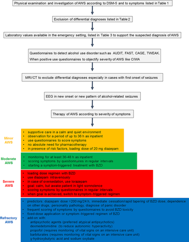
Figure 2 illustrates how to proceed in the clinical setting of suspected AWS to confirm the diagnosis and to start sufficient therapy.
10.2 Search Strategy and Selection Criteria
References for this review were identified by searches of PubMed between 1985 and 2016, and references from relevant articles. The search terms “alcohol withdrawal,” “alcohol withdrawal seizures,” “alcohol withdrawal diagnosis,” “alcohol withdrawal therapy,” “alcohol abstinence syndrome,” “abstinence treatment,” “delirium tremens,” “alcohol withdrawal EEG,” and “alcohol withdrawal MRI” were used. There were no language restrictions. The final reference list was generated on the basis of relevance to the topics covered in this review.
Acknowledgment
The authors have no acknowledgment to declare.
Declaration of Interests
None of the authors declare conflict of interests. There was no funding.


