Radiomorphometric analysis of the metapodial bones in the Scottish fold cats
Abstract
Scottish Fold cats (Felis catus, Linnaeus 1758) are one of the most well-known and popular cat breeds in the world, characterized by their folded ears attached to the head. Very frequently, cats fall prey of different trauma and accidents that can cause bone fractures especially in the metapodial bones. The method of radiometry is used in veterinary practice to visualize and measure different parts of the animal skeleton. The aim of this study was to assess the linear parameters derived from radiographic images of the metacarpals and metatarsals in Scottish Fold cats and additionally detecting potential sexual dimorphism. Radiographic images of 24 adult Scottish Fold cats (12 male and 12 females) of different ages and weights were analysed. Six linear measurements of the metapodial bones were evaluated to investigate any differences between the sexes. The linear radiometric measurements of the five metacarpals (MC1-5) and the four metatarsals (MT2-5) bones were larger in male metapodial bones than that of female cats. The maximum length (Ml) of the MC1 and MC2 was statistically different between sex, respectively, (p = 0.001) and (p = 0.05). The others metacarpal bones were different in mostly all linear parameters but not statistically significant. The most significant differences between sexes were observed in the parameter of width proximal end (Wp) of MC1-3 (p = 0.001) and MC4 (p = 0.05). More statistical different was MT2 and less MT3. The linear parameter of Bd of the MT4 was the most different statistically between sex (p = 0.001). The results of the study will be useful in function of comparative anatomy, in veterinary clinical practice, in zoo archaeology and in the veterinary forensic investigation.
1 INTRODUCTION
Scottish Fold cats (Felis catus, Linnaeus 1758) are one of the most known and popular cats in the world with round bodies and thick fur that makes them look stocky (Fournier, 2002; Wastlhuber, 1991). The folded ears attached to the head that bend forward and downward are the most distinctive feature of these cats. This morphological difference is caused by an autosomal dominant gene mutation affecting cartilage development and this mutation not only creates a simple morphological difference, but also causes a disease called hereditary osteochondrodysplasia (Chang et al., 2007; Malik, 2001; Malik et al., 1999).
Although cats are domestic animals that stay mostly in closed environments, they fall prey to road accidents, falling objects, falling from a height, fighting, crush injury and road accidents, or even dog attacks and bone fractures may include one or more metapodial bones (Phillips, 1979). These accidents cause serious damages to many parts of the cat's body, especially to the limbs skeleton.
Metacarpal (MC) and metatarsal (MT) fractures are reported to occur with an incidence of up to 5%–12% of all fractures in dogs and 3% in cats (Failing et al., 2014; Kornmayer et al., 2007; Kulendra & Arthurs, 2014; Minar et al., 2013; Polat et al., 2017). They are usually a result of trauma, most frequently road traffic accidents and falls (Muir & Norris, 1997; Phillips, 1979) even less than 10% of cat's fatalities recorded while falling from heights (Vnuk et al., 2004), most commonly affect the mid- or distal diaphysis of MC, and the proximal region of MT (De La Puerta et al., 2008; Fitzpatrick et al., 2011; Gomaa et al., 2016). The most of the fractures affecting multiple MC and MT bones (Rosselló et al., 2022).
The distal part of the hand and foot skeleton in cats include metapodial bones such as five metacarpal and five metatarsal bones which are named from medial to lateral as first, second, third, fourth and fifth metacarpal and metatarsal bone, respectively (Constantinescu, 2018; Getty, 1975; König & Liebich, 2020; Nickel et al., 1986), even the first metatarsal bone usualy is missing or is very small and does not have a phalanx at its tip (Dyce et al., 2017).
The first metacarpal bone is the shortest and thinnest, the third is the longest and the thickest of the metacarpal bones. From the metatarsal bones the third and fourth are the longest and thickest which suport the cat's body wight (Choi et al., 2021).
Metapodial bones have attracted the attention of the researchers in many different scientific fields and many studies are carried on them. These studies include obtaining morphometric data specific to animal breeds (Demiraslan et al., 2019), evaluation of bones obtained in archaeological excavations (Ince et al., 2018; Onar et al., 2015, 2018; Pazvant et al., 2015) and metapodial fractures in dogs (Muir & Norris, 1997).
In the practical veterinary clinic the method of radiometry is used to visualize and measured the different parts of the animal skeleton (Akbaş et al., 2023; Gunay et al., 2022; Gündemir et al., 2020; Gündemir, Hadžiomerović, et al., 2021). Many studies are made in this way to evaluate fetal head diameter in dogs and cats (Limmanont et al., 2019), the parameters of the foramen magnum in diferent cats breeds (Akbaş et al., 2023), carpal and phalanges morphometry in horses (Oheida et al., 2019), the morphometrical parameters of phalanges in arabian and throughbreeding horses (Gündemir, Szara, et al., 2021), variations of the distal phalanges in cattle (Manuta et al., 2024), linear paramters and angels on calcaneus in cats (Şenol et al., 2022) and horses (Erdikmen et al., 2021), description of the thoracic limbs in lions (Kirberger et al., 2005), pelvic limbs in New Zealand rabbit (Oryctolagus cuniculus) and domestic cat (Felis domestica) (El-Ghazali & El-Behery, 2018b), or to assess the sexual dimorphism in humans through calcaneus (Uzuner et al., 2016).
This method is useful, fast, costless and has resulted in an accurate method from the study of Güzel et al. (2022) and Koçyiğit and Demircioğlu (2024) where it has been proven that there is no statistical difference with the direct method of bone measurement with digital callipers. With the developments in computed tomography (CT), it has become easier to understand the differences between species such as cats dogs and birds or between sex: male and female (Demircioğlu et al., 2021; Szara et al., 2024).
The morphometric parameters of the metapodial bones in different breed cats as a reference guide are of great importance during the treatment of different fractures of the distal part of the limbs, in forensic medicine, in comparative anatomy studies and in zooarchaeology.
To date, there are very few data available in the literature regarding the linear morphometric parameters of metapodial bones in cats and especially in Scottish Fold cats.
The aim of this study was to first evaluate the parameters of linear measurements from radiographic images of the metacarpals and metatarsals of Scottish Fold cats and then investigate the presence of sexual dimorphism. Additionally, the study aimed to statistically assess the influence of age and weight of cats on the linear parameters of the metapodial bones.
These data will serve as a reference for numerous orthopaedic interventions in veterinary clinical practice, and the data of this study modestly fills the gap in the literature, which is also the main contribution of this study.
Moreover, the results of the study will be valuable for comparative anatomy studies and veterinary forensic investigations, providing valuable insights into clinical veterinary sciences.
2 MATERIALS AND METHODS
2.1 Cats and radiographic images
The study was performed on radiographic images of 24 adult Scottish Fold cats (12 male and 12 females) of different ages (from 1 to 10 years) without orthopaedic problems. The average of weight of cats were 3.5 kg for female and 5.59 kg for male. The average age of cats was 3.29 years and 2.58 years for female and male, respectively. After determining the body weight, sex and age of adult Scottish Fold cats, radiographic images of the metacarpal and metatarsal regions of both fore and hind limbs were taken from 30 cm in the dorso-ventral direction using a Fujifilm FCR Prima T2 X-ray reader and X-ray equipment X brand Ecoray Ultra HF100. Measurements of the images were taken in the Radiant DICOM program in millimetre unit (mm). In order to carry out the study, permission number 2021/5 was obtained from the Ethics Committee of Istanbul University-Cerrahpaşa, Faculty of Veterinary Medicine.
2.2 Radiometric measurements
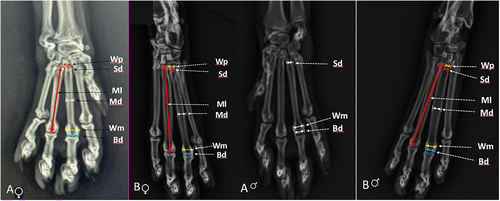
Six liner measurements of the cat metapodial bones of both sides were performed on radiographic images based on the studies of Von den Driesch (1976), Guintard and Lallemand (2003) and Pourlis et al. (2017) as below (Figure 1).

Maximum length (Ml): The distance in between the most proximal point of the base of the metacarpal or metatarsal bone and the most distal point of the head of the respective bone.
Width of proximal end (Wp): The distance between two most lateral points of the base of the metacarpal or metatarsal bone.
Smallest width of the diaphysis (Sd): The smallest width of the two most lateral points of the bone just under the proximal base of the metacarpal or metatarsal bone.
Middle width of the diaphysis (Md): The width in the middle point of the diaphysis of the metacarpal or metatarsal bone.
The greatest width of the metaphysis (Wm): The distance between the two most lateral points of the metacarpal or metatarsal head.
Width of distal end (Bd): The distance between the two most lateral points of the metacarpal or metatarsal trochlea.
The slenderness index (Si) of every metapodial bones was calculated based on the formula: Si = Ml/Md (Choi et al., 2021).
Given the possible effect of the weight differences between male and female cats a normalization was carried out. Namely, various measurements such as Ml, Sd, Wp, Bd and Wp were dividing by Md and therefore presented as ratios. The resulting data are presented in Tables 3 and 4 for the metacarpal and metatarsal bones, respectively. All measurement data were expressed as a mean ± STD and are presented in tables and figures for more detail evaluation and comparison.
2.3 Statistical analysis
The statistical analysis was performed using the SPSS 22 package program, which calculated mean values, standard deviations and p values. To determine if there were statistical differences between the sexes of the cats, the Wilcoxon signed-rank test was utilized, with significance set at p < 0.05. Additionally, correlation tests were conducted to examine the relationships and influences between the cats' sex, age and weight on the parameter results.
3 RESULTS
Radiographic images of the metapodial bones of each Scottish Fold cat were carefully examined, and none of the bones showed obvious signs of disease, fracture or orthopaedic intervention. None of the samples had the first metatrsal bone.
The results of six linear radiometric measurements conducted on the bones of the five metacarpals MC1-5 and the four metatarsals MT2-5 of both limbs (left and right) of 24 cats (12 females and 12 males) are presented as the mean value, standard deviation and p values in Tables 1 and 2. The values of all linear measurements of the left and right metapodial bones in all cats were nearly identical. However, for consistency and safety, they were recorded as the average value of both sides for the metacarpals and metatarsals bones.
| Measurements | Sex | MC1 | MC2 | MC3 | MC4 | MC5 | ||||||||||
|---|---|---|---|---|---|---|---|---|---|---|---|---|---|---|---|---|
| Mean | STD | P | Mean | STD | P | Mean | STD | P | Mean | STD | P | Mean | STD | P | ||
| Ml | F | 8.40 | 0.8 | *** | 25.60 | 1.9 | * | 29.44 | 2.63 | NS | 28.20 | 2.5 | NS | 23.70 | 2.1 | NS |
| M | 9.80 | 0.8 | 27.30 | 1.7 | 31.20 | 1.9 | 29.46 | 1.7 | 24.60 | 2.4 | ||||||
| Md | F | 2.10 | 0.3 | NS | 2.50 | 0.3 | ** | 2.77 | 0.40 | NS | 2.46 | 0.30 | NS | 2.24 | 0.30 | * |
| M | 2.30 | 0.3 | 2.83 | 0.2 | 3.10 | 0.30 | 2.73 | 0.30 | 2.45 | 0.30 | ||||||
| Sd | F | 2.30 | 0.3 | NS | 2.30 | 0.2 | * | 2.00 | 0.20 | * | 1.99 | 0.20 | * | 2.60 | 0.20 | NS |
| M | 2.30 | 0.4 | 2.38 | 0.3 | 2.21 | 0.20 | 2.21 | 0.10 | 2.42 | 0.40 | ||||||
| Wp | F | 3.10 | 0.3 | *** | 2.90 | 0.3 | *** | 2.70 | 0.40 | *** | 2.50 | 0.40 | * | 3.08 | 0.40 | NS |
| M | 3.70 | 0.2 | 3.30 | 0.4 | 3.20 | 0.20 | 2.79 | 0.30 | 3.20 | 0.50 | ||||||
| Bd | F | 2.30 | 0.3 | ** | 3.60 | 0.4 | * | 4.00 | 0.30 | ** | 3.66 | 0.40 | *** | 3.20 | 0.50 | NS |
| M | 2.70 | 0.2 | 4.00 | 0.3 | 4.38 | 0.30 | 4.10 | 0.30 | 3.50 | 0.40 | ||||||
| Wm | F | 2.70 | 0.3 | ** | 4.18 | 0.3 | ** | 4.59 | 0.30 | ** | 4.23 | 0.20 | *** | 3.78 | 0.30 | * |
| M | 3.10 | 0.3 | 4.70 | 0.3 | 4.98 | 0.20 | 4.60 | 0.30 | 4.23 | 0.40 | ||||||
| Si | F | 4.00 | 0.39 | * | 10.24 | 0.89 | NS | 14.50 | 1.06 | NS | 11.45 | 1.18 | NS | 10.57 | 1.13 | NS |
| M | 4.26 | 0.63 | 9.66 | 0.99 | 10.06 | 1.09 | 10.78 | 0.87 | 10.04 | 2.08 | ||||||
- * P < 0.05.
- ** P < 0.01.
- *** P < 0.001.
| Measurements | Sex | MT2 | MT3 | MT4 | MT5 | ||||||||
|---|---|---|---|---|---|---|---|---|---|---|---|---|---|
| Mean | STD | P | Mean | STD | P | Mean | STD | P | Mean | STD | P | ||
| Ml | F | 39.40 | 3.60 | * | 46.60 | 3.80 | NS | 46.50 | 4.30 | NS | 42.30 | 3.70 | NS |
| M | 42.70 | 3.20 | 49.10 | 3.20 | 49.60 | 3.20 | 44.50 | 3.20 | |||||
| Md | F | 3.00 | 0.30 | ** | 4.20 | 0.30 | * | 3.40 | 0.20 | NS | 2.60 | 0.20 | * |
| M | 3.40 | 0.30 | 4.60 | 0.30 | 3.80 | 0.30 | 2.90 | 0.30 | |||||
| Sd | F | 2.90 | 0.30 | NS | 2.50 | 0.40 | NS | 2.90 | 0.40 | NS | 3.20 | 0.50 | NS |
| M | 2.80 | 0.30 | 2.80 | 0.50 | 2.90 | 0.30 | 2.80 | 0.60 | |||||
| Wp | F | 3.40 | 0.20 | NS | 3.10 | 0.50 | NS | 3.80 | 0.50 | NS | 4.90 | 0.60 | ** |
| M | 3.30 | 0.40 | 3.20 | 0.60 | 4.20 | 0.60 | 5.50 | 0.40 | |||||
| Bd | F | 3.90 | 0.30 | ** | 4.70 | 0.40 | NS | 4.30 | 0.20 | *** | 3.50 | 0.40 | NS |
| M | 4.40 | 0.40 | 5.40 | 0.30 | 4.70 | 0.30 | 4.00 | 0.20 | |||||
| Wm | F | 4.60 | 0.40 | ** | 5.40 | 0.30 | NS | 4.90 | 0.30 | ** | 4.20 | 0.30 | NS |
| M | 5.10 | 0.40 | 6.00 | 0.30 | 5.30 | 0.30 | 4.70 | 0.30 | |||||
| Si | F | 13.13 | 1.81 | NS | 11.10 | 3.2 | NS | 13.68 | 3.58 | NS | 16.27 | 2.67 | NS |
| M | 12.56 | 2.11 | 10.67 | 3.67 | 13.05 | 2.56 | 15.34 | 3.46 | |||||
- * P < 0.05.
- ** P < 0.01.
- *** P < 0.001.
In general, all measurements of male metapodial bones were larger than that of female cats. From the data of metacarpal bones both in female and male cats, the longest and widest was MC3 followed by MC4, MC2, MC5 and the shortest and slenderness was MC1.
The longest and widest of metatarsal bones in female cats was MT3 and MT4 (almost same length, Table 1), MT5 and the shortest was MT2 but, in male cats the longest was MT4 and MT3 (almost same length, 46.6 mm and 46.5 mm, respectively; Table 2) followed by MT5 and the shortest was MT2, similar as in female cats.
From the comparison of the maximum length of the respective metacarpal and metatarsal bones, it was found that the MC2-5/MT2-5 ratio in female cats was 0.65; 0.63; 0.61 and 0.56 and in male cats, it was 0.64; 0.63; 0.59 and 0.55, respectively.
The data of the Table 1, show that the maximum length (Ml) of the MC1 and MC2 was statistically different between sex, respectively (p = 0.001) and (p = 0.05). The others metacarpal bones were different in mostly all linear parameters but not all were statistically different. The most significant differences between sex was the linear parameter of Wp of MC1-3 (p = 0.001) and MC4 (p = 0.05) but not statistically different at the MC5.
Differences between sex in metatarsal bones were less than metacarpal bones. More statistical different was MT2 (four parameters) and less MT3 (only one parameter). The linear parameter of Bd of the MT4 was the most different statistically between sex (p = 0.001).
Metapodial slenderness index was calculated for all metacarpal (MC1-5) and metatarsal bones MT2-5 and the values are given in Tables 1 and 2. From these results, there were differences between sexes but only the slender index of MC1 was statistically different.
The results of the Tables 3 and 4 show the ratio between the measurements like Ml, Sd, Wp, Bd and Wp dividing by Md in order to exclude the effect of the body weight of the cats to these metapodial measurements. The ratios of measurements were also different between female and males cats. The most different ratios were in the MC1 and MT2. The MC3 to MC5 and MT2, MT4 and MT5 had only one ratio statistically different. None of the MC2 and MT3 ratios were statistically different.
| Measurements | Sex | MC1 | MC2 | MC3 | MC4 | MC5 | ||||||||||
|---|---|---|---|---|---|---|---|---|---|---|---|---|---|---|---|---|
| Mean | STD | P | Mean | STD | P | Mean | STD | P | Mean | STD | P | Mean | STD | P | ||
| Ml/Md | F | 3.97 | 0.39 | ** | 10.31 | 0.89 | NS | 10.45 | 0.80 | NS | 11.50 | 0.82 | * | 11.10 | 1.22 | NS |
| M | 4.36 | 0.45 | 9.84 | 0.75 | 10.38 | 0.96 | 10.93 | 0.74 | 10.23 | 1.41 | ||||||
| Sd/Md | F | 1.08 | 0.13 | NS | 0.91 | 0.07 | NS | 0.72 | 0.09 | NS | 0.80 | 0.11 | NS | 1.21 | 0.20 | *** |
| M | 1.02 | 0.12 | 0.88 | 0.08 | 0.74 | 0.07 | 0.79 | 0.05 | 0.97 | 0.21 | ||||||
| Wp/Md | F | 1.47 | 0.16 | *** | 1.16 | 0.05 | NS | 0.96 | 0.09 | *** | 1.01 | 0.13 | NS | 1.45 | 0.21 | NS |
| M | 1.64 | 0.16 | 1.19 | 0.10 | 1.06 | 0.09 | 1.02 | 0.10 | 1.32 | 0.21 | ||||||
| Bd/Md | F | 1.08 | 0.12 | * | 1.42 | 0.12 | NS | 1.42 | 0.15 | NS | 1.50 | 0.14 | NS | 1.48 | 0.24 | NS |
| M | 1.18 | 0.13 | 1.43 | 0.10 | 1.45 | 0.09 | 1.53 | 0.14 | 1.43 | 0.13 | ||||||
| Wm/Md | F | 1.28 | 0.14 | * | 1.69 | 0.11 | NS | 1.66 | 0.21 | NS | 1.73 | 0.20 | NS | 1.78 | 0.29 | NS |
| M | 1.40 | 0.14 | 1.67 | 0.13 | 1.65 | 0.11 | 1.72 | 0.12 | 1.71 | 0.15 | ||||||
- * P < 0.05.
- ** P < 0.01.
- *** P < 0.001.
| Measurements | Sex | MT2 | MT3 | MT4 | MT5 | ||||||||
|---|---|---|---|---|---|---|---|---|---|---|---|---|---|
| Mean | STD | P | Mean | STD | P | Mean | STD | P | Mean | STD | P | ||
| Ml/Md | F | 13.31 | 1.46 | NS | 11.01 | 0.84 | NS | 13.88 | 1.33 | NS | 16.43 | 1.93 | NS |
| M | 12.74 | 1.31 | 10.65 | 0.96 | 13.10 | 1.44 | 15.48 | 2.06 | |||||
| Sd/Md | F | 0.96 | 0.06 | *** | 0.59 | 0.09 | NS | 0.88 | 0.14 | * | 1.24 | 0.18 | *** |
| M | 0.84 | 0.09 | 0.60 | 0.12 | 0.77 | 0.14 | 0.95 | 0.20 | |||||
| Wp/Md | F | 1.13 | 0.08 | ** | 0.74 | 0.13 | NS | 1.14 | 0.15 | NS | 1.91 | 0.23 | NS |
| M | 1.00 | 0.12 | 0.68 | 0.12 | 1.10 | 0.18 | 1.92 | 0.19 | |||||
| Bd/Md | F | 1.30 | 0.09 | NS | 1.12 | 0.10 | NS | 1.28 | 0.08 | NS | 1.34 | 0.15 | NS |
| M | 1.30 | 0.13 | 1.16 | 0.06 | 1.23 | 0.13 | 1.39 | 0.15 | |||||
| Wm/Md | F | 1.55 | 0.16 | NS | 1.28 | 0.10 | NS | 1.46 | 0.13 | NS | 1.63 | 0.14 | NS |
| M | 1.53 | 0.15 | 1.30 | 0.07 | 1.40 | 0.14 | 1.63 | 0.17 | |||||
- * P < 0.05.
- ** P < 0.01.
- *** P < 0.001.
The proximal base width of the metapodials (the sum of Wp parameter of every metacarpal MC1-5 or metatarsal bones MT1-4) of front and hind limbs were almost same size; the maximum width of the metacarpal base in female and male cats was 14.28 ± 0.36 mm and 16.19 ± 0.32 mm, respectively. The maximum width of the metatarsal base in female and male cats was 15.2 ± 0.45 mm and 16.2 ± 0.5 mm, respectively.
In female Scottish Fold cats, the metacarpal bones exhibited a greater slenderness index in the following order: MC4, MC5, MC3, MC2 and finally MC1. Conversely, in male cats, this range followed the order of MC4, MC3, MC5, MC2 and MC1.The slenderness index of metatarsal bones in both female and males cats were MT5 followed by MT4, MT2 and the last was MT3.
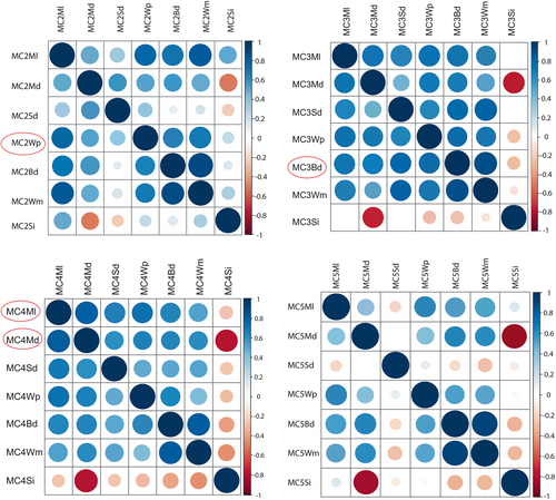
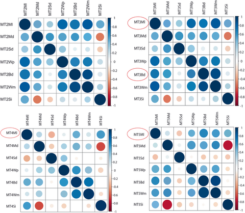
To see the influence of age and weight on the sexual dimorphism at the linear parameters of metapodial bones of Scottish Fold cats, the correlation test was used and it resulted that the linear parameters of metacarpal bones: MC2-Wp, MC3-Bd, MC4-Ml, MC4-Md and parameters of metatarsal bones: MT3-Ml, MT3-Bd, MT4-Ml and MT5-Ml were influenced by cat weight between 80% and 86% (Figures 2 and 3).


4 DISCUSSION
Scottish Fold cats are a popular and recognized breed worldwide. They are found in many countries across the globe, and their popularity continues to grow due to their unique appearance and friendly demeanour (Fournier, 2002; Malik, 2001; Wastlhuber, 1991). Clinical veterinary practice has shown that there are many cases of damage to different parts of the skeleton of cats as a result of car accidents, falls from a height or various diseases that affect the skeleton and mostly metapodial bones (Phillips, 1979). Fractures of metacarpals and metatarsals bones in cats are reported to occur with an incidence of up to 3% of all fractures (Kornmayer et al., 2007; Minar et al., 2013), mostly from road traffic accidents and falls (Muir & Norris, 1997; Phillips, 1979) and affect the mid or distal diaphysis of MC, and the proximal region of MT (De La Puerta et al., 2008; Fitzpatrick et al., 2011; Gomaa et al., 2016). The more detailed data on their linear morphometric parameters or its dimensions, the easier the intervention for immobilization and surgery of these fractures will be.
The metapodial skeleton in cats as in the other carnivores species is composed of five metacarpal and five metatarsal bones (Constantinescu, 2018; Dyce et al., 2017; Getty, 1975; König & Liebich, 2020; Nickel et al., 1986). However, in this study, not a single first metatarsal bone was present in any of the Scottish Fold cat samples.
The morphometric parameters of the metapodial bones in different breed cats as a reference guide are of great importance during the treatment of different fractures of the distal part of the limbs, in forensic medicine, in comparative anatomy studies and in zooarchaeology.
The primary goal of this study was to address the scarcity of literature on the linear morphometric parameters of metapodial bones in cats by providing detailed measurement results based on radiographic images of Scottish Fold cats' metapodial bones which also constitutes the novelty of this research.
The radiometry method as a useful, fast, cost-free and accurate method (Güzel et al., 2022: Koçyiğit & Demircioğlu, 2024) is used to visualize and measure different elements of skeletons from a large number of animal species like dog (Mutlu et al., 2023), dog and cats (Akbaş et al., 2023; Limmanont et al., 2019; Şenol et al., 2022), cattle, (Ince et al., 2015; Telldahl, 2015), sheep (Kahraman et al., 2022), horses (Erdikmen et al., 2021; Gündemir, Hadžiomerović, et al., 2021; Oheida et al., 2019), lions (Kirberger et al., 2005), New Zealand rabbit and cats (Oryctolagus cuniculus) (El-Ghazali & El-behery, 2018a, 2018b).
The results of this study carried out on the radiografic images of metapodium of Scottish Fold cats of differents ages and weights on both sexes show the six linear parameters of five metacarpals and four metatarsal bones in detail and aditionally the slendernes index for each of them. The mean values of all linear parameters of metapodial bones of male cats were usually larger than females. In metacarpals, the longest and widest bone was MC3 followed by MC4, MC2, MC5 and the shortest and slenderness was MC1. In metatarsal bones the longest and widest in female cats was MT3 and MT4, MT5 and the shortest was MT2 but, in male cats the longest was MT4 and MT3, followed by MT5 and the shortest was MT2. These results are a bit different with the study of Choi et al. (2021) conducted in domestic shorthair cats were the longest metacarpal bone was MC-2 and in the metatarsals it was MT-5.
The maximum length of MC3 as the longest bone of metacarpals was 29.44 ± 2.63 mm and 31.20 ± 1.9 mm, respectively in female and male cats. The maximum length of the MT3 as the longest bone of the metatarsals in female cats was 46.60 ± 3.8 mm and in males the longest bone was MT4 which was 49.6 ± 3.2 mm which were almost similar with the study of Day and Jayne (2007) which have calculated the length of the front and hind limbs in domestic cats as 24 ± 1 cm (22–26) and 33 ± 1 cm (30–35), and the metacarpals and metatarsals were calculated as 14.6 ± 0.3% (3.5 cm) of the total length of the front leg and 22.1 ± 0.3% (7.26 cm) of the hind legs, respectively.
Even the differences in all linear parameters between male and females of Scottish Fold cats were present not all were statistically significant. The maximum length (Ml) of the MC1 and MC2 was statistically different between sex, respectively (p = 0.001) and (p = 0.05). The most significant differences between sex was the linear parameter of Wp of MC1-3 (p = 0.001) and MC4 (p = 0.05). Differences between sex in metatarsal bones were less than metacarpal bones. More statistical different was the second metatarsal (MT2) in four linear parameters like Ml, Md, Bd and Wm and less MT3 (only the parameter—Md). The linear parameter of Bd of the MT4 was the most different statistically between sex (p = 0.001).
From the data of this study, the most significant different element in the determination of sexual dimorphism based on statistical data was Wm in all metacarpal bones but MC4 which was 4.1 ± 0.30 mm in female and 4.23 ± 0.20 mm in male cats was the most significant (p = 0.001) and in metatarsal bones was significant only in MT2 as 4.6 ± 0.40 mm in female and 5.1 ± 0.40 mm in male cats (p = 0.01) and MT4 as 4.9 ± 0.30 mm in female and 5.3 ± 0.30 mm in male cats, respectively.
In order to exclude the effect that the body size of male and female cats has on the different measurements, the ratios of the division of all measurements with the corresponding width of each metacarpal and metatarsal bone were calculated. From Tables 3 and 4 we see that the values of the measurements according to the sexes are different and 45 reports in total, only 9% or 16% of them are statistically different. Perhaps the not very large number of samples constitutes a shortcoming of the study, but still some ratios of measurements are truly statistically different (p = 0.001) such as Wp/Md (MC1 and MC3) and Sd/Md in MC5, MT2 and MT4.
In the study conducted in the cadaveric examinations of metapodial bones in shorthair cats by Choi et al. (2021) was proven the gender-specific variation related to the length and width of these bones. In the same study was shown that, the mean length of the metacarpals in female cats were MC2–19.46 ± 1.23 mm; MC3–22.53 ± 1.41 mm; MC4–21.60 ± 1.41 mm and MC5–17.83 ± 1.18 mm and in male cats were MC2–20.62 ± 1.06 mm; MC3–23.78 ± 1.13; MC4–22.90 ± 1.41 mm and MC5–19.01 ± 1.42 mm. The length of metatarsals in female cats were MT2–31.62 ± 1.97 mm; MT3–34.81 ± 2.04 mm, MT4–35.07 ± 2.2 mm and MT5–33.19 ± 2.09 mm and in male cats were MT2–34.09 ± 1.86 mm; MT3–37.43 ± 2.16 mm, MT4–37.94 ± 1.9 mm and MT5–36.21 ± 1.92 mm. The width of metacarpal bones varies from 1.79 ± 0.15 mm MC1 to 2.3 ± 0.15 mm MC3 and 1.95 ± 0.18 mm MT5 to 3.38 ± 0.23 MT3. The most significant difference was identified for the width of MC5 (p = 0.0008). These data on shorthair cats were smaller compare to the data of this study on Scottish Fold cats (Tables 1 and 2).
The metapodial slenderness index calculated for all metacarpal and metatarsal bones shown differences between sexes of Scottish Fold cats but only the slender index of MC1 was statistically different (p = 0.05). The greater slenderness index of metacarpal bones in female cats were at the MC3 (14.5 ± 0.8) and MT5 (16.27 ± 2.67) at the metatarsal bones of the females cats. The results of this study are comparable with the results of the Choi et al. (2021) which presents no significant differences in the slenderness index between sex and the females cats have more slender metapodial bones and regardless of the sex the most slender metacarpal bone was MC4 (10.81 ± 0.65) and the most slender bone was MT5 (17.15 ± 1.46). The most thickest metacarpal were MC2 and MC3 and maybe this is related with the stress that affecting these bones when the cats are landing from the different levels of height as it is confirmed by Xu et al. (2022), which discovered that the stress is mainly concentrated at the metacarpal and proximal phalanx especially the MP2 had the highest stress level which was mainly concentrated in the middle and rear of the MP2 and the MP5, and the distal end of the MP3. This can also explain with the slenderness index of the MC2 and MC5 which in this study were the thickest bones.
The linear parameter of the proximal base width of every metapodial bones especially in cats are very small and very important to evaluate in special cases for the treatment of fractures of these bones mostly with the open method like Kirschner wires (K-wire) that used intramedullary pins but the smallest width of every metapodial bones shaft is more important in order to make the proper calculation what intervention technique to choose (Karsli, 2022; Kulendra, 2014; Rosselló et al., 2022).
Scottish Fold cats have genetic features of bone damage as a result of genetic mutations during creation, they are very vulnerable to skeletal damage from diseases but also from trauma and accidents. The database with complete data on linear morphometric parameters for all metacarpals and metatarsals of Scottish Fold cats constitutes a valuable asset for comparative anatomical studies, studies in the field of clinical veterinary practice but also various findings in the field of veterinary forensics or even of zooarchaeology.
Since the Scottish Fold cats are suffering very often from the Osteochondrodysplasia disease or osteoarthritis in the veterinary clinical praxis are shown many cases with different symptoms like: inflexible tails and shortened splayed feet, signs of lameness, reluctance to jump, stiff and stilted gait (Allan, 2000; Chang et al., 2007; Malik et al., 1999), these patients are more susceptible to have fractures or need the specialization orthopaedic intervention and the reference data of linear parameters of the metapodial bones are very important as there are cases of size changes, shape disorders and loss of symmetry of the bones between the respective limbs.
In the radiological studies of El-Ghazali & El-behery, 2018a; El-Ghazali & El-Behery, 2018b in thoracic and hind limbs concluded in rabbits and in domestic cats of unidentified breed was shown the relative length of the manus region in cats (metacarpals) as about 6.70 ± 0.249 cm and also the relative length of the pes region (metatarsals) as about 11.160 ± 0.437 cm which are relatively larger than the results of this study.
The indirect distal width of the metapodials (the sum of Wm parameter of every metacarpal (MC1-5) or metatarsal bones MT1-4) of front and hind limbs were almost same size; the maximum width of the metacarpal base in female and male cats was 19.48 ± 0.28 mm and 21.62 ± 0.3 mm, respectively. The maximum width of the metatarsal base in female and male cats was 19.1 ± 0.33 mm and 21.1 ± 0.33 mm, respectively. The ratio between the respective width and the third metacarpal/metatarsal bone was about 0.6 and 0.4, respectively. This fact is also quite similar with the comparative study between clouded leopard (Neofelis nebulosa) and unknown breed of domestic cat (Felis catus) prepared by Hubbard et al. (2009) on radiographic images, shown the relative size of the manus and pes between these species. The width was measured as the distance between the lateral boarders of digits 2 and 5 with a line that passed between the metacarpal/metatarsal phalangeal joints of digits 3 and 4, and the length was the length of the third metacarpal/metatarsal bone. The width average of the manus in domestic cats was 1.67 cm and the length of the third metacarpal was 2.77 cm with the ratio 0.6 between them, but the pes width was 1.73 cm and the third metatarsal was 4.33 cm with the ratio about 0.4.
5 CONCLUSION
Based on the radiometric measurements extracted from the radiographic images, this study has provided comprehensive data on the six linear morphometric parameters of both the metacarpals and metatarsals in Scottish Fold cats. Our findings highlight the presence of sexual dimorphism, providing a valuable foundation for future research in areas such as comparative anatomy, forensics, zooarchaeology and clinical veterinary practice, particularly concerning surgical interventions for fractures.
ACKNOWLEDGEMENTS
This study was obtained from the results of Utku OĞUZ's doctoral thesis titled ‘Radiometric measurements analysis of metapodial bones in Scottish Fold breed cats’. The authors are very greateful to both referees of the paper for numerous suggestions and corrections. Moreover, the idea of the normalization to offset for weight differences was kindly suggested by one of the referees.
CONFLICT OF INTEREST STATEMENT
The authors declared no potential conflicts of interest with respect to the research, authorship and/or publication of this article.
Open Research
DATA AVAILABILITY STATEMENT
The data that support the findings of this study are available from the corresponding author upon reasonable request.


