Macroscopic and microscopic comparison of pecten oculi in different avian species
Abstract
The current study aims to present differences between the pecten oculi of different avian species through morphologic, macroscopic, light, and electron microscopic examinations. The study is a comprehensive research on seven avian species (sparrowhawk, hawk, magpie, swan, heron, pheasant, duck). The right eyes of the animals utilized in the study were removed for light microscopic examination, whereas their left eyes were removed for electron microscopic and macroscopic examinations. Morphometric analyses, as well as stereo and light microscopic measurements, were carried out on the pecten oculi of the animals. Given all these data, it was determined that the height of the pecten oculi did not differ among the species in the study; however, the pecten oculi were larger in birds with the highest value compared to the other species in the macroscopic measurements. Also, the pecten oculi vessels were larger, and the number of melanocytes was higher in keen eyesight, raptor, and migratory birds with large bulbus oculi. All these data suggest that the pecten oculi not only supplies nutrient to the retina but also contributes to sharp vision during migration and hunting, UV absorption from sunlight, as well as preservation of intraocular equilibrium.
1 INTRODUCTION
The intraocular structure of birds differs anatomically from that of mammals (Reese et al., 2009). Avian ocular anatomy shows many polymorphisms in itself; one of the anatomical formations in which this polymorphism appears is the pecten oculi (Dayan & Ozaydin, 2013; Korkmaz & Harem, 2021). The pecten oculi is an organ found only in the eyes of birds that extends from the retinal entrance of the optic nerve towards the vitreous body (Ince et al., 2017; Yilmaz et al., 2017). This anatomical structure is significant since it performs several tasks, such as supplying nutrients to the retina, which lacks vessels and nerves, eliminating metabolic wastes, regulating intraocular pH and pressure, and balancing the corpus vitreum (Bellhorn, 1997; Brach, 1975; Kiama et al., 2001; Tucker, 1975).
The pecten oculi has three distinct morphological structures: conical (in kiwis), vaned (in ostriches), and pleated (found in most the bird species; Baumel et al., 1993; Dayan & Ozaydin, 2013; Kiama et al., 2006; Meyer, 1977). Only histological and electron microscopic examinations can present the number of pleats and their structural differences. The structural variation of pecten oculi in different bird species is thought to depend on the hunting time. Pecten oculi has a smaller and simpler structure in the nocturnal birds compared to diurnal birds (Ince et al., 2017; Micali et al., 2012; Onuk et al., 2013; Rahman et al., 2010).
The pecten oculi, composed of neuroectoderm, is a melanocyte pigment-containing structure with abundant and different diameters of vessels (Gelatt, 2013). The number of melanocytes may vary across avian species. Melanocytes are mostly found in the apical part and have the task of protecting blood vessels from Ultraviolet rays (Kiama et al., 1994). The vitreo-pecteneal membrane surrounds the pecten oculi (Uhera et al., 1996). On this basal membrane, some avian species contain macrophage-like cells called as hyalocytes (Ince et al., 2017; Korkmaz & Kum, 2016; Yilmaz et al., 2021).
This study analysed the pecten oculi samples of different avian species (sparrowhawk, hawk, magpie, swan, heron, pheasant, duck) through macroscopic, stereo, light and electron microscopic examinations. The differences were established between the pecten oculi of the species as a result of the assessment and literature comparisons, and migration, hunting, eating habits, and sizes of the bird species were compared with this difference. Based on all these data, an explanation for the differences in the pecten oculi among bird species was sought.
2 MATERIALS AND METHODS
2.1 Sample collection
The samples of pecten oculi from the eyes of different bird species [sparrowhawk (Common sparrowhawk) (Accipiter nisus), hawk (Common buzzard) (buteo buteo), magpie (Common magpie) (Pica pica), swan (Black swan) (Cygnus atratus), heron (Common little bittern) (Ixobrychus minutus), pheasant (Common pheasants) (Phasianus colchicus), duck (Common teal) (Anas crecca)] that were brought to Harran University Veterinary Faculty Animal Hospital for treatment but could not be rescued or were brought as dead for necropsy were utilized in the study. Three adults from each species (the age of maturity varies by species) were used in the study. The right eyes of the animals were removed for light microscopic examination, whereas their left eyes were removed for electron microscopic and macroscopic examinations. The ethical approval (date: 20/06/2019 and number: 51025321-050.05.04) was obtained from the Harran University Animal Experiments Local Ethics Committee.
2.2 Morphometric analysis
Morphometric data of bulbus oculi of all animals included in the study were measured using a stereomicroscope (Olympus-SZX7, Olympus Optical) with an attached camera (Olympus Cam-SC50). The eyeballs were cut from the equatorial region and the pecten oculi was uncovered after dorsoventral (CDV) and laterolateral diameter (CLL) of the cornea, dorsoventral diameter of bulbus oculi (BODV), and laterolateral diameter of bulbus oculi (BOLL) were measured (Table 1). The basal border length and heights of pecten oculi of the uncovered pecten oculi were measured to determine the number of pleats (Table 2). For the study's nomenclature, Nomina Anatomica Avium (Baumel et al., 1993) was utilized.
| Groups | CDV | CLL | BODV | BOLL |
|---|---|---|---|---|
| SPARROWHAWK | 8.39d | 9.02d | 15.26ab | 16.50d |
| HAWK | 11.39b | 11.46b | 21.67a | 20.76b |
| MAGPIE | 7.56g | 8.17f | 19.31ab | 13.64f |
| SWAN | 10.83c | 10.80c | 19.28ab | 19.17c |
| HERONS | 12.11a | 12.67a | 22.33a | 21.67a |
| PHEASANT | 8.31e | 8.54e | 13.17b | 15.05e |
| DUCK | 7.82f | 7.77g | 12.62b | 12.18g |
| SE | 0.390 | 0.386 | 1.091 | 0.752 |
| p | * | * | * | * |
- Abbreviations: BODV, dorsoventral diameter of bulbus oculi; BOLL, laterolateral diameter of bulbus oculi; CDV, dorsoventral diameter of cornea; CLL, laterolateral diameter; SE, standard error.
- a–gThe differences in mean represented by different letters in the same column are significant, *p < 0.05.
| Groups | Height | Length |
|---|---|---|
| Sparrowhawk | 8.400 | 2.933d |
| Hawk | 9.533 | 4.100c |
| Magpie | 8.700 | 3.100d |
| Swan | 6.170 | 4.933b |
| Herons | 8.600 | 5.933a |
| Pheasant | 8.267 | 3.933c |
| Duck | 6.400 | 2.300e |
| SE | 0.404 | 0.261 |
| p | ns | * |
- Abbreviation: SE, standard error.
- a–eThe differences in mean values represented by different letters in the same column are ns: non-significant. *p < 0.05.
2.3 Histological analyses
Tissue samples taken for light microscopic examinations were fixed in 10% neutral buffered formalin solution for 24 h and then washed for 24 h. Following routine tissue follow-up procedures, tissue samples were embedded in paraffin after being processed through graded alcohols (50%–70%–80%–96%–100%) and xylol. Serial sections of 5 μm thickness at 50 μm intervals were cut from these paraffin blocks and stained using Crossman's modified triple staining method to examine the general structure (Crossman, 1937; Denk et al., 1989). The stained preparations were examined and photographed under an Olympus Cover (018 model) microscope (Olympus DP71 Olympus Optical). The cellsens image software analysis program was used for the vessel measurements in serial sections (from three randomly selected regions for each animal; Olympus BX53). The mean vessel diameters of the animals in each group were determined using standard deviations (Table 3).
| Groups | LV | SV |
|---|---|---|
| Sparrowhawk | 45.9861c | 20.1850c |
| Hawk | 67.2900a | 35.9336a |
| Magpie | 41.2825d | 13.9019c |
| Swan | 40.0503d | 21.8167b |
| Heron | 46.5594c | 18.4925c |
| Pheasant | 42.0131d | 19.3236c |
| Duck | 51.0347b | 19.6075c |
| SE | 0.70448 | 0.46542 |
| p | * | * |
- Abbreviation: SE, standard error.
- a–eThe differences in mean values represented by different letters in the same column; *p < 0.05.
2.4 Scanning electron microscopic (SEM) examinations
Tissue samples taken for electron microscopic examinations were washed twice with phosphate-buffered solution (0.1 M, pH 7.4) and fixed in 2.5% glutaraldehyde for 48 h. The fixed tissue samples were kept in 1% osmium tetroxide for 1 h for the second fixation, dehydrated through a series of increasing concentrations of graded acetone (25%, 50%, 75%, and 100%) and dried using a critical point drier (Dunlap & Adaskaveg, 1997). These tissue samples were coated with gold–palladium, examined with SEM (Scanning Electron Microscope-Zeiss, Evo 50) at Harran University Central Laboratory, and photographed at different magnifications.
2.5 Statistical findings
The data were statistically analysed using the spss package program and the One-way anova model. The Duncan Test was applied to determine the difference between the groups and check the significance of this difference (Heiberger & Neuwirth, 2009).
3 RESULTS
3.1 Macroscopic results
When examined macroscopically, it was determined that dark brown pecten oculi extended from the retina towards the vitreous body, where nervus opticus (NO) entered the eye in all animal species. The pecten oculi had three main structures: the basal, apical, and pleats.
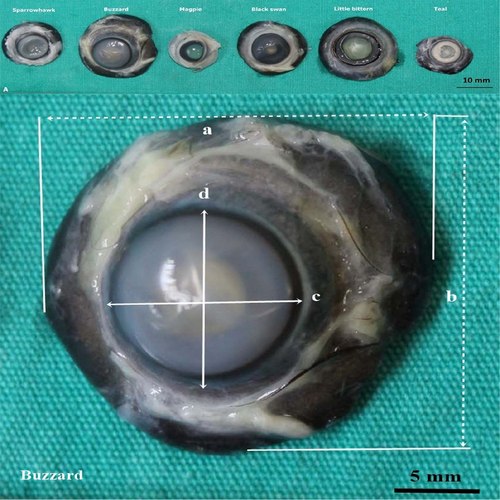
CDV, CLL, BODV, and BOLL were measured macroscopically in sparrowhawk, hawk, magpie, swan, heron, pheasant, and duck, respectively (Table 1; Figure 1).
The basal length of the pecten oculi and height of the pecten oculi were measured in sparrowhawk, hawk, magpie, swan, heron, pheasant, and duck in stereomicroscopic examinations (Table 2; Figure 2).
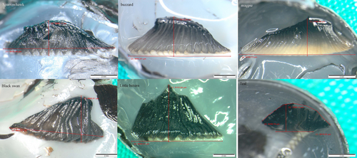
When examined macroscopically, the number of pleats in sparrowhawk, hawk, magpie, swan, heron, pheasant and duck was counted as 14–16, 15–17, 27–30, 10, 11–12, 20–21 and 12, respectively.
3.2 Microscopic results
3.2.1 Sparrowhawk (common sparrowhawk)
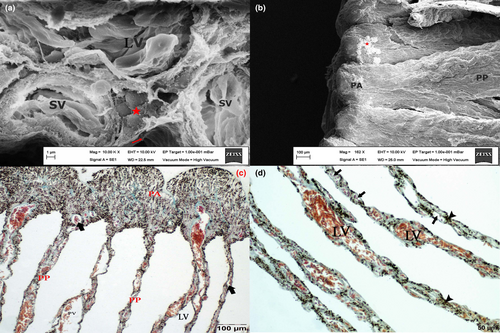
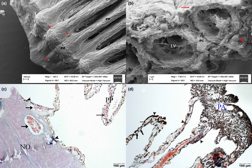
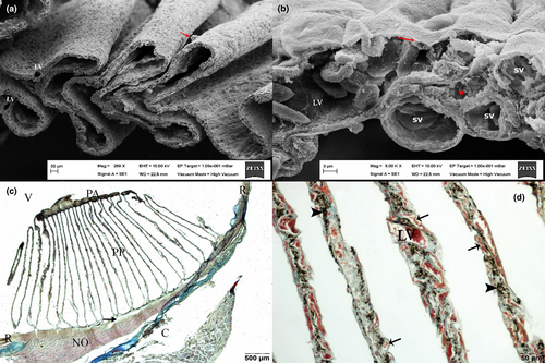
When examined at small magnifications, it was observed that the origin of the pecten oculi was not limited to the optic nerve, but continued on the retina. Large vessels and melanocytes were found at the basal part of the pecten oculi. The pleats of pecten oculi that extended towards the vitreous body were folded and returned in the apical part (Figure 3c). Extensive accumulation of melanocytes was remarkable at the apical part of the pleats (Figure 3c,d). Large vessels were located in the core of the pleats of the pecten oculi, while small vessels and melanocytes were located on the pleat periphery (Figure 3a–d). In the basal part of the pecten oculi, in addition to the vessels, an extensive accumulation of melanocytes also drew attention. The vessels with large diameters were found extensively in the pleats of the pecten oculi. Light microscopic examinations revealed no hyalocytes on the pecteneal membrane, but in SEM a substantial number of hyalocytes was remarkable (Figure 3b).
3.2.2 Hawk (common buzzard)
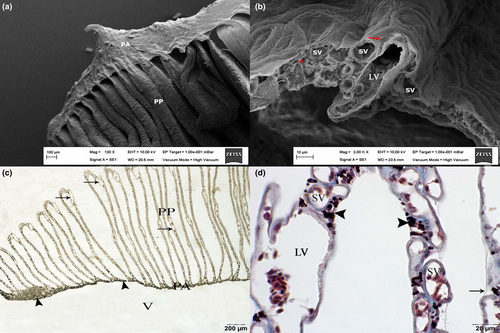
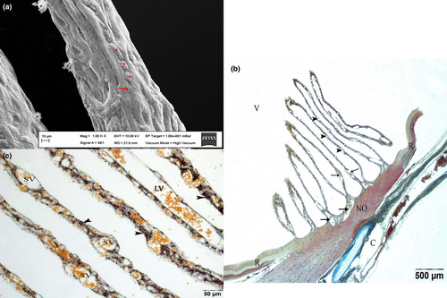
The optic nerve, located between the open ends of the retina, was identified as the entrance point of the pecten oculi of the hawk to the vitreous body (Figure 4c). The vessels with large diameters and melanocytes were found extensively in the basal part of the pecten oculi (Figure 4c,d). In the pleats of the pecten oculi, in addition to the large and small vessels, an extensive accumulation of melanocytes also drew attention (Figure 4b). As in the sparrowhawk, it was observed in the hawk that large-diameter vessels were concentrated in the core of the pleats of the pecten oculi, whilst small vessels and melanocytes were located on its periphery. As with the sparrowhawk, it was discovered that pleats of the pecten oculi extended towards the vitreous body, curved at the apical part, and then returned to the basal part (Figure 4d). Extensive accumulation of melanocytes was observed at the apical part. Pecteneal hyalocytes were identified in the hawk in both light and electron microscopic (Figure 4a).
3.2.3 Magpie (common magpie)
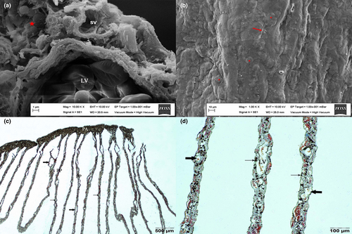
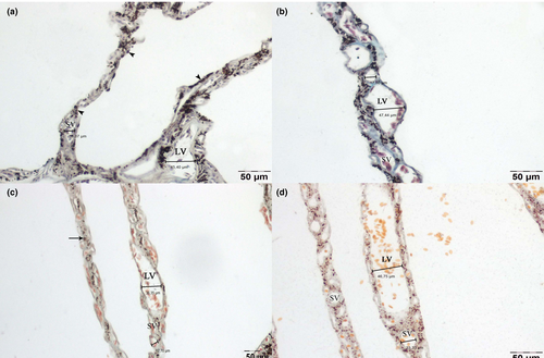
Although the NO appeared to be the origin point of the pecten oculi, it was observed that the pecten oculi continued in the ruptured retina, which is located to the right and left of the NO. The retinal pigmented epithelium was found underneath the basal part of the pecten oculi in these regions. Unlike the sparrowhawk and hawk, no melanocytes were identified in the basal part of magpie's pecten oculi, despite the presence of blood vessels. Again, whereas the vessels with small and moderate diameters were concentrated in the pleats of pecten oculi, melanocytes were found in extremely limited quantities (Figure 5b–d). However, pleats were rich in melanocytes in the apical part of the pecten oculi (Figure 5c), which are bound to each other by bridges. However, subjectively, it was observed that this richness was not as high as that of a hawk or sparrowhawk. Neither light nor electron microscopic examinations distinguished the hyalocytes (Figure 5a).
3.2.4 Swan (black swan)
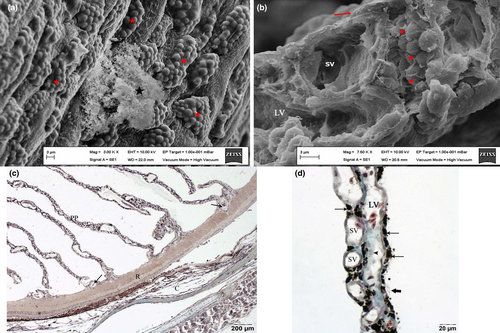
It was found that the pecten oculi not only arose from the optical nerve but also continued on the retina. The large vessels and pigments from the choroidea were observed to be the sources of vessels and melanocytes at the basal part of the pecten oculi. In the basal part of pecten oculi, in addition to the large vessels, an extensive accumulation of melanocytes also drew attention (Figure 6a–d). Extensive melanocytes were identified around the blood vessels in pleats of pecten oculi extending from the basal part towards the vitreous body (Figure 6d). In the swan, as was the case with the sparrowhawk and hawk, it was observed that the pleats folded back after forming a bridge in the apical (Figure 6c), and the presence of extensive melanocytes were observed in this part of the swan. While no hyalocytes were found in light microscopic examinations, electron microscopic examinations drew attention to hyalocytes on the vitreo-pecteneal membrane in pleats of pecten oculi (Figure 6b).
3.2.5 Heron (common little bittern)
The pecten oculi was observed to arise from the area of the optical nerve and continued on the retina on both sides of the optic nerve (Figure 7c). Blood vessels and melanocytes were found in the basal part of the pecten oculi (Figure 7b). In the pleats of pecten oculi of the heron, it was found that large vessels were located at intervals, and rich small vessels and melanocytes were around these vessels (Figure 7d). Melanocytes mostly concentrated in the apical part of the pleats (Figure 7d), extending towards the vitreous body and returning. Pecteneal hyalocytes were observed in light microscopic examinations (Figure 7d) and electron microscopic examinations (Figure 7a).
3.2.6 Pheasant (common pheasants)
It was observed that the pecten oculi arose from the optic nerve and extended towards the vitreous body and was settled in a very short area over the retina. The large vessels in the pecten oculi's basal part originated from the choroidea (Figure 8c). Blood vessels and melanocytes were found in the basal part of the pecten oculi (Figure 8b). Many small vessels and melanocytes encircled large vessels that were settled in the middle of the pleats of pecten oculi (Figure 8d). Melanocytes were concentrated in the apical part (Figure 8c). While hyalocytes could not be distinguished in light microscopic examinations, many hyalocytes were observed in electron microscopic examinations (Figure 8a).
3.2.7 Duck (common teal)
It was observed that the pecten oculi originated from the NO and also continued on the retina. Melanocytes were not identified in the pecten oculi's basal part, despite large blood vessels (Figure 9b). Small and medium vessels were located around the large vessels in the middle of pleats of the pecten oculi (Figure 9c). Melanocytes were observed to be concentrated in the apical part. Although hyalocytes could not be distinguished in light microscopy, they could be detected through electron microscopy (Figure 9a).
3.3 Statistical results
When analysed statistically, the avian species having the highest CDV, CLL, BODV and BOLL values was determined to be the heron. Besides, the avian species having the lowest CDV, CLL, BOLL, and BODV values were magpie and duck. Table 1 shows the statistical differences between the other groups.
When analysed statistically, no difference was identified among the groups in terms of heights of the pecten oculi. However, the species with the longest basal length of the pecten oculi was the heron, followed by swan, hawk, pheasant, magpie, and sparrowhawk, respectively. The duck was the species having the shortest basal length.
Large and small vessel diameters of sparrowhawk, hawk, magpie, swan, heron, pheasant, and duck were put forth by microscopic measurements. The data indicated a statistical difference in both large and small vessel diameters among the species. As a result of the analyses, the hawk had the largest large vessels diameter (67.29 μm), followed by the duck (51.03 μm), the heron (46.55 μm), and the sparrowhawk (45.98 μm). The diameters of a pheasant, magpie, and swan were measured as 42.01, 41.28, and 40.05 μm, respectively. When small vessel diameters were examined, the hawk again had the largest vessel diameter (35.93 μm), followed by the swan (21.81 μm). Small vessel diameters measured in hawk, duck, pheasant, heron, and magpie were 20.18, 19.60, 19.32, 18.49, and 13.90 μm, respectively (Figure 10).
4 DISCUSSION
According to the studies conducted by The World Bird Database (Avibase) since 1992, there have been roughly 10,000 species and 20,000 subspecies of birds worldwide (Lepage, 2014). Many factors, such as their survival conditions and feeding habits, have been considered when classifying birds. Today, studies in various fields are being conducted to identify class differences.
Avian is a different class of animals, there are some differences in basic organ structures (Baumel et al., 1993). The avian retina is one of these differences. The avian retina, unlike mammals, is fully avascular, and it is supported by a unique structure known as the pecten oculi. The pecten oculi, also known as the fundus oculi when initially noticed in 1917 (Wood, 1917), is a structure that many researchers have studied until today (Braekevelt, 1994; Elghoul et al., 2022; Mishra & Meshram, 2019; Smith et al., 1996).
Previous studies identified the pecten oculi with different sizes and numbers of pleats in many different animal species. On the other hand, all pecten oculi are composed of the pecten vessels, pericytes surrounding the vessels, and melanocytes. The pecteneal limiting membrane surrounds all of these structures (Orhan et al., 2011; Pourlis, 2013; Rahman et al., 2010; Yilmaz et al., 2017). Nevertheless, some species, like the Australian Galah (Breakevelt & Richardson, 1996), the budgerigar (Micali et al., 2012), and the chicken (Korkmaz & Kum, 2016), mention the presence of pecteneal hyalocytes, which are estimated to be macrophages, on this limiting membrane, these cells have not been reported at all in species, such as ostrich (Korkmaz et al., 2017), quail (Orhan et al., 2011), and Barred owl (Smith et al., 1996). In the present comparative study, electron microscopic examination confirmed the presence of hyalocytes that could not be identified by light microscopic examination in sparrowhawk, swan, pheasant, and duck. However, hyalocytes identified in hawk and heron using both light and electron microscopic examinations, were not found in magpie using either light or electron microscopic examinations. Three studies report that hyalocytes may be a special subtype of macrophages in birds (Korkmaz & Kum, 2016; Llombart et al., 2009; Uhera et al., 1996). In mammals, however, hyalocytes have been described as irregularly shaped granular cells that express many tissue macrophage markers and are particularly distributed in the vitreous body cortex (Qiao et al., 2005). Given all these data, the following conclusions have been drawn. Hyalocytes, which are reported to be situated on the pecteneal limiting membrane, can be found free in the vitreous body in birds. Hyalocytes identification may be difficult in light microscopy because of their separation from the pecten oculi during histological preparation. However, since hyalocytes are a species of macrophage, their numbers might be expected to grow under certain pathological conditions such as inflammation. This might explain why it is identified in some species but never mentioned in others.
It was observed that there were significant differences among species in the number of pleats forming pecten oculi. A large number of pleats were found in birds such as ostrich (Korkmaz & Harem, 2021; 20–24 pleats), American crow (Braekevelt, 1994; 22–25 pleats) and Australian galah (Breakevelt & Richardson, 1996; 20–25 pleats), while the birds such as the Barred owl (Smith et al., 1996; 8–10 pleats), duck (Moselhy & El-Hady, 2019; 10–14 pleats), and Black kite (Kiama et al., 1994; 12 pleats) had few pleats. As a result of the present comparative study, the number of pleats in sparrow, hawk, magpie, swan, heron, pheasant and duck was determined to be 14–16, 15–17, 27–30, 10, 11–12, 20–21 and 12, respectively.
When the study results were examined, the magpie, a small bird from the Corvidae family and included in the Pica pica family (Kaplan, 2019), had a small eye. Magpie had pecten oculi similar to the other bird species in height and mean basal length. However, it had the most pecten pleats. The heron, often known as the little bittern, is a migratory bird in the Ardeidae (pelicans) family (Wallace, 2013). Despite having a relatively large eye, the little bittern was determined that it had pecten oculi of similar height to other bird species. The basal part length of heron pecten oculi was longer than that of other species, and pecten oculi had the least pleats. The diurnal raptor hawk (Buteo buteo), which is related to the same family as the sparrowhawk (Accipiter), is considered to have a UV refracting keen eyesight and migrates by detecting the earth's magnetic field (Lind et al., 2013). The pecten oculi in the hawk, which had larger eyes than any other species observed in the study, was of similar height, mean basal length, and mean pleat count as the other birds. Given all these data, it was determined that the live weight and the size of the eye of the animal did not show parallelism with the size of the pecten oculi and the number of pleats. However, it was determined that all bird species had pecten oculi of similar sizes, but the basal length of pecten oculi was longer in species with large bulbus oculi.
When the animal species were compared in the study, the pecten oculi of magpie had the minimum number of melanocytes, while the heron had the maximum. An extensive accumulation of melanocytes was noticed in the pecten oculi of the sparrowhawk, hawk, and swan, particularly in the apical part. The studies have reported that uveal melanocytes provide homeostasis in the human eye, play roles in light absorption, regulate oxidative stress, and participate in immunological regulation, angiogenesis, and inflammation (Sitiwin et al., 2019). The data suggested that melanocytes, uncommon in non-migratory birds such as magpies, are prevalent in species such as hawks, herons, and swans that make long migration flights, which may be correlated to the light absorption and homeostasis required during migration. This might account for the difference in the number of melanocytes.
In studies, the vessel diameters were reported as 20.23 μm in ostrich (Dayan & Ozaydin, 2013), 15 μm in chickens (Dietrich et al., 1973), 11.78 μm in pigeons and 12.78 μm in starlings (Dayan & Ozaydin, 2013). In some literature blood vessels were called ascending and descending blood vessels or large and small blood vessels (Kiama et al., 1994). In the present comparative study, vessels were classified and measured as large and small. This terminology was made in order to understand the vessels detected in different sizes in microscopes. Hawks had the largest vessel diameters, (67.29 μm in large and 35.93 μm in small), whereas pheasants (42.01–19.32 μm), magpies (41.28–13.90 μm), and swans (40.05–21.81 μm) had the smallest diameters. When the diameter measures were compared with the literature, it was determined that there were differences. We believe this is associated with using of different microscopes and counting programs. Therefore, it is believed that drawing a conclusion based on measurements made in the present study would provide more accurate information.
5 CONCLUSION
Given all these data in this study, it was determined that the height of the pecten oculi did not differ across animal species in the study. However, the pecten oculi was larger in birds with the highest macroscopic value in measurements made in the present study compared to the other species. Also, the vessels of pecten oculi were larger and the number of melanocytes were higher in keen-sighted, raptor, and migratory birds with large bulbus oculi. All these data suggest that the pecten oculi not only supply nutrients to the retina but also contribute to a keen vision during migration and hunting, UV absorption from sunlight, and preservation of intraocular equilibrium.
Consequently, the study examined comparatively macroscopic, light, and electron microscopic features of pecten oculi from many different bird species, revealed differences and similarities and sought an answer for the reasons for these differences and similarities. This aspect of the study is thought to provide the researchers with a broad perspective.
ACKNOWLEDGEMENTS
The authors would like to thank Harran University Veterinary Faculty Animal Hospital.
FUNDING INFORMATION
This study is not supported.
CONFLICT OF INTEREST STATEMENT
The authors declare that they have no conflicts of interest with regard to the work presented in this report.
Open Research
DATA AVAILABILITY STATEMENT
The data set supporting the conclusions of this article are included within the article.


