A comparison of embalming fluids on the structures and properties of tissue in human cadavers
Abstract
Cadaveric material has long been used to teach anatomy and more recently to train students in clinical skills. The aim of this study was to develop a systematic approach to compare the impact of four embalming solutions on the tissues of human cadavers. To this end, a formalin-based solution, Thiel, Genelyn and Imperial College London soft-preservation (ICL-SP) solution were compared. The effect of these chemicals on the properties of the tissue was assessed by measuring the range of motion (ROM) of joints and measuring the dimensions of different structures on computed tomography (CT) images before and after embalming. The mean changes in the ratio (angle to ROM) differed statistically between embalming methods (Welch Statistic 3,1.672 = 67.213, p = 0.026). Thiel embalmed cadavers showed an increase in range of motion while ICL-SP cadavers remained relatively the same. Genelyn and formalin embalmed cadavers registered a notable decrease in range of motion. Furthermore, investigation into the impact of the embalming chemicals on the dimensions of internal organs and vessels revealed that Thiel embalming technique leads to a decrease in the dimension of the cardiovascular system alone while formalin-based solutions maintain the shape of the organs and vessels investigated. Our findings suggest that the joints of cadavers’ embalmed using ICL-SP technique may faithfully mimic that of unembalmed cadavers and that formalin is necessary to retain shape and size of the organs and vessels investigated in this study. Despite this, a study with larger numbers of cadavers is required to confirm these findings.
1 INTRODUCTION
The use of human cadavers for learning and research purposes has a long history (Balta, Cronin, Cryan, & O'Mahony, 2015). The use of human cadavers to teach anatomy has been described as the most superior method to learn about the human body, even though technology has introduced many new teaching tools (Patel & Moxham, 2008). Cadavers are also widely used for training in different medical specialties such as surgery, radiology and clinical skills (Tsui, Dillane, Pillay, Ramji, & Walji, 2007; Turner, Mellington, & Ali, 2005). Moreover, medical equipment companies use human cadavers to test new instruments and develop new tools that can improve the practice of medicine (Eisma & Wilkinson, 2014).
Embalming is the process of introducing chemicals into a subject post-mortem to prevent decay (Balta, Cronin, et al., 2015). Different chemicals are used as part of the embalming solution including formaldehyde, glutaraldehyde, phenol, glycerin, bronopol, ethanol and glycol. Studies have reported changes in the quality of tissue after embalming, which can be considered a disadvantage. Ideally, an embalmed cadaver should present the same features as that of an unembalmed cadaver (Balta, Cronin, et al., 2015). However, this is not possible with most of the current chemicals being used during the embalming process. For example, cadavers embalmed using formaldehyde are inflexible and produce some features that are not representative of an unembalmed cadaver (Hubbell, Dwornik, Alway, Eliason, & Norenberg, 2002). Nevertheless, some of these chemicals such as those used in Thiel embalming are able to improve the quality of imaging post-mortem, producing a better model to facilitate training in certain procedures such as ultrasound-guided nerve blocks (Balta, Twomey, et al., 2017; Benkhadra et al., 2009). Other disadvantages include expenses involved in the embalming process and potential health risks that could be encountered when working with certain chemicals (Brenner, 2014). Formaldehyde has been reported in several studies as a potential carcinogen, and phenol is an exposure hazard that is corrosive to the throat and stomach among other health complications (Balta, Cronin, et al., 2015; Brenner, 2014).
Thiel embalming was introduced in 1992 by Walter Thiel, initially as a method of colour preservation (Thiel, 1992). This technique set the basis for a new photographic atlas of practical anatomy (Thiel, 1992). Thiel embalmed cadavers have been described as more representative of the human body compared to formalin embalmed cadavers (Balta, Lamb, & Soames, 2015; Fessel et al., 2011). Several studies have reported the benefits of using Thiel embalmed cadavers for training in surgical and radiological procedures (Balta, Cronin, et al., 2015). For example, Thiel embalmed cadavers were described as an ideal tool to train on flap raising and microvascular suturing (Balta, Cronin, et al., 2015).
Genelyn embalming solution is a commercial product that produces a stiff and more brittle cadaver compared to that of an unembalmed cadaver (Norton-old et al., 2013). This was observed when Genelyn embalmed cadavers were used to train on three-/four-dimensional ultrasound for epidural catheter insertion (Balta, Cronin, et al., 2015).
An understudied solution is the Imperial College London soft-preservation (ICL-SP) solution (Prof. Ceri Davies- Imperial College London, personal communication) which was first described in 2009 for a cadaver-based workshop (Barton et al., 2009). In this study, ICL-SP cadavers were dissected to improve the surgical anatomy knowledge of gynaecological oncologists’ trainees (Barton et al., 2009).
No studies have been published comparing the quality of tissue, of the same cadavers, before and after being embalmed using different techniques. A recent study compared the joint flexibility of cadavers embalmed using Thiel technique, Genelyn and Dodge solutions to that of an unembalmed cadaver (Jaung, Cook, & Blyth, 2011). Measuring the joint flexibility provides a comprehensive idea on the effect of the embalming chemicals on the muscles, tendons and joints of embalmed cadavers. It was noted that the joints of Thiel embalmed cadavers were more flexible than that of a different unembalmed cadaver (Jaung et al., 2011). Another study compared the range of motion of cadavers embalmed using a new saturated salt solution (SSS), to that of Thiel and formalin solutions. This study concluded that the joints of SSS-embalmed cadavers are softer when compared to formalin embalmed cadavers, but harder than that of Thiel embalmed cadavers (Hayashi et al., 2014, 2016 ).
Measurements of organs and tissue of human cadavers have been reported in literature (Ishida et al., 2015; Okuma et al., 2013; Takahashi et al., 2013). Changes have been reported such as a decrease in the size of the aorta after death and bilateral adrenal glands (Ishida et al., 2015; Takahashi et al., 2013).
The aim of this study was to develop a systematic approach to assess embalming solutions used to preserve human cadavers. This approach was used to compare the effect of Thiel, ICL soft-preservation, Genelyn and formalin-based solutions on the structure and properties of tissue in human cadavers. In this study we also aimed to assess the use of the understudied ICL soft-preservation solution for the embalming of human cadavers.
2 MATERIALS AND METHODS
The study was carried out under the auspice of the “License to Practise Anatomy”, granted by the Irish Medical Council under the Anatomy Act 1832. For the purpose of the study, eight human cadavers were embalmed using four different embalming techniques; Thiel, formalin, Genelyn and ICL-SP solution. Donors premorbidly signed written consent to use their bodies for education and research by the Department of Anatomy and Neuroscience. All cadavers were admitted into the department 24–48 hr after death and stored at −20⁰C. Cadavers were thawed for 5 days after which data was collected and followed by embalming on the same day. Cadavers treated using the same technique were embalmed at the same time, in the same department and by the same embalmer. Table 1 includes information on the eight donors used in the study.
| Donor | Gender | Race | Age | Height (cm) | Cause of death |
|---|---|---|---|---|---|
| Thiel 1 | M | Caucasian | 83 | 168 | Pneumonia |
| Thiel 2 | M | Caucasian | 69 | 184 | Uraemia, kidney cancer |
| Formalin 1 | M | Caucasian | 82 | 170 | Cardiac failure |
| Formalin 2 | F | Caucasian | 64 | 150 | breast cancer, secondary in the spine |
| Genelyn 1 | F | Caucasian | 84 | 167 | Pneumonia, myasthenia gravis and heart disease |
| Genelyn 2 | M | Caucasian | 69 | 173 | Bladder cancer |
| ICL-SP 1 | M | Caucasian | 88 | 183 | Respiratory infection |
| ICL-SP 2 | M | Caucasian | 74 | 178 | Pancreatic cancer with gall bladder metastasis |
Appendix Tables 1-3 are the chemicals used for the embalming solutions for formalin, Thiel and ICL-SP solution, while the Genelyn solution was based on the commercial products provided by the European Embalming Products Company Ltd.
2.1 Range of motion
The range of motion of several joints was measured before and after embalming using a standard goniometer (BaselineTM15cm) and was carried out as previously described (Jaung et al., 2011) in collaboration with a senior physiotherapist, Ms. Orla Duggan.
Before recording any measurements, the cadaver was moved into a supine anatomical position. When recording the measurements, the joints were in zero position and the proximal joint components were stabilized. Bony landmarks were identified and palpated to accurately position the goniometer. The stationary arm of the goniometer was aligned at the proximal component of the joint, the body of the goniometer was positioned at the junction of the joint and the free arm was aligned at the distal component of the joint. The investigator passively moved the joint to end of range of available motion without exerting any overpressure. The goniometer was read by two investigators blinded to the embalming method.
The joints and orientation measured were cervical flexion, shoulder (abduction and flexion), elbow flexion, wrist flexion, second metacarpophalangeal joint flexion, second proximal interphalangeal joint flexion, hip (flexion and abduction), knee flexion, ankle plantarflexion, metatarsophalangeal and interphalangeal joint flexion of the hallux. All measurements were carried out on the right side of each of the cadavers before and after embalming.
2.2 Measurement of components of organs and vessels
Whole body multi-detector scanning including the brain, thorax, abdomen and pelvis was performed before and after embalming by two specialist CT radiographers using a 128-slice Discovery HD 750 (GE Healthcare GE Medical systems, Milwaukee WI, USA). Cadavers were scanned 2–6 hr before embalming, and post-embalming scans were performed 24–48 hr after the completion of the embalming process. The cadavers were scanned in a supine position, cranio-caudally, and with their arms by their sides. The following CT parameters were used in conjunction with automatic exposure control: tube voltage: 120kVp; gantry rotation time: 0.8 s; collimation: 40 x 0.62 mm; and pitch factor: 0.98. Images were reconstructed from an acquisition thickness of 0.625 mm to a final slice thickness of 1.25 mm. All images were reconstructed from the raw-data acquisitions using the standard departmental protocol employing hybrid iterative reconstruction (60% filtered back projection and 40% ASiR, adaptive statistical iterative reconstruction (GE Healthcare, GE Medical Systems, Milwaukee, USA).
Quantitative analysis was performed independently by two radiographers who were blinded to both the embalmment status and the embalming agent used. Images were analyzed on a picture archiving and communication system (PACS) (Impax 6.3.1; Agfa Healthcare, Mortsel, Belgium).
Size measurements of various organs were taken before and after embalming as follows: Brain: pons (maximum transverse diameter), lateral ventricles (maximum transverse and anteroposterior diameters); heart: left and right cardiac ventricles (maximum diameter orthogonal to the long axis of the heart at the level of the mitral and tricuspid valves respectively including the chamber wall), respiratory system: left and right lungs (maximum craniocaudal length); liver: (maximum craniocaudal length in the mid-clavicular line, maximum anteroposterior and transverse dimensions); spleen: (maximum craniocaudal length), left and right kidney (maximum craniocaudal length) and reproductive structures: prostate or uterus (maximum transverse dimension).
The long and short axis dimensions of the ascending, descending and abdominal aorta in the transverse plane were measured at the level of the tracheal bifurcation and superior mesenteric artery origin, respectively. The maximum diameter was taken as the long axis and the short axis was orthogonal to it as observed on CT scans of the ascending, descending and abdominal aorta. To evaluate the shape of the aorta, the long axis-short axis ratio was calculated using the following equation: Long axis-short axis ratio =length of long axis x 100/length of short axis.
2.3 Statistical analysis
Statistical analysis was performed using SPSS statistical package version 22 (IBM Corp., Armonk, NY). In order to combine the data from different joints into an overall score to give a whole body indication, a ratio was determined using a denominator, which is the average range of motion provided by the American Academy of Orthopaedic Surgeons. The assumption of homogeneity of variance could not be tested due to the small sample size, and therefore ANOVA Welch's test for equality of differences was used in order to test if the change (pre-minus post-embalming) ratio was different for the four embalming methods. A post-hoc test was not used due to the small sample size. Instead the mean changes are described.

An ANOVA with the Welch test for equality of group means was applied to the composite score differences to test if the change (pre-minus post-embalming) in score was different for the four embalming methods. Fourteen out of the 19 structures were included in the statistical analysis.
3 RESULTS
3.1 The range of motion of joints from the different embalming techniques
Both cadavers embalmed using Thiel technique had an increased range of motion after embalming, while cadavers embalmed using ICL soft-preservation technique maintained a relatively similar range of motion. On the other hand, both cadavers embalmed using a formalin-based solution demonstrated a substantial decrease in the range of motion compared to a smaller decrease for cadavers embalmed using Genelyn solution. Figure 1 shows a scatterplot of the range of motion (angles) of all joints measured from cadavers before and after being embalmed using the different techniques with a line of equality (y = x) superimposed.
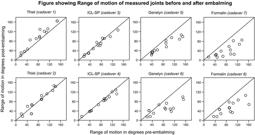
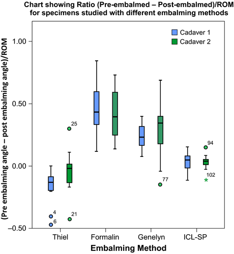
Figure 2 summarizes the ratio of pre-post embalming angle to the average range of motion provided by the American Academy of Orthopaedic Surgeons for all joints by method and donor. Thiel embalmed cadavers show negative values while ICL-SP cadavers closer to zero followed by Genelyn embalmed cadavers and formalin embalmed cadavers with the highest positive ratio reflecting a marked decrease in range of motion.
The ratios for all of the joints measured for each of the cadavers are represented in Table 2. The mean changes (pre-minus post-embalming) in the ratio (angle to ROM) differed statistically between embalming methods (Welch Statistic 3,1.672 = 67.213, p = 0.026) (Table 3). Thiel embalmed cadavers showed an increase in range of motion while ICL-SP cadavers remained relatively the same. Genelyn and formalin embalmed cadavers registered a notable decrease in range of motion.
| Method | Donor | Pre-Embalming Ratio (Angle to ROM) | Post-Embalming Ratio (Angle to ROM) |
Difference (Pre - Post) |
|---|---|---|---|---|
| Thiel | 1 | 0.706 | 0.873 | −0.168 |
| Thiel | 2 | 0.807 | 0.849 | −0.042 |
| Formalin | 1 | 0.861 | 0.399 | 0.463 |
| Formalin | 2 | 0.958 | 0.543 | 0.416 |
| Genelyn | 1 | 0.886 | 0.647 | 0.239 |
| Genelyn | 2 | 0.834 | 0.534 | 0.3 |
| ICL-SP | 1 | 0.67 | 0.637 | 0.033 |
| ICL-SP | 2 | 0.819 | 0.784 | 0.035 |
| Method | N of Donors | Pre-Mean Ratio (Angle to ROM) | Post-Mean Ratio (Angle to ROM) | Difference (Pre - Post) | Fa(3,1.672) | Sig.b |
|---|---|---|---|---|---|---|
| Formalin | 2 | 0.910 | 0.471 | 0.439 | 67.213 | 0.026 |
| Thiel | 2 | 0.756 | 0.861 | −0.105 | ||
| Genelyn | 2 | 0.860 | 0.590 | 0.270 | ||
| ICL-SP | 2 | 0.744 | 0.710 | 0.034 |
Note
- a asymptotically F distributed.
- b Welch statistic.
Supporting information Table S1 includes the raw data of all measured joints before and after embalming using the four techniques.
3.2 The measurements of internal organs and vessels
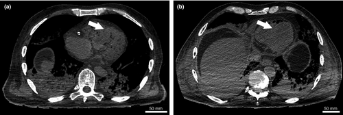
An axial CT scan of the left heart ventricle of a Genelyn embalmed cadaver shows an increase in size post-embalming (white arrows) (Figure 3). The low attenuation fluid containing chamber can be distinguished from the chamber wall. While an axial CT scan of a Thiel embalmed cadaver shows that the splenic and portal veins have collapsed post-embalming (white arrows) (Figure 4), while the volume of fluid in the retroperitoneal fat has reduced. It is clearly visible that the renal shape is maintained post-embalming.

All the measurements taken from all different organs and structures are reported in Supporting information Table S2. There was no difference between rates for 67.4% of the measurements, 98.7% agreed to within ±0.01 mm and the rest were within ±0.02 mm. The intraclass correlation coefficient (ICC) average measure using a two-way mixed model with absolute agreement was 0.99998.
Composite scores per donor were determined for pre- and post-embalming and their difference as described in the statistical analyses section above.
The composite score (average of z-scores for the 14 structures) for each cadaver before embalming and after embalming is represented in Table 4. The difference determined was post – pre therefore negative values express a decrease in dimension. Fourteen out of 19 structures were included in the statistical analysis. The uterus and the prostate glands were excluded as not all cadavers had these structures given that we included both male and female cadavers. The short diameter of the ascending, descending and the abdominal aorta were excluded as their values were markedly higher making it inaccurate to include in the overall composite score. Analysis indicates that the change in composite score was trending towards significance only (p = 0.058) for the four embalming methods (Table 5).
| Method | Donor |
Pre-Embalming Composite Score |
Post-Embalming Composite Score |
Difference Post–Pre |
|---|---|---|---|---|
| Thiel | 1 | 0.038 | −0.36 | −0.397 |
| Thiel | 2 | 1.226 | 0.737 | −0.488 |
| Formalin | 1 | 0.084 | 0.178 | 0.094 |
| Formalin | 2 | −0.491 | −0.579 | −0.089 |
| Genelyn | 1 | −0.12 | −0.214 | −0.095 |
| Genelyn | 2 | 0.076 | −0.076 | −0.152 |
| ICL-SP | 1 | −0.397 | −0.348 | 0.049 |
| ICL-SP | 2 | 0.131 | 0.116 | −0.015 |
| Method | N of Donors | Pre-Mean Composite Score | Post-Mean Composite Score | Difference (Post-Pre) | Fa(3,2,133) | Sig.b |
|---|---|---|---|---|---|---|
| Thiel | 2 | 0.632 | 0.189 | −0.443 | 14.44 | 0.058 |
| Formalin | 2 | −0.204 | −0.201 | 0.003 | ||
| Genelyn | 2 | −0.021 | −0.145 | −0.124 | ||
| ICL-SP | 2 | −0.133 | −0.116 | 0.017 |
Note
- a asymptotically F distributed.
- b Welch statistic.
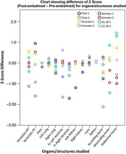
The difference in dimension between organs varied among each cadaver and between cadavers embalmed by the same technique as represented by the Z-score difference (Figure 5) indicating that more cadavers are required per technique to determine the outcome.
4 DISCUSSION
As the repertoire of embalming techniques increases, it is important to evaluate the differences between them. Here, we show the relative utility of four different embalming techniques and solutions. Although most anatomy departments still use one embalming technique for their teaching, in the past decade some departments have started using different methods for postgraduate courses or clinical training (Balta, Cronin, Cryan, & O'Mahony, 2017; Benkhadra et al., 2009). The results from this study highlight the importance of considering the pre-embalming conditions of cadavers used for embalming research. This was used to show that the understudied ICL soft-preservation technique faithfully mimics the range of motion of an unembalmed cadaver. Moreover, being able to measure the difference in the dimension of various organs and vessels show that this could be a potential non-invasive method to assess the internal organs of embalmed cadavers.
Different anatomical and pathological factors influence the range of motion of cadavers making the comparison between cadavers embalmed with the same solution, without considering its pre-embalming status, inaccurate. In this study, we look at the condition of cadavers before and after embalming and here, we show using different modalities the benefits of such comparison. A clear example of anatomical influences in the current study can be seen in one of the cadavers embalmed with Thiel technique, which had an unusually long neck that lead to neck flexion of 110° while the second cadaver embalmed with Thiel had only 56° of neck flexion. If post-embalming values were only considered, this would show an inconstant effect of the chemicals on cadavers embalmed by the same technique. Another example is the first cadaver embalmed with ICL technique which had a smaller range of motion when flexing the hip due to bilateral hip replacement compared to that of the second cadaver embalmed with the same embalmment. Measuring the range of motion of cadavers before embalming adds some technical difficulties, as cadavers are usually embalmed promptly after being admitted into the mortuary but adds to the accuracy of the comparison process.
Many of the characteristics of Thiel embalmed cadavers have been studied (Benkhadra et al., 2009; Hayashi et al., 2014; Jaung et al., 2011). In this study, the range of motion of joints from the first cadaver embalmed with Thiel technique are above the y = x line. This means that the range of motion increased post-embalming due to the Thiel technique. These results do not exactly match those of the second cadaver embalmed with Thiel, though some joints did show increased range of motion post-embalming. Thiel cadaver number 1 was fully embalmed after 3 months of being submerged in the tank, which is the average period (Eisma, Lamb, & Soames, 2013), while Thiel cadaver number 2 required 6 months. However, Thiel embalming induced an increase in the range of motion in both cadavers. These findings match those by Jaung et al.in 2011 and by Hayashi et al. in 2014, though a different assessment method has been used. This could mean that the soft tissue within the joint, the tendon attached to the joint or the muscular tissue has been influenced by the chemicals used in the Thiel solution (Fessel et al., 2011; Liao, Kemp, Corner, Eisma, & Huang, 2015).
ICL soft-preservation technique is a novel strategy and both cadavers that were embalmed using this technique had relatively similar pre- and post-embalming range of motion. The difference between pre- and post-embalming values were the closest to zero. This means that the embalming solution did not affect the joint flexibility and therefore faithfully mimics the range of motion of an unembalmed cadaver. This could be due to the alcohol present in the solution and lack of formaldehyde.
Meanwhile, cadavers embalmed by the Genelyn solution had joints with a smaller range of motion post-embalming. These findings corroborate with other studies that describe Genelyn embalmed cadavers as stiff and lack flexibility (Belavy, Ruitenberg, & Brijball, 2011; Norton-old et al., 2013). This is due to formaldehyde being present in the solution (Balta, Cronin, et al., 2015), though the quantity of formaldehyde present is considered to be low compared to that of formalin-based solution.
Formaldehyde causes a notable decrease in the range of motion post-embalming. This is due to the swelling of soft tissue in the joints and fixing effect on muscles and tendons (Balta, Cronin, et al., 2015; Entius, Rijn, Zwamborn, Kleinrensink, & Robben, 2004). The change in the ratio (angle to the average ROM as described by the American Academy of Orthopaedic Surgeons) from pre- to post-embalming was significantly different between the embalming methods. Formalin produced the largest change followed by Genelyn, with ICL-SP solution showing little change and Thiel leading to a modest increase. Using these measurements, we show that Genelyn embalmed cadavers are not statistically different to formalin embalmed cadavers. The ratio of pre-post embalming angle over the range of motion of all joints shows that both Thiel and ICL soft-preservation techniques are closer to zero than Genelyn and significantly different to that of formalin embalmed cadavers.
The most recently published embalming solutions claim to mimic the tissue of the living human body. This is very important considering the recent increase in the use of cadavers for clinical training and the fact that they are used as commercial products (Balta, Cronin, et al., 2015). Medical doctors from different specialties are continually researching a high-fidelity training tool to train junior doctors in order to reduce clinical error (Balta, Cronin, et al., 2015). With the lack of quantitative methods to assess the differences in the quality of tissue, it is nearly impossible to objectively compare different embalming solutions. For this reason, this study aimed to quantify the change in the dimension of internal organs and structures before and after embalming with different embalming techniques.
After measuring different structures before and after embalming on CT images, several values were excluded when calculating the composite score. The uterus and the prostate glands were excluded as not all cadavers had these structures. The short diameter of the ascending, descending and the abdominal aorta were excluded as the difference between pre- and post-embalming values were substantially higher than the others making it inaccurate to include in the overall z-score.
When looking at CT scans taken from cadavers before and after embalming, a general observation could be made. Embalming fluids circulate in the body through blood vessels and this affects tissues differently potentially due to the different chemicals within the solution. When chemicals are first injected, they will expand blood vessels and highly vascularized structures, primarily the heart and lungs and then other structures such as the liver. If the embalming solution contains fixing agents such as formaldehyde, this will lead to fixing the tissue in its enlarged state as seen in our study. Meanwhile, if the solutions do not have strong fixing agents the tissue will collapse as we observed with the other solutions.
It was observed that changes in dimension mainly occurred in the thoracic region (see Supporting information Table S2). This supports the concept that the dimension of highly vascularized structures is most affected by the embalming process. The overall composite score of Thiel embalmed cadavers decreased in size compared to other embalming techniques. This change was not statistically significant and therefore we cannot conclude that the effect of embalming on the dimensions of organs and vessels measured differed among the methods. This may be related to our small number size per embalming method.
This study suggests that using CT images to assess the influence of embalming techniques on the tissue of human cadavers is an effective method. Further research should include different parameters such as bone density and tissue reconstruction. Moreover, different surgical approaches should be used to dissect these embalmed cadavers and assess their suitability for different techniques.
Different limitations restrict the findings of this study in specific and any cadaver-based studies. With an average of 20 donations admitted per year, it was difficult to recruit a larger number of cadavers for the study. A multicenter study could help in overcoming this limitation. While different structures were measured to increase the statistical power, caution needs to be exerted when interpreting statistical results based on a sample of eight cadavers, with two per embalming solution. Definitive conclusions are not possible without larger studies. Another limitation is the unknown medical history of the donor as we do not have access to the donors’ medical background which could possibly affect the embalming process. Moreover, inter-cadaveric variations could affect the embalming process and hence the quality of tissue.
5 CONCLUSION
Different embalming techniques are used for different purposes and it is important to initiate the process of assessing these techniques using quantitative methods for the benefit of education and research. This is the first comparative study that uses a systematic approach to assess different embalming solutions by comparing cadavers before and after embalming. Moreover, we were able to highlight the benefits of using the alcohol-based solution ICL soft-preservation technique for the embalming of human cadavers.
Our findings confirm that joints of cadavers embalmed using ICL soft preservation solution faithfully mimics joints of unembalmed cadavers. This may support its use for cadaver-based surgical courses such as orthopaedic surgery and rheumatology.
In conclusion, there is no ideal embalming technique as many factors need to be taken into consideration such as period of preservation and antimicrobial abilities. Other factors include the elimination of any hazardous chemicals and their use in embalming procedures. This project comes to support the effort of pushing embalming into a modern-day science with research at its heart.
ACKNOWLEDGEMENT
We would like to thank those who generously donated their bodies for education and research whom without their contribution, we wouldn't have been able to conduct this study. We would like to thank Prof. Ceri Davies for his guidance on using the ICL-SP. We would like to thank Prof. Sue Black for her guidance on using the Thiel embalming technique. We would also like to acknowledge both Ms. Mary-Jane Murphy and Ms. Niamh Moore who performed the CT imaging. We also want to thank Ms. Bereniece Riedewald for her assistance with the image and figure formatting.
CONFLICT OF INTEREST
None.
APPENDIX A
| Chemical | Percentage |
|---|---|
| Industrial Methylated Spirits | 62.5% |
| Phenol | 5% |
| Formaldehyde | 7.5% |
| Glycerine | 17.5% |
| Water | 7.5% |
| Arterial infusion | Venous infusion | Tank fluid | Moistening fluid | |
|---|---|---|---|---|
| Hot tap water | 6.8 L | 1.45 L | 1250 L | 20 L |
| Boric acid | 250 g | 80 g | 45 kg | 600 g |
| Ammonium nitrate | 1680 g | 520 g | 150 kg | — |
| Potassium nitrate | 420 g | 130 g | 75 kg | — |
| Sodium sulphite | 700 g | 190 g | 105 kg | 1 kg |
| Propylene glycol | 2.5 L | 780 mL | 150 L | 1 L |
| Stock II | 500 mL | 190 mL | 30 L | 200 mL |
| Formal. Sol. (8.9%) | 2.1 L | 1.5 L | 125 L | — |
| Morpholine | 150 mL | 110 mL | — | — |
| Alcohol | 1 L | 1.1 L | — | — |
| Total Vol [L] ca. | 13 | 5.5 | 1720 | 22 |
| 80% Aqueous phenol |
| Glycerol |
| Industrial methylated spirits |
| Water |


