Atypical Chordae Tendineae of the Canine (Canis familiaris) Right Atrioventricular Valve
Summary
The canine right atrioventricular valve cusps are anchored to papillary muscles by chordae tendineae. During ventricular systole, these tendineae keep the cusps from being pushed into the atrium. While this is the general description for chordae tendineae, several researchers have briefly documented chordae tendineae in animal and human hearts that do not attach to papillary muscles. In the 39 canine hearts examined, atypical chordae tendineae were observed in two hearts. In both dogs, a single stranded chordae tendineae extended from the free edge of the parietal cusp of the right atrioventricular valve to the ventricular free wall. While the discovery of these atypical tendineae provides additional information on canine cardiac anatomy, their presence may also be clinically significant. A review of the veterinary and biomedical literature showed entanglement in normal chordae tendineae can be a complication during cardiac catheterization or pacemaker lead placement. Given this issue with normal chordae tendineae, it seems logical to propose that these atypical tendineae could also cause catheter or pacemaker lead entanglement and therefore warrant further study and documentation.
Introduction
In the canine heart, the atrioventricular valves are irregular serrated cusps that are located in the atrioventricular ostia. These cusps attach to the fibrous ring that separates the musculature of the right atrium from that of the right ventricle. According to two veterinary anatomy texts (Nickel et al., 1981 and Evans, 1993), this valve can consist of two to three cusps (septal, parietal and angular). The septal cusp (cuspis septalis) originates from the fibrous ring that is adjacent to the inter-ventricular septum, and the parietal cusp (cuspis parietalis) originates from the portion of the fibrous ring adjacent to the parietal wall (Nickel et al., 1981; Evans, 1993; International Committee on Veterinary Gross Anatomical Nomenclature, 2005). The third and smallest cusp, the angular cusp (cuspis angularis), when present is situated in the angle between the other two cusps (Nickel et al., 1981; Evans, 1993; International Committee on Veterinary Gross Anatomical Nomenclature, 2005).
During ventricular systole, the cusps of the canine right atrioventricular valve are kept from being pushed into the atrium by chordae tendineae. These chordae tendineae are fibromuscular cords that extend from the valve cusps to the apices of the papillary muscles (Nickel et al., 1981 and Evans, 1993).
While this description is the general pattern for chordae tendineae, Evans (1993) and other researchers (Fratter and Ellis, 1961; Costa et al., 1999, 2000; Williams, 1999; Loukas et al., 2008; Wafae et al., 2008) indicate that some chordae tendineae in both the domestic dog and human hearts may not attach to papillary muscles. In his work on the domestic dog, Evans (1993) describes small chordae tendineae that extend directly from the valvular cusp to smaller blunter elevations on the ventricular wall. Similarly, Fratter and Ellis (1961) describes mitral valve chordae tendineae that are found between the ventricular wall and the posterior cusp (cuspis parietalis) of this valve. Then, two recent works on the canine heart by Costa et al., 1999, 2000 describe anomalous chordae tendineae of the canine right ventricle. In this first study, Costa et al. (1999) documents chordae tendineae that extend from a valve cusp to the ventricular free wall. Then, the second study documents chordae tendineae that attach to the fibrous ring of the atrioventricular valve or the intermediate semilunar valve of the pulmonary trunk and then insert onto an associated papillary muscle (Costa et al., 2000).
In human hearts, anomalous chordae tendineae have been described in Gray's Anatomy-The Anatomical Basis of Medicine and Surgery as solitary structures that pass from the ventricular wall to a valve leaflet and are referred to as basal chordae (Williams, 1999). Additionally, studies by Loukas et al. (2008) and Wafae et al. (2008) also describe chordae tendineae that do not attach to papillary muscles. In his work, Loukas et al. (2008) describes chordae tendineae that connect the anterior cusp of the tricuspid valve to the right ventricular free wall. In the second study, Wafae et al. (2008) describes tendinous cords that extend from the tricuspid valve to the interventricular septum.
While the variation in the points of attachment of the chordae tendineae may be considered a minor anatomical difference, there is the possibility these variations can be clinically significant and warrant further study. As described, the literature reveals articles documenting this variation in the domestic dog and human, but the review also shows normal chordae tendineae may be entangled during catheterization or pacemaker lead placement (Schregel et al., 1991; Winrow et al., 1996; Saunders et al., 2007). Because the literature indicates catheter or pacemaker lead entanglement involves the normal chordae tendineae, it seems reasonable to assume the presence of atypical chordae tendineae might exacerbate this problem or impede disentanglement. Therefore, the purpose of this study was to describe the morphology of these chordae tendineae and document their prevalence within the canine heart.
Material and Methods
Canine Hearts–A total of 39 canine hearts were examined. These canine hearts were collected from pit bull mixes, beagles and tricolour hounds with no known history or symptoms of cardiac disease. All of the hearts used in this study are the same hearts used in a previous study by Cope (2015). The method of collection of these organs and opening of the right ventricles is described in this publication.
Results
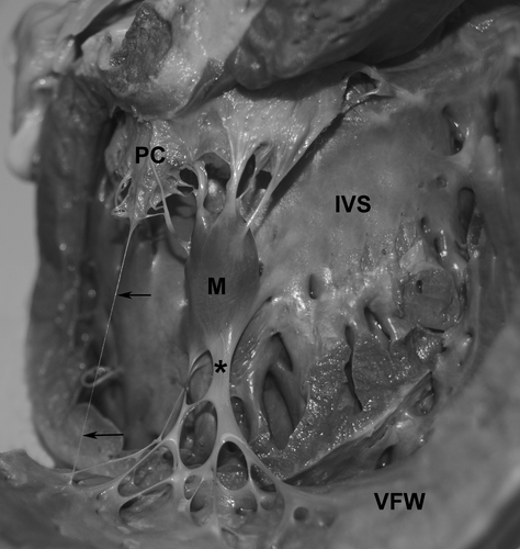
Two (one male and one female) of the 39 dog hearts showed a single atypical chordae tendineae extending from the free edge of a cusp of the right atrioventricular valve to the ventricular free wall.
In the male canine heart, the right atrioventricular valve had three distinct cups; parietal, angular and septal. The single chordae tendineae extended from the free edge of the parietal cusp to the right ventricular free wall (Fig. 1).

In the female canine heart, the right atrioventricular valve consisted of parietal and septal cusps. The single chordae tendineae in this heart also extended from the free edge of the parietal cusp to the right ventricular free wall (Fig. 2).

In both canine hearts, this single chordae tendineae was located to the right of the trabeculae septomarginalis and as previously described, this structure can be classified as one of four types (Cope, 2015). In the male canine heart, the trabecula septomarginalis was classified as type IB, and in the female canine heart, this trabecula was classified as type IC (Figs 1 and 2).
Discussion
In veterinary anatomy text, the chordae tendineae are described as cords that attach to the ventricular surfaces of the valve cusp and insert into associated papillary muscles (Nickel et al., 1981; Evans, 1993). Previous work on these strands in the dog indicate the strongest cords can be followed as ridges under the endocardium to their attached valve base, whereas the weakest cords go to the points (free edge) of the valvular cusp serrations and disappear. Then, the intermediate cords go as far as the middle part of the ventricular surfaces of the valve cusp (Evans, 1993).
While this description of the chordae tendineae in the canine heart is the most commonly observed pattern, previous work on canine and human hearts shows there is variability in the points of attachment of these cords. The chordae tendineae, which do not consistently extend between a papillary muscle and an atrioventricular valve cusp, are referred to as basal chordae, 3rd order chordae, anomalous chordae, false tendons and tendinous cords (Fratter and Ellis, 1961; Ettinger and Buergelt, 1969; Williams, 1999; Loukas et al., 2008; Wafae et al., 2008).
In the domestic dog, Fratter and Ellis (1961) and Evans (1993) appear to be the first researchers to describe atypical chordae tendineae. Both of these early researchers describe chordae tendineae that extend from a valve cusp to the ventricular free wall, and in their work, Fratter and Ellis (1961) refer to these strands as 3rd order chordae. Subsequent studies on these structures do not follow until 1999 and 2000 when Costa et al. provide additional information about the variability in chordae tendineae attachment in the right ventricle. In the first of these two studies, Costa et al. (1999) describes that of 50 canine hearts, 20% have a single stranded anomalous chordae tendineae extending from the parietal cusp of the right atrioventricular valve to a small pillar of muscle on the right ventricular free wall. Then in the second study, Costa et al. (2000) documents two other variations in chordae tendineae anatomy in the hearts of 40 dogs. In this study, Costa et al. (2000) first describes a chordae tendineae that originates from the muscular tissue of the fibrous annulus of the right atrioventricular valve and then blends with tendon-like strands from the musculus papillaris magnus. The second pattern that Costa et al. (2000) describes in this study is a chordae tendineae that extends from a semilunar valve of the pulmonary trunk to the musculus papillaris subarteriosus.
In the human literature, the 2008 study by Loukas et al. on the right ventricle shows that of 35 hearts, 14.5% had a false tendon connecting the anterior leaflet of the tricuspid valve to the right ventricular free wall. A second 2008 study by Wafae et al. also examined tendinous cords that extend from the inter-ventricular septum to the septal, anterior or posterior cusp of the tricuspid valve in 50 human hearts (25 male and 25 female). In this study, tendinous cords that originate from the septum and insert onto the tricuspid valve cusp were observed in 98% of the hearts. From this research, results show that the majority of these cords (76.7%; 158 of 206 cords) are attached to the septal cusp followed by the anterior cusp (21.3%) and then the posterior cusp (1.0%). This study also shows the main point of attachment for these tendinous cords is the free edge (62%) of the cusp followed by the ventricular surface (area between the free edge and basal surface) (32%) and then the basal surface (6%) (Wafae et al., 2008).
From these previous studies in the canine and human heart, the most commonly described variation in chordae tendineae is between the cusp of an atrioventricular valve and the ventricular free wall (Fratter and Ellis, 1961; Costa et al., 1999; Williams, 1999; Loukas et al., 2008; Wafae et al., 2008).
In this current study, the atypical chordae tendineae of the canine right ventricle extend from the parietal cusp of the atrioventricular valve to the ventricular free wall. This pattern is similar to previous observations made in the right ventricle of both the canine and human heart by Costa et al. (1999), Williams (1999) and Loukas et al. (2008). Additionally in the two canine hearts with atypical chordae tendineae, these strands inserted into the free edge of the valve cusp similar to the 3rd order chordae described by Fratter and Ellis (1961) in the canine mitral valve, and the basal chordae and tendinous cords described by Williams (1999) and Wafae et al. (2008) in the right ventricle of the human heart.
Given the previous work on these chordae tendineae, it is easy to see why the researchers assume these strands could be anomalous. Wafae et al. (2008), however, proposes that because these unusual chordae tendineae have been documented in studies as early as 1889, it might indicate the strands are a constant structure but the frequency of their presence is variable and requires further study.
While the discovery of these atypical chordae tendineae in canine and human hearts is unique and provides additional information on cardiac anatomy, their presence may also carry an important clinical and biomedical significance. At this time, some of the research in the human heart suggest these chordae tendineae may be involved in several cardiac pathologic processes or lead to catheter and/or pacemaker lead entanglement during right-sided heart cardiac catheterization (Winrow et al., 1996; Moreno et al., 2003; Loukas et al., 2008).
In the dog, the available veterinary literature does not describe these atypical chordae tendineae in association with underlying cardiac pathologies. Instead, chordae tendineae that are fused, ruptured or malformed are found in dogs with dysplasia of the right atrioventricular valve (Liu and Tilley, 1976; Kirk and Bonagura, 1995), chordae tendineae rupture (Ettinger and Buergelt, 1969) and/or mitral valve degeneration (Liu and Tilley, 1975; Serres et al., 2007).
At this time, because these atypical chordae tendineae in the domestic dog are not directly associated with a specific cardiac pathology, it is believed they can be significant in the biomedical field. Animal models continue to be used for the study of cardiovascular diseases in human and veterinary medicine. These animals are often used for pre-clinical assessment of pharmaceuticals, mechanical devices, therapeutic procedures and/or continuation therapies. In terms of the dog, this animal model is used in myocardial ischaemia studies, as a heart transplantation model for studies on organ preservation, reperfusion injury, rejection and/or post-transplant organ monitoring. More recently, though, the dog is being used as an animal model in stem cell research in terms of cellular cardiomyoplasty in which stem cells are delivered to the heart by an endocardial injection using radiographically guided stem cell injection catheters (Iaizzo, 2009).
Given the use of the dog in biomedical research, a review of the literature shows chordae tendineae entrapment can be a problem in cardiac catheter development and testing. Winrow et al. (1996) and Schregel et al. (1991) both document cases of entanglement of pigtail and Swan-Ganz catheters within the chordae tendineae of the canine tricuspid valve. Then in 2007, Saunders et al. describes entrapment of tined endocardial pacing leads within the chordae tendineae of the tricuspid valve.
According to the veterinary literature entanglement of cardiac catheters and endocardial pacing leads is a rarely discussed complication and when this complication occurs the catheter or leads can often be untangled (Saunders et al., 2007). However, when successful detanglement cannot be accomplished complications associated with this attempted removal can be chordae tendineae or papillary muscle rupture (Saunders et al., 2007).
Because it is proposed these atypical chordae tendineae may be a normal feature of the ventricle, it is plausible to believe these strands may increase the chance of catheter or pacemaker lead entanglement (Wafae et al., 2008). While the purpose of this study was to document the presence of these atypical chordae tendineae, it has also illustrated the clinical and biomedical significance of normal chordae tendineae. The literature shows damage to normal chordae tendineae during detanglement of catheter or pacemaker leads may cause tendineae rupture and subsequent valve insufficiency, cardiac compromise or failure. Therefore, the presence of these atypical chordae tendineae in the canine heart may further challenge cardiac catheterization, catheter development and testing if they are not adequately documented for veterinarians and biomedical researchers.
Acknowledgement
The author extends sincere gratitude to Maura Farley Pipkins, MS Preclinical Sciences and 1st year medical student for her ‘sharp’ eye during dissection of these hearts and conscientious help with this research.


