Root and root canal morphology of mandibular second permanent molars in the Gansu province population: A CBCT study
Abstract
To study the anatomical characteristics of the root and root canal system of the mandibular second molars in the population of Gansu province, and to provide theoretical and clinical references for improving the success rate of root canal therapy (RCT) of mandibular second molars. The number of roots and root canals, root canal type and pulp chamber height of mandibular second molars were determined by observing cone-beam computed tomography (CBCT) images of people living in Gansu. The most common type of mandibular second molars in the Gansu province population was a double root with three root canals (47.55%), followed by a C-shaped root (35.56%). There were more females than males with a C-shaped root (p < 0.05). The most common root canal subtype of the C-type root was C3 (13.91%). Most of the population (77.11%) had bilateral mandibular second molars with symmetrical root canal morphology. With an increase in age, the height of the pulp chamber decreased significantly. The incidence of root canal variation of the mandibular second molars is relatively high in the population of Gansu province. Preoperative examination with CBCT is essential for mandibular second molars that need RCT to avoid root canal treatment failure and decrease the occurrence of postoperative pain as much as possible.
INTRODUCTION
The results of the Fourth National Oral Health Epidemiological Survey in Gansu [1, 2] have shown that the incidence of dental caries and periapical disease is relatively high in the Gansu province population. The Gansu province is an underdeveloped area where ethnic minorities gather, and most of the population has low awareness of oral health care. Mandibular second molars are prone to periapical disease owing to the influence of mandibular third molars and the difficulty of tooth cleaning. Considering its limited operation space and anatomical variations, the mandibular second molar has the lowest success rate of root canal therapy (RCT) among all teeth [3].
Familiarity with the anatomy of the root canal is essential for the success of RCT. At present, cone-beam computed tomography (CBCT) plays a very important role in the diagnosis and treatment of endodontic diseases. It can display the number and morphology of tooth roots and root canals in detail in three dimensions [4]. Dentists can use CBCT to evaluate the difficulty of RCT, such as the degree of root bending, the number of root canals (which helps them to avoid missing one) and root canal variation.
It is necessary to improve the success rate of RCT of the mandibular second molar because it is difficult to insert an artificial tooth in this position; thus, it is best to preserve one's own tooth. To study the number of roots, number of root canals, incidence of a C-shaped root, gender distribution of the incidence of a C-shaped root, symmetry of root canal morphology and ageing changes in the pulp chamber height of mandibular second molars, we examined CBCT images of patients from the Gansu province obtained over the past 2 years.
MATERIAL AND METHODS
Data collection
A total of 284 CBCT images of patients from the Gansu province eligible for inclusion from August 2019 to August 2021 from Lanzhou Stomatological Hospital were collected, and a total of 568 mandibular second molar images were obtained. There were 134 males (47.18%) and 150 females (52.82%). The age range was 16–73 years, and the average age was 42.52 ± 3.22 years. The inclusion criteria for mandibular second molars were as follows: complete image information; a well-developed apical foramen; no RCT or other endodontic treatments; no obvious caries or fillings, or only enamel caries; and the presence of bilateral mandibular second molars that met the above criteria.
Equipment and methods
KaVo 3D eXam CBCT (KaVo, Shangai, China) equipment was used for full dentition scanning, and the parameters used were as follows: voltage, 120 kV; current, 5 mA; irradiation time, 14 s; pixel size, 0.15 mm and minimum layer thickness, 0.3 mm. Continuous images of all teeth were observed from the sagittal, coronal and horizontal planes, and the number of roots, number and morphology of root canals and height of the pulp chamber were recorded.
Observation and classification
The root morphology was divided into the following three types: (1) a single root that contained a C-shaped root or tapered root; (2) double roots that consisted of one root in the mesial direction and one root in the distal direction and (3) three roots that consisted of two roots in the mesial direction and one root in the distal direction, or one root in the mesial direction and two roots in the distal direction (Figure 1).

The root canal morphology classification was based on the Vertucci method [5], as follows: type I (type 1-1), type II (type 2-1), type III (type 1-2-1), type IV (type 2-2), type V (type 1-2), type VI (type 2-1-2), type VII (type 1-2-1-2), type VIII (Type 3-3) and other types (not belonging to the abovementioned subtypes).
C-shaped root canals were classified according to the Melton method [6], as follows: type C1 (continuous band-like form or C-shaped form), type C2 (root canal orifice form like “;”) and type C3 (several independent root canal orifices connected by an isthmus, so that these independent orifices are lined up in a C shape).
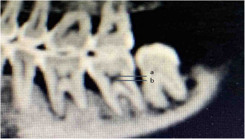
Measurement of the height of pulp chamber: the distance between the highest point of the bottom of the pulp chamber and the lowest point of the roof of the pulp chamber was considered to be the height of the pulp chamber when the tooth was in the sagittal view (Figure 2).

Statistical analysis
The data are expressed as mean ± SD. Differences among groups were analysed with the chi-square test using SPSS v16.0 software (SPSS Inc., Chicago, IL, USA). p values <0.05 were considered statistically significant.
RESULTS
Number of roots and root canals of the mandibular second molar
Among the 568 CBCT images of mandibular second molars, the detection rates of a tapered root, C-shaped root, double root and triple root were 1.06% (6/568), 35.56% (202/568), 63.03% (358/568) and 0.35% (2/568), respectively. Quadruple roots and other root types were not detected. Double root was the main root form of mandibular second molars. The detection rates of a double root with two canals, three canals and four canals were 14.44% (82/568), 47.55% (270/568) and 1.06% (6 /568), respectively. The detection rate of a triple root with three canals was 0.35% (2/568). Table 1 summarises the number of roots and root canals of mandibular second molars.
| Total (%) | C-shaped root canal | One root canal | Two root canals | Three root canals | Four root canals | |
|---|---|---|---|---|---|---|
| Single root | 208 (36.62) | 202 (35.56) | 6 (1.06) | 0 (0) | 0 (0) | 0 (0) |
| Double root | 358 (63.03) | - | - | 82 (14.44) | 270 (47.55) | 6 (1.06) |
| Triple root | 2 (0.35) | - | - | - | 2 (0.35) | 0 (0) |
Root canal morphology of a double-rooted mandibular second molar
The detection rate of two root canals in the mesial root was 77.09% (276/358) among the 358 double-rooted mandibular second molars. As for the root canal morphology classification, there were 43.30% (155/358) with type IV, 29.61% (106/358) with type II, 1.12% with type III (4/358) and 3.07% with type V (11/358). The detection rate of type IV was significantly different compared with that of the other root canal types (p < 0.05). The detection rate of one root canal in the mesial root was 22.91% (82/358). The detection rate of one root canal in the distal root was 98.32% (352/358), and that of two root canals was 1.68% (6/358). The detection rate of type I root canals in the distal roots was significantly different from that of the other root canal types (p < 0.05). Table 2 summarises the root canal morphology of double-rooted mandibular second molars.
| Type I | Type II | Type III | Type IV | Type V | Type VI | Type VII | Type VII | |
|---|---|---|---|---|---|---|---|---|
| Mesial root | 82 (22.91) | 106 (29.61) | 4 (1.12) | 155 (43.30) | 11 (3.07) | 0 (0) | 0 (0) | 0 (0) |
| Distal root | 352 (98.32) | 1 (0.28) | 0 (0) | 5 (1.40) | 0 (0) | 0 (0) | 0 (0) | 0 (0) |
Root canal morphology of a C-shaped root of the mandibular second molar
The morphology of the root canal was C shaped in 202 C-shaped roots. According to the Melton method, there were 11.44% (65/568) with the C1 subtype, 10.21% (58/568) with the C2 subtype and 13.91% (79/568) with the C3 subtype. The incidence of the C3 subtype was significantly different compared with that of the other subtypes of C-shaped root canals (p < 0.05). Table 3 summarises the root canal morphology of a C-shaped root of the mandibular second molar.
| Non-C-shaped root | C-shaped root | |||
|---|---|---|---|---|
| C1 | C2 | C3 | ||
| Quantity | 366 (64.44) | 65 (11.44) | 58 (10.21) | 79 (13.91) |
Incidence of a C-shaped root of mandibular second molars in different genders
Among the 284 CBCT scans, 51 males and 72 females were shown to have a C-shaped root (C-shaped root on one or both sides). The detection rate of a C-shaped root in females was significantly different than that in males (p < 0.05). Table 4 summarises the incidence of a C-shaped root in different genders.
| Male | Female | |
|---|---|---|
| Incidence of a C-shaped root | 51 (17.96) | 72 (25.35) |
Study of the root symmetry of mandibular second molars
Among the 284 CBCT scans, the incidence of bilateral tooth with root morphology and number symmetry was 86.62% (246/284), and that of the root canal morphology and number symmetry was 77.11% (219/284). The incidence of bilateral symmetric C-shaped roots was 27.82% (79/284). The incidence of left C-shaped roots alone was 7.39% (21/284), and that of right C-shaped roots alone was 8.10% (23/284). There was no significant difference in the incidence of C-type roots between the left side and right side (p > 0.05). Table 5 summarises the incidence of a C-shaped root on both sides.
| Bilateral symmetric C-shaped roots | Left C-shaped roots | Right C-shaped roots | |
|---|---|---|---|
| Quantity | 79 (27.82) | 21 (7.39) | 23 (8.10) |
Age-related changes in the height of the pulp chamber of the mandibular second molar
The results from 568 cases showed that the maximum value of the measured pulp chamber height was 3.66 mm, and the minimum value of the measured pulp chamber height was 0.51 mm. The average height of the pulp chamber was 2.64 mm in the 16–35-year-old group, 1.71 mm in the 36–55-year-old group and 1.33 mm in the 56–73-year-old group. The difference in the height of the pulp chamber among all age groups was statistically significant (p < 0.05). Table 6 summarises the average height of the pulp chamber in each age group.
| 16–35-year-old group | 36–55-year-old group | 56–73-year-old group | |
|---|---|---|---|
| The average height of the pulp chamber (mm) | 2.64 | 1.71 | 1.33 |
DISCUSSION
Our data revealed that the main root form of mandibular second molars in the Gansu province population was a double root (63.03%), followed by a single root (36.62%), and the detection rate of a triple root was 0.35%. Quadruple roots and other root types were not detected. The most common mesial root canal morphology of double-rooted mandibular second molars was type IV (type 2–2) (43.30%), followed by type II (type 2–1) (29.61%) and type I (type 1–1) (22.91%). In addition, four cases of type III (type 1–2-1) and 11 cases of type V (type 1–2) mesial root canals were detected. Therefore, attention should be paid to the variation of two mesial root canals when performing RCT of mandibular second molars. A total of 98.32% of distal root canals of double-rooted mandibular second molars were of type I (type 1–1); hence, preparation and filling of the distal root canal during RCT are less difficult than the preparation and filling of the mesial root. The most common root and root canal morphology of mandibular second molars in the Gansu province population was a double root with three root canals (47.55%), followed by a C-shaped root (35.56%). A tooth with complex and variable root canals increases the difficulty of RCT and the probability of instrument separation, thereby increasing the failure rate of RCT. Therefore, it is essential to examine the root canal of the mandibular second molar by CBCT before performing RCT.
The most common root variation of the mandibular second molar is a C-shaped root with C-shaped root canal. There are different reports on the incidence of C-shaped root canals in various nations. The incidence of C-shaped roots in Western people is about 6.8%–11.3% [7], while the average incidence of C-shaped roots in the Chinese population is 39%, which is significantly higher than that in the Western population [8]. The incidence of C-shaped roots in Shandong, Kunming, Lingnan and Sichuan provinces is about 32.84%–41.64% [9-12], while the incidence of C-shaped roots in Qinghai, Ningxia and Xinjiang provinces is about 22.4%–28.3% [13-15]; the latter provinces are in northwest China, where there are ethnic minority settlements. According to the Melton method, the results of C-shaped root canals in the apex showed that the proportion of C3 root canals was relatively high, which is similar to the results of a previous study [9-15]. The occurrence of C-shaped roots may be related to the degradation of mandibular bone mass caused by refined diet. As a result, the roots of mandibular second molars are fused incompletely, and the incidence of C-shaped roots in ethnic minorities in northwest China is lower than that in inland people, which may be related to the large difference in their diets. In this study, the detection rate of C-shaped roots was 35.56%, which was lower than the national average rate and higher than that in ethnic minorities in northwest China; this may have been caused by the fact that there are a large number of ethnic minorities in Gansu province. In this research, more women than men had C-shaped roots, which is similar to the results of previous studies [9-15]. The existence of a C-shaped root canal increases the difficulty of root canal preparation and root canal filling. The operator may undermine the isthmus and cause band penetration of the side wall of the root canal. It is difficult to remove the infection of the isthmus completely via conventional mechanical preparation; hence, ultrasonic irrigation and chemical preparation are required. Close obturation of the irregular parts of the root canal with vertical condensation using hot gutta percha can ensure the curative effect of RCT.
Most of the population (77.11%) had bilateral mandibular second molars with symmetrical root canals and shapes, while 27.82% of the population had bilateral mandibular second molars with C-shaped roots. Therefore, C-shaped roots often occurred symmetrically. The position, number and shape of the root canals of a mandibular second molar can be applicable to the contralateral same tooth when performing RCT.
With an increase in age, the dental pulp undergoes age-related changes, which presents as secondary dentin formation and a decrease in the pulp chamber and root canal. Therefore, it is more difficult to perform RCT in older than in young individuals. The results of this study show that the height of the pulp chamber decreases significantly with an increasing age. Therefore, dentists must take the age of the patient into consideration while performing unroofing and avoid perforation of the pulp floor. Comprehensive and detailed evaluation before the operation can avoid the occurrence of pain after RCT as much as possible.
AUTHORS' CONTRIBUTIONS
Q.G., QX.W. and YL.Y. performed the research. DM.G designed the research study. Q.G. performed the statistical analysis. Q.G. wrote the paper. All authors have read and approved the final manuscript.
ACKNOWLEDGEMENTS
The present study was supported by Grants from the Development of Science and Technology Guidance Project of Lanzhou (No.2020-ZD-163), Talents of Innovative and Entrepreneurial of Lanzhou (No.2021-RC-127) and Science and Technology Planning Project of Lanzhou (No.2021-1-126). We thank LetPub (www.letpub.com) for its linguistic assistance during the preparation of this manuscript.
CONFLICT OF INTEREST
The authors have no competing interests to declare that are relevant to the content of this article.


