Volume 60, Issue 4 pp. 506-513
Medical Imaging—Pictorial Essay
Free Access

Malignant hyperechoic breast lesions at ultrasound: A pictorial essay

Stephen Tiang

Stephen Tiang

Radiology Department, Royal Perth Hospital, Perth, Western Australia, Australia

Search for more papers by this author
Cecily Metcalf

Cecily Metcalf

PathWest, Royal Perth Hospital, Perth, Western Australia, Australia

Search for more papers by this author
Deepthi Dissanayake

Deepthi Dissanayake

Radiology Department, Royal Perth Hospital, Perth, Western Australia, Australia

Search for more papers by this author
Elizabeth Wylie

Corresponding Author

Elizabeth Wylie

Radiology Department, Royal Perth Hospital, Perth, Western Australia, Australia

University of Western Australia, Crawley, Western Australia, Australia

Correspondence

Dr Elizabeth Wylie, Radiology Department, Royal Perth Hospital, Wellington Street, Perth 6000, WA, Australia.

Email: [email protected]

Search for more papers by this author
First published: 24 May 2016
Citations: 6
S Tiang MBBS; C Metcalf MBBS, FRCPA, FIAC; D Dissanayake MBBS, FRANZCR; E Wylie MBBS, FRANZCR.
Conflict of interest: None declared.

Summary

Malignant breast lesions are typically hypoechoic at sonography. However, a small subgroup of hyperechoic malignant breast lesions is encountered in clinical practice. We present a pictorial essay of a number of different hyperechoic breast malignancies with mammographic, sonographic and histopathologic correlation. Suspicious sonographic features in a hyperechoic lesion include inhomogeneity in echogenic pattern, an irregular margin, posterior acoustic shadowing and internal vascularity. A hyperechoic lesion at ultrasound does not discount the need to undertake histological assessment of a mammographically suspicious lesion.

Introduction

On ultrasound, a hyperechoic breast mass is defined as a lesion that is of increased echogenicity compared to the subcutaneous adipose tissues. Approximately 0.6–5.6% of breast masses are hyperechoic.1-3 In 1995, Stavros et al.1 described lesions that were uniformly hyperechoic with no isoechoic or hypoechoic areas as benign, reporting a negative predictive value of 100% after 42 biopsies of hyperechoic nodules. One of the most common benign hyperechoic breast lesion is fat necrosis from previous breast trauma. Other benign entities include lesions containing adipose tissue (lipoma), lesions containing fibrotic tissue (pseudoangiomatous stromal hyperplasia and others), vascular lesions (haemangioma) and a combination of tissue types such as a hamartoma and angiomyolipoma. A number of authors have published data indicating that a very small proportion of hyperechoic lesions are malignant. Linda et al.2 reported 9 (0.5%) hyperechoic malignant lesions out of 1849 biopsied malignancies. Nam et al.3 reported 103 hyperechoic lesions out of 16416, of which 27 were biopsied, five (4.9%) were malignant, and Soon et al.4 found two (0.5%) hyperechoic nodules among 393 screen detected breast cancers.

Background

The cases in this pictorial essay are patients that were referred to the breast assessment centre at Royal Perth Hospital from 2011 to 2015.

Primary breast carcinoma

Invasive ductal carcinoma

Invasive ductal carcinoma (IDC) is the most common invasive primary breast malignancy. Clinically, the carcinoma may present as a palpable mass with features such as skin retraction and nipple discharge, or as an assymptomatic screen detected lesion.

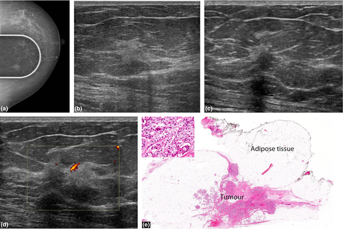
The characteristic mammographic appearances are of an irregular, spiculated mass (Fig. 1a) with or without pleomorphic calcifications and architectural distortion. The typical sonographic appearance of a primary breast malignancy is that of a hypoechoic mass with irregular margins and variable posterior acoustic shadowing.
Details are in the caption following the image
Invasive ductal carcinoma. A 68-year-old woman presented with a palpable lump in the left breast. (a) An irregular mass is shown at mammography. (b) Ultrasound showed an 8.4 mm hyperechoic and irregular mass with parenchymal distortion. (c) Examining the lesion from another plane reveals a hypoechoic component and posterior acoustic shadowing, further suspicious features. (d) The lesion has internal vascularity. (e) Excision biopsy of breast shows infiltrating duct carcinoma within stroma and fat. The tumour forms ill-defined nodules imparting a vague multilobulated appearance. Original magnification ×1.0. Haematoxylin & Eosin. The insert shows a solid sheet of large, pleomorphic tumour cells with abundant pale cytoplasm. Original magnification ×40. Haematoxylin & Eosin.

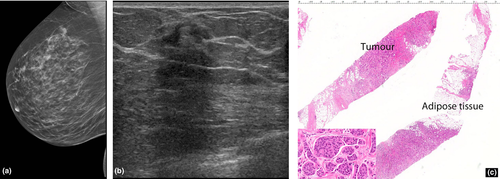
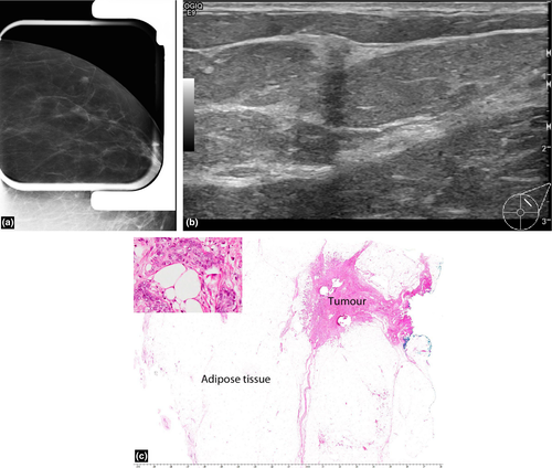
Malignant hyperechoic lesions are more likely than benign lesions to have an irregular shape (Fig. 1-3), non-parallel orientation and a hypoechoic component.2, 3 A lesion that is not completely hyperechoic and has small hypoechoic areas should be regarded as suspicious.1, 2 Other predictive features of malignancy include a spiculated shape, posterior shadowing and distortion of surrounding tissues3 (Fig. 1-3).
Details are in the caption following the image
Invasive ductal carcinoma. (a) This 65-year-old woman presented with a screen detected opacity which persists on compression in the left upper outer quadrant of breast. There is no prior history of trauma. (b) Ultrasound shows a 6 mm predominantly hyperechoic lesion with small central hypoechogenicity and posterior shadowing. (c) Excision biopsy of breast shows an infiltrating duct carcinoma within stroma and fat and extending close to the surgical margin (blue ink). Original magnification ×1. Haematoxylin & Eosin. The insert shows several solid nests of large, pleomorphic tumour cells surrounding several fat cells. Original magnification ×40. Haematoxylin & Eosin.
Details are in the caption following the image
Invasive duct carcinoma. This 63-year-old woman with a screen detected, non-palpable solid nodule in the right upper outer quadrant of breast. There is no history of trauma. (a) Mammogram left breast, mediolateral oblique (MLO). (b) Ultrasound shows a heterogenous hyperechoic and hypoechoic mass with posterior acoustic shadowing. (c) Several core biopsies of breast tissue show infiltration of adipose tissue and stroma by tumour. Original magnification ×1.3. Haematoxylin & Eosin. The insert shows small and large solid nests of large, pleomorphic tumour cells separated by small amounts of fibrous stroma. Original magnification ×40. Haematoxylin & Eosin.

Nam et al.3 reported that all five hyperechoic carcinomas had corresponding suspicious mammographic findings such as spiculated margins, interval enlargement, suspicious microcalcification and lymphadenopathy.

Invasive lobular carcinoma

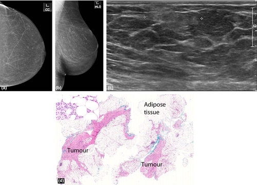
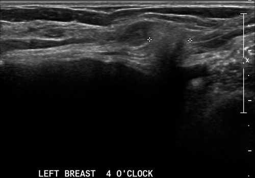
Invasive lobular carcinoma (ILC) is a tumour composed of small, usually bland cells arranged in poorly cohesive aggregates or in single files surrounding ducts and not destroying normal parenchyma, with minimal desmoplastic response. The commonest appearance on mammography is a spiculated mass. However, many present as an isolated architectural distortion or focal asymmetry or are seen on one mammographic view (Fig. 5) only or are radiographically occult. On ultrasound, they are usually hypoechoic with typical suspicious features of irregular margins and posterior acoustic shadowing (Fig. 4, 5).
Details are in the caption following the image
Invasive lobular carcinoma. This 60-year-old woman has bilateral retropectoral silicone implants. She presented with a palpable mass in the left breast. Ultrasound of the left breast (transverse view) shows a small hyperechoic mass with ill-defined margins superficial to the implants. At the time of examination, this was felt to be silicone extravasation due to the “snowstorm” appearance of the mass. This prompted a silicone implant exchange. At surgery the mass was excised and subsequently shown to be a lobular breast carcinoma
Details are in the caption following the image
Invasive lobular carcinoma. This 68-year-old woman with a history of prior breast reduction surgery presented with a left sided palpable lump. (a) Craniocaudal (CC) mammogram of the left breast does not show any abnormality. (b) Mediolateral oblique (MLO) mammogram of the left breast also does not show any abnormality. The lesion was occult on both mammographic views. (c) The ultrasound showed a 20 mm region of increased echogenicity in the subcutaneous adipose tissues. The lesion has ill-defined margins. (d) Excision biopsy of breast shows a lobular carcinoma widely infiltrating adipose tissue and stroma. Original magnification ×1. Haematoxylin & Eosin. The insert shows small, regular tumour cells infiltrating around fat cells. Original magnification ×40. Haematoxylin & Eosin.

The literature suggests that hyperechoic malignancies may be more commonly associated with ILC.4-6 In a series by Cawson et al.6, 21 (57%) out of the 37 ILCs were at least partly hyperechoic, concluding that ILC was 9.94 times more likely to be hyperechoic than IDC. Waterman et al.6 who studied sonographic appearances of 406 invasive breast carcinomas reported that a hyperechoic or isoechoic pattern was more frequent in ILC. Jones et al.7 conducted a retrospective review of 509 ILCs and concluded that 27 (5%) were hyperechoic. The periductal infiltration by ILCs with formation of concentric rings may act as acoustic reflectors5 causing the bright hyperehoic halo around the small hypoechoic tumour nidus to become the predominant ultrasound finding.

Mucinous (colloid) carcinoma

This is an uncommon breast malignancy which comprises 0.5–3% of breast carcinomas.8 These mucin producing tumours can be pure or mixed, with the pure subtype conferring a better prognosis. Microscopically, the tumour cells occur within large mucinous lakes which may result in a core biopsy finding of mucinous pools with a paucicellular tumour.

On mammography, the tumour may be round, oval, irregular or dense with circumscribed or partially indistinct margins (Fig. 6a). On sonography (Fig. 6b), the tumour is frequently isoechoic or hypoechoic to surrounding adipose tissues but may occasionally be hyperechoic.8 Because of their wide ranging appearances and occasional well-defined margins, they may mimic a fibroadenoma or hamartoma. Posterior enhancement is a common association and posterior shadowing is rare (Fig. 6b). A homogeneous appearance to the lesion is usually associated with a pure subtype and conversely lesional heterogeneity at ultrasound is more predictive of a mixed subtype.8

Details are in the caption following the image
Mucinous (colloid) carcinoma. This 79-year-old woman presented with a palpable lump. (a) Mammogram of the left breast, craniocaudal (CC) view shows a non-calcified mass in the upper inner quadrant. (b) Ultrasound examination found a corresponding lesion measuring 2.5 cm at the left upper inner quadrant at 10 o'clock. It is well-defined, heterogenous, with hypo- and hyperechoic components. There is posterior enhancement. (c) Excision biopsy of breast shows a large well circumscribed mucinous carcinoma. Most of the tumour consists of mucin with central haemorrhage and with necrosis of the tumour cells but a small component in the lower part of the tumour shows viable tumour cells admixed with mucin. Original magnification ×0.6. Haematoxylin & Eosin. The insert shows small aggregates of tumour cells floating in mucin. Original magnification ×40. Haematoxylin & Eosin.

Metastasis

Metastases to the breast are uncommon but should be considered as a differential diagnosis for multiple breast masses in the setting of a known malignancy. The commonest malignancies that metastasize to the breast are melanoma (skin) and lung adenocarcinoma.9 Other primaries to consider include small cell carcinoma, sarcoma, neuroendocrine tumours and squamous cell carcinomas. Rarely metastases may originate from a contralateral breast primary.

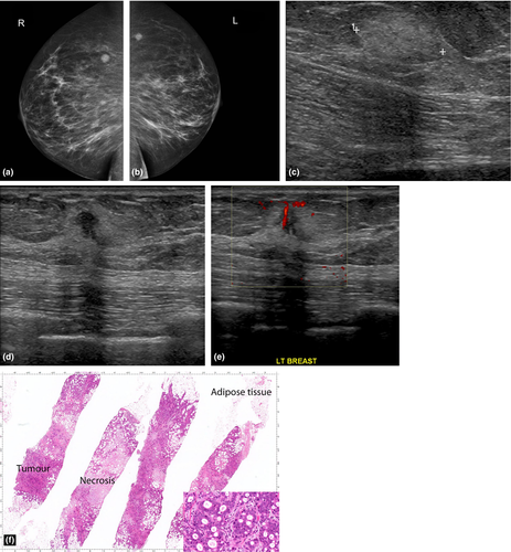
When the tumour spread is haematogeneous, the mammographic findings include round, circumscribed, non-calcified masses (Fig. 7a). If the spread is lymphangitic, there may be breast enlargement, skin thickening, asymmetric density and axillary adenopathy. Although metastases to the breast are most commonly hypoechoic, they can rarely present as hyperechoic masses (Fig. 7c). Lesional hypervascularity is seen in the majority of melanoma metastases.8
Details are in the caption following the image
Metastatic adenocarcinoma. This 63-year-old woman had a screen detected non-calcified opacity in both breasts. Soon after the mammogram, she was admitted to hospital with obstructive jaundice, where a CT abdomen showed a common bile duct stricture. During ERCP, the stricture was felt to be malignant, although formal brushings were not performed. Core biopsies of the breast lesions revealed well-differentiated adenocarcinoma, considered metastatic and consistent with a tumour of foregut origin. (a) Mammogram right breast, craniocaudal (CC) view shows a round, well-defined mass with no calcification or stromal distortion. (b) Mammogram left breast, carniocaudal (CC) view shows a solitary mass similar to the right breast. (c) Ultrasound of the left breast shows a 17 mm lesion which appears completely hyperechoic but with suspicious features including an ill-defined margin and posterior acoustic shadowing. (d) Ultrasound of the same lesion in a different plane reveals a hypoechoic area with posterior shadowing. (e) Doppler ultrasound of the lesion demonstrates internal vascularity. (f) Several cores of breast tissue showing tumour infiltrating adipose tissue and stroma. Large areas of necrosis are also present. Original magnification ×2.0. Haematoxylin & Eosin. The insert shows small nests of intermediate, pleomorphic tumour cells arranged around small glandular spaces and separated by small amounts of fibrous stroma. Original magnification ×40. Haematoxylin & Eosin.

Lymphoma

Breast lymphoma is the second most common (after melanoma) non-mammary malignancy of the breast. Breast lymphoma may be primary or secondary. The diagnostic criteria for primary breast lymphoma include the breast as the site of clinical presentation of the disease, absence of prior lymphoma or absence of widespread disease at diagnosis, close association of mammary tissue with lymphomatous infiltrate and disease limited to the breast and/or ipsilateral lymph nodes at the time of diagnosis.10 Secondary breast lymphoma occurs in setting of systemic lymphoma.

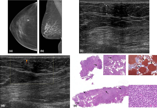
The mammographic appearance is usually a solitary, non-calcified mass with no overt spiculation (Fig. 8a,b). In secondary breast lymphoma, axillary adenopathy is a common mammographic presentation, with asymmetry of the breast and skin thickening. On sonography, the appearance is variable, ranging from hypoechoic, mixed echogenicity to near completely hyperechoic lesions11 (Fig. 8c). There is usually marked vascular flow9 in the lymphoma mass.
Details are in the caption following the image
Secondary breast lymphoma. This 60-year-old had a screen detected solitary nodule on mammogram. There is a prior history of low grade follicular lymphoma. (a) Digital tomosynthesis of the left breast, craniocaudal (CC) view shows a solitary, well-defined, lobulated nodule, without calcification and stromal distortion. (b) The lesion is also identified on the mediolateral oblique (MLO) left breast mammogram. No evidence of axillary lymphadenopathy. (c) Ultrasound shows a heterogeneous lesion with hypoechoic area centrally and a hyperechoic halo. (d) There is a vascular flow within the lesion. (e) Core biopsy of breast consisting of lymphoid cells with a small amount of residual fat infiltrate Original magnification ×2.4. Haematoxylin & Eosin. The upper middle insert shows diffuse infiltrating lymphocytes in small ill-defined nodules (arrows). Original magnification ×10. Haematoxylin & Eosin. The lower right hand insert shows a mixture of small cleaved cells and large lymphocytes. Original magnification ×40. Haematoxylin & Eosin. The upper right hand insert show tumours cells labelled with a B cell marker. Original magnification ×8. CD20 immunohistochemistry.

Pitfalls for detection

Stavros et al.1 recommended that a lesion should be homogeneous in echogenicity and more hyperechoic than adipose tissue for it to be considered benign on ultrasound.

Some infiltrating malignancies can have a small hypoechoic central nidus surrounded by a larger hyperechoic halo. Scanning through the thick hyperechoic halo and not appreciating the hypoechoic central focus can cause such lesions to be misinterpreted as purely hyperechoic.12

In conclusion, approximately 0.5%2, 11 of malignant breast lesions appear hyperechoic. Correlation with mammographic findings and clinical history should determine the need for biopsy. A hyperechoic lesion in the absence of a preceding history of trauma and characteristic mammographic radiolucent appearances of fat necrosis may ultimately require a biopsy for a definitive assessment.

    The full text of this article hosted at iucr.org is unavailable due to technical difficulties.