Skin acidification with a water-in-oil emulsion (pH 4) restores disrupted epidermal barrier and improves structure of lipid lamellae in the elderly
Abstract
The pH of the skin surface increases with age and thus reduces epidermal barrier function. Aged skin needs appropriate skin care to counterbalance age-related pH increase and improve barrier function. This confirmatory randomized study investigated the efficacy of water-in-oil (w/o) emulsions with either pH 4 or pH 5.8 in 20 elderly subjects after 4 weeks of treatment. After the treatment, the skin was challenged with a sodium dodecyl sulphate (SDS) solution in order to analyze barrier protection properties of both formulations. The pH 4 w/o emulsion resulted in a significantly lower skin pH compared with the pH 5.8 w/o emulsion and an improved skin hydration after 4-week treatment. Further, the pH 4 emulsion led to more pronounced improvements in length of intercellular lipid lamellae, lamellar organization as well as lipid levels than the pH 5.8 emulsion. Following SDS-induced barrier damage to the skin, the pH of all test areas increased, but the area treated with the pH 4 emulsion showed the lowest increase compared with baseline. In addition, even after the SDS challenge the skin area treated with the pH 4 emulsion still maintained a significantly increased length of intercellular lipid lamellae compared with the beginning of the study. This study provides evidence that topical application of a w/o emulsion with pH 4 reacidifies the skin in elderly and has beneficial effects on skin moisturization, regeneration of lipid lamellae and lipid content. Application of a pH 4 emulsion can improve the epidermal barrier as well as the stratum corneum organization in aged skin.
Introduction
The acidic pH of the stratum corneum (SC) is a key factor for a fully functioning skin barrier. The skin's pH is involved in maintenance of SC integrity, epidermal barrier function and desquamation of the SC as well as antimicrobial defense.1-4 Different mechanisms contribute to the SC acidification (among others generation of free fatty acids, breakdown of filaggrin and lamellar body secretion).5 The secretion of lamellar bodies also delivers precursor molecules to the SC required for the generation of the interstitial lipid matrix. The extracellular processing of these lamellar body-derived lipid precursors is in turn regulated by skin pH.6 Processing of lipid precursor molecules is vital for homeostasis of epidermal permeability as the lipid matrix is a crucial element of skin barrier function.7 The lipid matrix of the SC mainly consists of lipids from three distinct classes: cholesterol, free fatty acids and ceramides. They adopt a highly ordered, 3-D structure of stacked and densely packed lipid layers, the lipid lamellae.8 These sheets are critical for the mechanical and cohesive properties of the SC and their organization is influenced by the composition of the lipids.9, 10 Importantly, the overall lipid content as well as the lipid quality of the human skin decreases with age.9-11
With increasing age, skin pH becomes less acidic.4, 5, 12, 13 The elevated pH in aged skin impacts the barrier function of the SC in different ways, including decreased lipid processing, disturbed organization of the lipid bilayers and increased serine protease activity.2, 5, 10 Functional consequences include: (i) disturbed barrier homeostasis; (ii) impaired SC cohesion; and (iii) reduced antimicrobial activity, leading eventually to xerosis cutis and skin infection.5, 12-14
Due to effects of an increased pH in aged skin, it has been suggested to acidify aged skin by using appropriately buffered skin care products with an acidic pH to improve physiological skin function and thus promoting skin health.5, 10, 15 To investigate the effects of a skin care product with a buffered acidic pH of 4 on functional and structural parameters of the skin barrier in aged skin, measurements of skin pH, skin hydration and transepidermal water loss (TEWL) together with analysis of SC lipid lamellae structure and lipid content were performed. The long-term efficacy and barrier protection properties of acidic water-in-oil (w/o) emulsion (pH 4) and the corresponding w/o emulsion with a less acidic pH (pH 5.8) on the volar forearms of elderly (50+) volunteers after 4 weeks of treatment were compared. In addition, the impact of the two w/o emulsions on barrier protection was investigated after challenge with sodium dodecyl sulphate (SDS) solution.
Methods
Study design
This was a confirmatory randomized study with open label for negative controls and double-blinding for cosmetic test products. Two w/o emulsions with different pH (pH 4 [WO 3741] vs pH 5.8 [WO 4081-1]) were tested in elderly (aged 63.4 ± 6.8 years) subjects with Fitzpatrick skin type II and III. The pH of the emulsions was chosen as the lowest and highest pH of the skin reported by Segger et al.16 Mixture of glycolic acid and ammonia buffered pH to 4, while pH 5.8 emulsion did not contain the mix (difference for glycolic acid and ammonia was balanced with water). The remaining ingredients were identical (aqua, isohexadecane, cetearyl isononanoate, dicaprylyl ether, sorbitan oleate, glycerin, hydrogenated vegetable oil, polyglyceryl-3 polyricinoleate, sucrose polystearate, magnesium sulfate, parfum, tocopherol, limonene, helianthus annuus seed oil, linalool, BHT, citral). Participants had to provide written informed consent. Exclusion criteria included: any skin condition or skin disease at the test area that could influence the investigation; any topical medication within the last 5 days prior to the start of the study; treatment with antibiotics within the last 2 weeks or systemic therapy with immunosuppressive drugs or antihistamines within the last 7 days prior to the start of the study; and participation or being in the waiting period after participation in similar cosmetic and/or pharmaceutical studies. The study was performed according to the Declaration of Helsinki and was approved by the Freiburg Ethics Commission International (016/1581).
Investigation of epidermal barrier restoration after 4-week application of the test formulations
Two test areas (4 cm × 4 cm) were selected on each volar forearm. The test areas for the test emulsions were located on the same volar forearm while the respective control areas were located on the contralateral volar forearm. Emulsions were applied to the test area twice daily over a period of 4 weeks. For each application, a pea-sized amount of the test emulsions was used. Control areas were left untreated. Subjects had to follow instructions prior to and throughout the study, for example, pertaining to water exposure and the use of detergents and cosmetics in the test area several hours prior to measurements. All measurements were performed on study days 1 (baseline), 29 (after treatment) and 30 (after SDS challenge).
Instrumental measurements were used to determine the skin pH (Skin pH meter pH 900 PC; Courage + Khazaka, Cologne, Germany), TEWL (Tewameter®; Courage + Khazaka) and the capacitance of the skin surface (Corneometer®; Courage + Khazaka). All instrumental measurements were performed in an air-conditioned room at a temperature of 21 ± 1°C and at 50 ± 5% relative humidity. Before measurements, the subjects stayed in the climatized room for at least 30 min.
A non-invasive skin sampling technique (Lipbarvis®; Microscopy Services Dähnhardt, Flintbek, Germany) was employed to determine SC lipid lamellae structure and lipid content, as previously described.17-19 Briefly, corneocytes were removed from the skin surface using the adhesive Lipbarvis and a special carrier. Samples were then prepared for analysis of intercellular lipid lamellae (ICLL) organization in the SC by transmission electron microscopy (TEM CM 10; FEI, Eindhoven, the Netherlands).20 Lipids were extracted from the strips using n-hexane and ethanol (95:5, v/v).17, 21 After removal of the carrier, samples were dried under nitrogen gas and subsequently resolved in a small amount of n-hexane/ethanol (95:5, v/v). For the chromatographic analysis, Nano-Sil 20 plates (10 cm × 10 cm; Macherrey-Nagel, Duren, Germany) were used with commercially available standards. Finally the high-performance thin-layer chromatographic (HPTLC) plates are densitometrically measured and quantitatively analyzed (with focus on cholesterol, free fatty acids [FFA], ceramides EOS, NP and NH).21 All Lipbarvis samples were obtained from a region of the test areas not used for instrumental measurements. Lipbarvis sampling was performed on test areas with applied formulation only and not on control areas.
Barrier recovery after SDS-induced skin barrier damage
After 4 weeks of treatment, a SDS challenge was performed to test the barrier protection efficacy of the emulsions. Patches with 0.5% SDS were applied to the test areas on day 29 of the study and left on the test areas for 24 h. After removal on day 30 of the study, test areas were patted dry with a paper towel. After a waiting period of 4 h, including 30 min of acclimatization in the climatized room, instrumental measurements and Lipbarvis sampling were performed as described above.
Statistical analysis
The data are presented as mean ± standard deviation. For statistical analysis of instrumental measurements, a significance level of 0.05 (α = 5%) was chosen. No adjustment for multiplicity was performed due to the explorative character of the study. Comparisons of treatments were done separately for each treatment using paired t-tests on raw data. A paired t-test on calculated values was used for the comparisons of post-treatment assessment time points. The computation of the statistical data was carried out with SAS for Windows (SAS Institute, Cary, NC, USA).
Lipbarvis data were tested for normal distribution using the Shapiro–Wilk test. Measurements between time points were compared using repeated-measures anova for global effects and Bonferroni's corrected post-hoc matched samples t-test for pairwise comparisons in the parametric way (normal distribution of data) or with Friedman's test and post-hoc comparison by Wilcoxon's matched pairs test, which was also used for non-parametric comparison of formulations. All tests were two-sided with significance level of 5%. In case of multiple testing an alpha adjustment was carried out by the method of Bonferroni. SPSS Statistics 24 (IBM SPSS and IBM Company, Chicago, IL, USA) was used for statistical analysis of Lipbarvis data.
Results
Twenty subjects were enrolled in this study. One subject was excluded from data analysis for reasons not related to the test product. Nineteen subjects were analyzed (14 female, five male) with a mean age of 63.4 ± 6.8 years. Lipbarvis analysis was performed on 14 of these subjects on test areas with applied formulation only and not on control areas.
Application of pH 4 w/o emulsion for 4 weeks lowers skin pH and increases skin hydration
At baseline, the mean skin pH of all test areas was above 5 (range, 5.02–5.12) and the values for skin pH in the four test areas were homogeneous (Table 1). A significant decrease of the mean skin pH was observed after 4 weeks of treatment with the pH 4 emulsion (baseline vs day 29, 5.08 ± 0.51 vs 4.62 ± 0.50, P < 0.001) while treatment with the pH 5.8 emulsion did not have any effect on skin pH (baseline vs day 29, 5.12 ± 0.52 vs 5.13 ± 0.60, P = 0.918) (Fig. 1a). The pH 4 emulsion was statistically significantly different to both the negative control (P < 0.001) and the pH 5.8 formulation (P < 0.001) at day 29. No statistical significance was found between application of pH 5.8 emulsion and untreated control areas (Table 1).
| pH | TEWL | Hydration | ||||
|---|---|---|---|---|---|---|
| Day 1 | Day 29 | Day 1 | Day 29 | Day 1 | Day 29 | |
| Control pH 4 | 5.02 ± 0.48 | 4.97 ± 0.45*† | 7.94 ± 1.81 | 8.44 ± 1.73 | 35.09 ± 6.35 | 32.72 ± 6.38*† |
| pH 4 | 5.08 ± 0.51 | 4.62 ± 0.50*‡§ | 7.82 ± 2.87 | 9.24 ± 2.76*‡ | 34.52 ± 5.83 | 37.82 ± 5.57*‡§ |
| Control pH 5.8 | 5.04 ± 0.47 | 4.98 ± 0.53*† | 8.26 ± 3.02 | 8.78 ± 3.37 | 35.04 ± 5.36 | 32.93 ± 4.53*‡§ |
| pH 5.8 | 5.12 ± 0.52 | 5.13 ± 0.60*† | 7.56 ± 2.52 | 8.54 ± 2.10*‡ | 35.07 ± 6.94 | 36.12 ± 5.29 |
- Absolute values, mean ± standard deviation (n = 19). *P < 0.05, †compared with pH 4, ‡compared with day 1, §compared to pH 5.8. TEWL, transepidermal water loss.
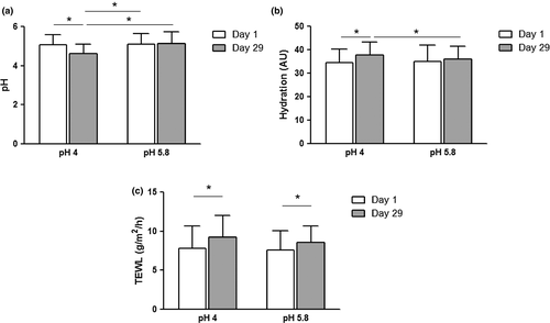
The influence of the applied emulsions on skin hydration was determined by measuring skin capacitance. At baseline, the values for skin hydration were homogeneous in the four test areas (Table 1). After 4 weeks of treatment, the overall skin hydration was increased for both pH 4 (from 34.52 ± 5.83 to 37.82 ± 5.57) and pH 5.8 (from 35.07 ± 6.94 to 36.12 ± 5.29) emulsions but not for the respective control areas. The observed increase was only significant for the pH 4 emulsion (pH 4 vs control, P = 0.002; pH 5.8 vs control, P = 0.294). Moreover, treatment with the pH 4 emulsion resulted in a significantly higher skin hydration compared with the pH 5.8 emulsion at day 29 (P = 0.031) (Fig. 1b, Table 1).
A slight increase in mean TEWL values compared with baseline was observed for both formulations (pH 4, 7.82 ± 2.87 to 9.24 ± 2.76 g/m2 per h; pH 5.8, 7.56 ± 2.52 to 8.54 ± 2.10 g/m2 per h) after 4 weeks of treatment. A similar trend was also seen on control fields (7.94 ± 1.81 to 8.44 ± 1.73 g/m2 per h and 8.26 ± 3.02 to 8.78 ± 3.37 g/m2 per h). Nevertheless, the differences to baseline did not differ significantly between each of the emulsions and the respective control areas (pH 4, P = 0.232; pH 5.8, P = 0.459) or between the skin areas treated with the pH 4 or the pH 5.8 emulsion (P = 0.463) (Fig. 1c, Table 1).
Application of pH 4 emulsion for 4 weeks increases lipid content and improves lipid lamellae organization
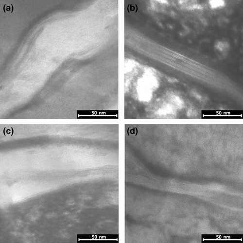
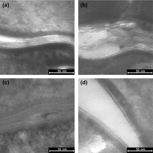
Analysis of the transmission electron microscopy (TEM) images showed that the 4-week treatment with both emulsions significantly increased the mean length of ICLL (pH 4, P < 0.001; pH 5.8, P < 0.001) from approximately 40 ± 8 nm/1000 nm2 at baseline to 135 ± 42 nm/1000 nm2 with the pH 5.8 emulsion and 184 ± 22 nm/1000 nm2 with the pH 4 emulsion (Fig. 2). The increase for the pH 4 emulsion was significantly higher than for the pH 5.8 emulsion (P = 0.002). According to published work, baseline values of ICLL indicate very dry skin while the values after treatment correspond to dry skin (pH 5.8) and normal skin (pH 4), respectively.17 In addition, the TEM images revealed that treatment with the pH 4 emulsion led to a clear improvement of ICLL organization in the SC compared to pH 5.8 emulsion (Fig. 2, Table 2).
| pH 4 | pH 5.8 | |||
|---|---|---|---|---|
| Day 1 | Day 29 | Day 1 | Day 29 | |
| TEM (nm ICLL/1000 nm2 ICS) | 41.01 ± 7.99 | 184.39 ± 22.08*† | 43.17 ± 10.45 | 134.78 ± 42.05*†‡ |
| Cholesterol (μg/slide) | 2.86 ± 0.79 | 2.93 ± 0.73 | 2.80 ± 0.76 | 2.56 ± 0.63*‡ |
| FFA (μg/slide) | 2.07 ± 0.79 | 2.46 ± 0.96 | 1.91 ± 0.83 | 1.95 ± 0.99*‡ |
| Ceramide EOS (μg/slide) | 2.92 ± 1.02 | 6.49 ± 1.37*† | 2.87 ± 0.82 | 5.60 ± 1.06*†‡ |
| Ceramide NP (μg/slide) | 3.59 ± 1.04 | 6.87 ± 1.34*† | 3.54 ± 0.90 | 5.70 ± 1.42*†‡ |
| Ceramide NH (μg/slide) | 6.26 ± 1.99 | 7.96 ± 1.95 | 6.10 ± 1.90 | 6.25 ± 1.16*‡ |
| Sum of lipids (μg/slide) | 17.70 ± 2.77 | 26.72 ± 3.52*† | 17.22 ± 2.25 | 22.07 ± 2.64*†‡ |
- Absolute values, mean ± standard (n = 14). Comparison of time points on raw data by matched pairs t-test (with Bonferroni's correction) on day 1 and day 29. *P < 0.05, †compared with day 1, ‡compared with pH 4. FFA, free fatty acid; ICLL, intercellular lipid lamellae; ICS, intercellular space; TEM, transmission electron microscopy.
The changes in the lipid lamellae structure induced by the two test emulsions were accompanied by changes in the lipid content as determined by HPTLC. For both the pH 4 and the pH 5.8 emulsion, the overall lipid content was significantly higher than at baseline (pH 4, 26.72 ± 3.52 vs 17.70 ± 2.8 μg/slide, P < 0.001; pH 5.8, 22.07 ± 2.64 vs 17.22 ± 2.25 μg/slide, P < 0.001). The increased lipid content was mainly due to a higher content of the ceramides EOS and NP. Compared with the pH 5.8 emulsion, the pH 4 emulsion resulted in significantly increased amounts of ceramides EOS and NP (P = 0.023 and P = 0.004, respectively) as well as a significantly higher overall lipid content (P = 0.003) (Table 2).
Skin treated with pH 4 emulsion shows lowest pH after SDS challenge
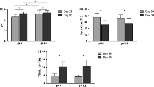
To investigate the skin barrier properties after 4 weeks of application, the skin was challenged by application of SDS. The SDS challenge significantly increased the pH of all test areas compared with the value immediately before challenge (i.e. after 4 weeks of treatment). The lowest mean skin pH after the SDS challenge was observed for the pH 4 emulsion (pH 4, 5.15 ± 0.36; pH 5.8, 5.36 ± 0.39; control areas, 5.23 ± 0.39 and 5.32 ± 0.39) (Fig. 3a). Moreover, the mean pH difference to baseline (i.e. before treatment/day 1) was significantly lower for the pH 4 emulsion compared with the untreated control (0.06 ± 0.39 vs 0.21 ± 0.39, P = 0.009) and also compared with the pH 5.8 emulsion (0.06 ± 0.39 vs 0.25 ± 0.40, P = 0.047). A comparison of treatments showed that there was no significant difference for the change in skin capacitance (as a measure of hydration) between the pH 4 and pH 5.8 emulsion (mean Δcapacitance, −11.74 ± 6.18 vs −8.53 ± 7.54, P = 0.166) or between either emulsion and the respective control area (Fig. 3c). Furthermore, the application of SDS to the skin for 24 h led to a significant increase in ΔTEWL for all test areas, due to the detergent-induced damage of the skin barrier. The SDS-induced changes in TEWL from day 29 to day 30 did show a lower TEWL for the pH 4 emulsion compared with the pH 5.8 emulsion (mean ΔTEWL, 11.58 ± 5.26 vs 13.35 ± 6.60 g/m2 per h, P = 0.175), but missed statistical significance. The increased TEWL was accompanied by significant decreases in skin hydration compared with day 29 (Fig. 3c, Table 3).
| pH | TEWL | Hydration | ||||
|---|---|---|---|---|---|---|
| Day 29 | Day 30 | Day 29 | Day 30 | Day 29 | Day 30 | |
| Control pH 4 | 4.97 ± 0.45 | 5.23 ± 0.39*†‡ | 8.44 ± 1.73 | 21.00 ± 7.01*† | 32.72 ± 6.38 | 24.30 ± 5.80*† |
| pH 4 | 4.62 ± 0.50 | 5.15 ± 0.36*†§ | 9.24 ± 2.76 | 20.82 ± 6.22*† | 37.82 ± 5.57 | 26.09 ± 6.01*† |
| Control pH 5.8 | 4.98 ± 0.53 | 5.32 ± 0.39*† | 8.78 ± 3.37 | 24.26 ± 8.21*† | 32.93 ± 4.52 | 26.70 ± 6.77*† |
| pH 5.8 | 5.13 ± 0.60 | 5.36 ± 0.39*†‡ | 8.54 ± 2.10 | 21.89 ± 7.60*† | 36.12 ± 5.30 | 27.60 ± 7.98*† |
- Absolute values, mean ± standard deviation (n = 19). *P < 0.05, †compared with day 29, ‡compared with pH 4, §compared to pH 5.8. TEWL, transepidermal water loss.
Lipid lamellae of skin treated with pH 4 emulsion are more resistant to SDS challenge
Determination of lipids after the SDS challenge revealed that the overall lipid content as well as the amount of ceramides EOS and NP had returned to values comparable with the baseline situation, namely before treatment (Table 4). The structure of lipid lamellae was assessed after SDS challenge. A significant reduction in the mean length of ICLL was observed compared with day 29 (P < 0.001 for both pH 4 and pH 5.8 emulsion). However, the skin area treated with the pH 4 emulsion showed a significantly greater mean length of ICLL than the emulsion with pH 5.8 (94 ± 36 vs 53 ± 11 nm/1000 nm2, P = 0.001). When compared with baseline (day 1), the skin area treated with the pH 4 emulsion displayed a significantly larger mean ICLL length (94 ± 36 vs 41 ± 8 nm/1000 nm2, P < 0.001). In contrast, the skin area treated with the pH 5.8 emulsion did not show significantly larger mean ICLL length compared with baseline (day 1) (Fig. 4).
| pH 4 | pH 5.8 | |||
|---|---|---|---|---|
| Day 29 | Day 30 | Day 29 | Day 30 | |
| TEM (nm ICLL/1000 nm2 ICS) | 184.39 ± 22.08 | 94.22 ± 36.33*† | 134.78 ± 42.05 | 53.08 ± 10.87*†‡ |
| Cholesterol (μg/slide) | 2.93 ± 0.73 | 3.83 ± 0.99 | 2.56 ± 0.63 | 3.67 ± 0.55*† |
| FFA (μg/slide) | 2.46 ± 0.96 | 2.84 ± 1.21 | 1.95 ± 0.99 | 2.76 ± 1.79 |
| Ceramide EOS (μg/slide) | 6.49 ± 1.37 | 3.97 ± 1.23*† | 5.60 ± 1.06 | 3.32 ± 0.68*† |
| Ceramide NP (μg/slide) | 6.87 ± 1.34 | 3.00 ± 1.35*† | 5.70 ± 1.42 | 2.56 ± 0.99*† |
| Ceramide NH (μg/slide) | 7.96 ± 1.95 | 6.24 ± 1.51*† | 6.25 ± 1.16 | 5.26 ± 1.02*‡ |
| Sum of lipids (μg/slide) | 26.72 ± 3.52 | 20.19 ± 3.24*† | 22.07 ± 2.64 | 17.56 ± 1.66*†‡ |
- Absolute values, mean ± standard deviation (n = 14). Comparison of time points on raw data by matched pairs t-test (with Bonferroni's correction) on day 29 and day 30. *P < 0.05, †compared with day 29, ‡compared with pH 4. FFA, free fatty acid; ICLL, intercellular lipid lamellae; ICS, intercellular space; TEM, transmission electron microscopy.
Discussion
Application of the emulsion with a pH of 4 significantly decreased skin's pH in elderly subjects. While it may not be regarded as a major change, due to the logarithmic nature of pH scale, a decrease of the pH as little as 0.3 units reflects half the concentration of H+. The emulsion with pH 4 was superior regarding moisturizing efficacy, regeneration of lipid lamellae and lipid content. Moreover, although SDS challenge after the 4-week treatment period negatively affected all assessed skin parameters, skin that had been treated with the pH 4 emulsion still showed the lowest pH of all test areas and, more importantly, the pH 4-treated area had the best structure of the lipid lamellae suggesting faster barrier regeneration.
Not every product with low pH is capable of restoring skin pH and physiology.22 This is because not only the type of emulsion, but also the buffer capacity/buffer system used in a cosmetic product is of utmost importance. In this study, we used w/o emulsions with an almost identical formulation, but with differently adjusted pH (either 4 or 5.8) using a buffering system based on glycolic acid for pH 4 emulsion, because a more acidic pH (e.g. pH 4) of cosmetic products for aged skin is considered beneficial.5 A w/o emulsion was chosen according to textbook knowledge because it shows prolonged skin hydrating effects and being most appropriate for the skin of elderly people.23 The observed acidification through treatment with the pH 4 formulation is in accordance with other observations in the elderly reported in the published work. For instance, a study on 20 nursing home residents aged 80 years and over found that treatment with a pH 4.0 formulation significantly improved SC integrity and SC recovery 24 h after perturbation compared with a formulation with a pH of 6.0.15 These beneficial effects on the SC coincided with a significantly lowered skin pH, improved skin hydration and a slightly higher TEWL.15 A reduction of skin pH and increased skin hydration was also observed after treating 15 elderly patients (≥80 years) with an o/w emulsion at pH 4.0 for 4 weeks, pointing to improved epidermal barrier integrity.24 Similar results were also obtained by Behm et al.23 on elderly subjects receiving a 4-week treatment with a pH 4 w/o emulsion. The treatment led to increased skin hydration and reduced skin pH. While the mean age of subjects in these studies was 70 years and older, this study showed that acidification with the pH 4 emulsion already benefits subjects between 60 and 70 years of age as well as 50+ as shown in this study.
It has been demonstrated that both the skin barrier function and the lipid organization in the SC are influenced by the lipid chain length.8 In particular, a better barrier function of the skin is related to an increase in ICLL length, particularly owing to ceramides and free fatty acids.8, 18 Furthermore, healthy skin is characterized by a unique lamellar arrangement of the lipid matrix in the SC.7, 25 Seasonal changes, race, body site and aging significantly influence the lipid lamellar architecture and composition.9, 26-29 The age group studied here (63.4 ± 6.8 years) showed a disturbed lipid lamellar organization which is in line with published work.9, 27, 29 Using Lipbarvis sampling in addition to standard instrumental measurements, we were able to show that lamellar organization clearly improved after 4-week treatment with the pH 4 emulsion. Detailed analysis showed that the length of ICLL had more than quadrupled and coincided with increased levels of ceramides EOS and NP. As a result, the epidermal barrier was similar to that of healthy skin. Importantly, the improvements achieved with the pH 4 emulsion were clearly different and superior to those for the pH 5.8 emulsion, where the skin did not show a complete repair at the end of treatment. In addition, for the skin area treated with the pH 4 emulsion the lamellar organization was significantly less disturbed after the SDS challenge compared with the skin area treated with the pH 5.8 emulsion. Hence, treatment of aged skin with the pH 4 emulsion improved ICLL length and lamellar organization, both of which are important for the barrier function of the skin, as well as the resistance to exogenous stress.
The decrease of ceramide EOS induced by SDS challenge is less distinctive after treatment with pH 4 emulsion compared with treatment with pH 5.8 emulsion. Ceramide EOS is partially associated with the cornified envelope (CE) of the corneocytes.30 The smaller decrease of ceramide EOS could be a result of stronger anchoring to the CE induced by the 28-day treatment with pH 4 emulsion. According to the published work, an enhanced ceramide biosynthesis may be associated with pH 4 or lower pH.31 In addition, many studies demonstrated that ceramide production is increased following ultraviolet B irradiation or other oxidative stressors and results in increasing ceramide-induced apoptosis in keratinocytes.32-35 It is possible to speculate that an acidic pH may also act as stressor on keratinocytes and thus increase ceramide synthesis.
Furthermore, lipid synthesis enzymes (β-glucocerebrosidase and acidic sphingomyelinase) catalyze the last step in ceramide synthesis and activity of those enzymes is pH dependent.36, 37 Thus, restoring a physiological pH of the skin may optimize the microenvironment of these enzymes and ultimately levels of ceramides will change.38, 39
The lipid lamellae could not be extracted in the same range as after treatment with pH 5.8 emulsion, indicating an improved epidermal barrier. Although the Lipbarvis method has clear advantages, there are some limitations, which we are aware of. Due to different ways of sampling the skin (second tape strip) as well as a different isolation procedure, measuring of skin lipids (either quantitatively or qualitatively) is not standardized.40, 41 The Lipbarvis sampling technique allows investigating the region in the SC from 5th till 9th layer. Lipids derived from the SC surface lipid film or from deeper regions in the epidermis were not analyzed with this method. Other methods for lipid extraction from skin samples (e.g. spilt skin, parts of biopsies) or rinsing the SC with solvents often include lipids of other regions in the epidermis and even lipids from sebaceous glands. This can lead to different ratios of cholesterol : fatty acid : ceramide. Even though all lipids were extracted and used for HPTLC analysis, only cholesterol, FFA, EOS, NP and NH were quantified because only for these commercial standards are available.18, 21
Despite clear improvements in skin pH, skin hydration and lamellar organization upon treatment with the pH 4 emulsion, the TEWL did not decrease as perhaps expected. However, it should be noted that no clear consensus on the influence of age on TEWL exists in the published work. Both unchanged values for TEWL42, 43 and subnormal values for TEWL44 in aged skin have been reported. In line with the observation in this study, Blaak et al.15 reported a slight, non-significant TEWL increase in a group of elderly patients after a 7-week treatment with a pH 4 formulation. Moreover, a recent meta-analysis on TEWL in individuals aged 65 years or more found a consistently lower TEWL for this age group compared with the group of 18–64-year-old individuals, indicating a subnormal TEWL in elderly individuals.45 For the mid-volar forearm area, this meta-analysis included data of over 2000 patients.45 The implication from this meta-analysis is that TEWL is generally subnormal in older individuals possibly due to reduced microcirculation. Therefore, an increase in TEWL after treatment with an acidic formulation, as observed in this study and by Blaak et al.,15 could well be a result of a restored epidermal barrier, and therefore in good agreement with the other beneficial effects. It may be speculated that skin moisturization contributes to this effect, as the 4-week application of the pH 5.8 emulsion also resulted in improved skin hydration and a significant increase in TEWL. However, although not significant, the increase in TEWL was higher for the pH 4 emulsion, suggesting that acidification of the SC may also play a role. In line with results of this study, our previous study46 with the same cosmetic products utilizing two different models (a short-term chemical damage and mechanical damage of epidermal barrier after long-term treatment) could clearly show that pH is important and an independent factor influencing epidermal barrier.46, 47 Finally, it is also important to note that not only aging skin, but also diseased skin (atopic dermatitis, psoriasis or ichthyosis vulgaris) have been linked to increased pH.48
Normalization of skin pH following exogenous acidification is improving epidermal permeability barrier homeostasis, SC integrity/cohesion and anti-inflammatory function12, 13 and reduces damage to exogenous stress (SDS) as shown in this study. Moreover, normalization of an increased pH improves the activity of pH-dependent enzymes involved in epidermal differentiation2, 49 and promotes recruitment of stored Ca2+ which in turn inhibits proliferation and induces differentiation.50-52 Nevertheless, exact mechanisms leading to increased lipid production as well as improved barrier integrity are still not fully elucidated. Further studies are required to characterize those exact mechanisms and how lower pH improves barrier function.
This study preformed in subjects with Fitzpatrick skin type II and III provides further evidence that acidifying the skin of elderly with an appropriately buffered pH 4 emulsion improves not only the epidermal barrier as well as the SC organization, but also reduces exogenous damage to the SC. Thus, age-appropriate skin care for older people should exhibit a buffered acidic pH of 4. Additional studies are needed to confirm these effects in younger age groups, other race or Fitzpatrick skin types and in other indications.
Acknowledgments
Medical writing support was provided by Dr Alexander Boreham (co.medical/co.faktor GmbH). The study was funded by Dr August Wolff GmbH & Co. KG Arzneimittel, Bielefeld, Germany.
Conflict of Interest
D. D. and S. D.-P. have no conflicts of interest to declare. They are employees of Microscopy Services Dähnhardt GmbH in Flintbeck. A. K., C. M., H. R., U. K. and C. A. are employees of Dr August Wolff GmbH & Co. KG Arzneimittel.


