Effects of Suture Tension and Surgical Approach During Unilateral Arytenoid Lateralization on the Rima Glottidis in the Canine Larynx
No reprints available.
Abstract
Objectives— To evaluate the effect of abduction suture tension and cricothyroid (CT) joint disarticulation on the area, height, and width of the rima glottidis (RG) during unilateral arytenoid lateralization.
Study Design— Experimental study.
Animals— Nine canine cadaver larynges.
Methods— Left arytenoid lateralization was performed with high or low abduction suture tension. RG area, height, and width were measured by computerized planimetric analysis with the epiglottis in an open and closed position. The experiment was performed with the CT joint intact and disarticulated. The effects of suture tension, CT disarticulation, and their interaction on RG area with the epiglottis closed or open were evaluated by repeated measures analysis of variance (ANOVA).
Results— RG area increased by 82% and 129% (P < .0001) with low and high suture tension, respectively. The aperture not covered by the epiglottis in a closed position was 467% larger with high suture tension than with low tension (P < .0001). CT disarticulation had no significant effect on RG geometry with either low or high suture tension (P= .4970).
Conclusions— Low suture tension increased RG area when the epiglottis was in an open position without increasing RG aperture when the epiglottis was closed. Suture tension had a significant effect on RG opening when the epiglottis was closed. CT disarticulation did not modify the geometry of the RG.
Clinical Relevance— Use of a low-suture tension should be considered during arytenoid lateralization because it has the potential to reduce the risk of aspiration pneumonia.
Laryngeal paralysis is a common upper airway disease that primarily affects older, large-breed dogs.1 A congenital form occurs in some breeds including Bouvier des Flandres and Siberian huskies.2 Acquired laryngeal paralysis is caused by damage to the recurrent laryngeal nerve or intrinsic laryngeal muscles from polyneuropathy, polymyopathy, iatrogenic or accidental trauma, or intrathoracic or extrathoracic masses, or it can be idiopathic.3 In laryngeal paralysis, the only intrinsic abductor muscle of the larynx, the cricoarytenoid dorsalis muscle, fails to contract during inspiration.1,3 The vocal folds and arytenoid cartilage remain in a paramedian position physically obstructing airflow at the rima glottidis (RG), the narrowest part of the laryngeal airway.1,2,4 This increases resistance to airflow and creates turbulence, which is manifested as inspiratory laryngeal stridor. During exercise, the speed of airflow through the larynx increases, resulting in an intraglottic pressure decrease. The arytenoid cartilages and vocal folds are then pulled across the lumen increasing resistance to inspiratory gas flow (Venturi effect),2,3,5,6 exacerbating the upper airway obstruction. Concurrent laryngeal inflammation, edema, tonsillitis, and weakening of laryngeal cartilages can induce a life-threatening condition.2,4,5,7–14
Surgery is considered the most appropriate treatment for long-term management of canine laryngeal paralysis. The aim of surgical treatment of laryngeal paralysis is to reduce airway resistance and to prevent dynamic collapse of the arytenoid cartilage during exercise.1,3,8,15–19 Unilateral arytenoid lateralization has been used successfully to achieve these goals.3,15,20,21 Arytenoid lateralization increases RG cross-sectional area and stabilizes the arytenoid cartilage by placing a prosthetic suture from the cricoid cartilage or thyroid cartilage to the muscular process of the arytenoid cartilage.16–19,21 Disarticulation of the cricothyroid (CT) joint has been recommended to increase surgical exposure.19,20 Complications of arytenoid lateralization include coughing and aspiration pneumonia because of pharyngeal dysfunction or over-abduction of the arytenoid cartilages.1,19 Excessive tension on the prosthetic suture might open the RG beyond the edges of the epiglottis, thus increasing the risk for aspiration pneumonia. In dogs, it has been recommended not to over-abduct the arytenoid cartilages17; similar recommendations have been made for horses.19 Reduction of suture tension might not abduct the arytenoid cartilage beyond the edges of the glottis but still open RG enough to decrease airway resistance.
The first purpose of our study was to evaluate the effect of the tension applied to the abduction suture on RG area during unilateral arytenoid lateralization in normal cadaveric canine larynges. Our second purpose was to evaluate the effect of CT disarticulation on RG area, height, and width during unilateral arytenoid lateralization in cadaveric canine larynges.
Materials and methods
Larynges were harvested from cadavers of male Walker hounds killed at completion of a research project on lung resection. All larynges appeared to be anatomically normal. Needles were placed through peripheral laryngeal tissues, without impeding laryngeal motion, and secured to a board so that the larynx was stabilized in a dorsoventral position. This resulted in a laryngeal view similar to laryngeal examination in a live dog.
Polypropylene (3–0) suture placed through the mucosa at the rostral point of the epiglottis was passed through the tracheal lumen so that the epiglottis could be moved from a resting (open) position to close the RG (closed position). Apposition of the epiglottis to the corniculate and cuneiform processes of arytenoid cartilages was achieved without distortion of the shape of the glottidis (1, 2). For each larynx, we measured RG area before and after arytenoid lateralization with a suture placed under either low or high tension. Measurements were performed with the epiglottis in a closed and open position. Measurements were repeated before and after CT disarticulation.
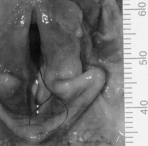
Rima glottidis with epiglottis open in a normal cadaveric larynx. A suture has been placed through the epiglottic mucosa to facilitate movement of the epiglottis into a closed position.
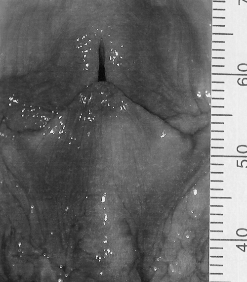
Rima glottidis aperture not covered by epiglottis when it was closed in a normal cadaveric larynx.
Arytenoid lateralization was performed on the left side with an intact CT articulation. The muscular process of the arytenoid cartilage was palpated under the junction of the dorsal cricoarytenoid muscle and thyroarytenoid muscle to localize the cricoarytenoid articulation. The dorsal cricoarytenoid muscle was transected midbody with scissors after blunt dissection. The cricoarytenoid articulation was opened using sharp dissection being careful not to damage the muscular process. The cranial part of the joint capsule of the cricoarytenoid articulation was maintained. A simple interrupted 2–0 polybutester suture was placed through the center of the articular surface of the muscular process of the arytenoid cartilage and through the caudodorsal edge of the cricoid cartilage to achieve arytenoid abduction. For “low” tension, the suture was tied until resistance from the cranial part of the joint capsule was felt. After digital images (Coolpix 990; Nikon, Tokyo, Japan) were taken, the suture was tightened as much as possible to achieve “high” tension. After images were recorded, the suture was removed and the CT articulation was separated with scissors. A new suture was placed using the same holes in the muscular process of the arytenoid and the cricoid cartilage. The suture was tightened to achieve abduction with low, then high tension.
Photographs of the glottic opening for each specimen at each step of the experiment were taken with a digital camera positioned in a plane parallel to the glottic opening. A ruler was placed next to the larynx for image calibration. Computerized planimetric analysis (Image 1,61/PPC; National Institutes of Health, Bethesda, MD) was used to measure RG height, width, and cross-sectional area on each image. RG was defined by the medial side of the corniculate process, vocal folds, and the base of the epiglottis ventrally with the epiglottis in an open position (1, 3, 4). With the epiglottis in a closed position, RG was defined by the glottic opening not covered by the epiglottis (2, 4-6). When a lateral opening was visible with the epiglottis closed, another image was taken of this opening with the camera in a plane parallel to the opening. Images were taken and analyzed by S.B.
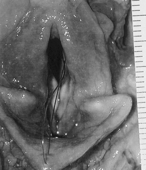
Rima glottidis with epiglottis open after a low tension abduction suture has been placed.
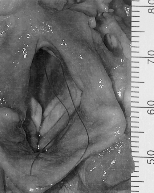
Rima glottidis with epiglottis open after a high tension suture was placed.
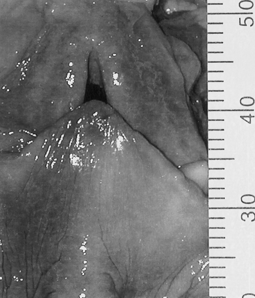
Rima glottidis aperture not covered by the epiglottis when it was closed after a low tension suture was placed.
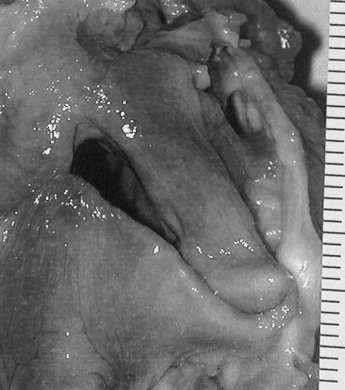
Rima glottidis aperture not covered by the epiglottis when it was closed, after a high tension suture was placed.
Analysis of variance for repeated measures was used to evaluate the effect of different suture tensions, CT disarticulation, and their interaction on RG area with the epiglottis closed or open. A value of P < .05 was considered significant. Data are presented as a mean ± standard error (SE).
Results
Mean dog weight was 28.6 ± 3.2 kg. Suture tension had a significant effect on RG area (P < .0001), with RG area being significantly increased with the high tension on the suture compared with the low tension on the suture (P= .0025) and the resting position (P= .0001; Fig 7). Epiglottis position and suture tension had a significant interaction on RG area (P < .0001). With the epiglottis open, RG area was 1.82 times larger with low suture tension and 2.2 times larger with high suture tension than with the larynx in the resting position. With the epiglottis closed, RG area was 2.28 times larger with low tension and 7.39 times larger with high tension than with the larynx in the resting position. CT disarticulation had no significant effect on RG area (P= 0.497) and no significant interaction with either low or high suture tension (P= .401), with the epiglottis closed or open (P= .815), or with the epiglottis closed or open and low or high tension (P= .4419).
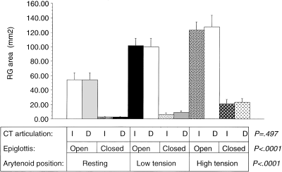
Mean (±SE) rima glottidis (RG) area (mm2) for 9 cadaveric canine larynges before (resting) and after unilateral arytenoid lateralization with 2 different suture tensions (low tension, high tension), with crycothyroid (CT) articulation intact (I) or disarticulated (D), and with the epiglottis in an open and closed position. P values are reported for each of the effects (CT disarticulation, suture tension, and epiglottis position).
Suture tension had a significant effect on RG height (P= .0318); RG height increased significantly with low tension compared with the resting position (P= .0107; Fig 8). RG width was significantly affected by suture tension (P= .001; Fig 9) with width being significantly increased by high tension compared with the resting position (P < .0001) and low tension (P= .0021). CT disarticulation had no significant effect on RG height (P= .25) or width (P= .78). The interaction between suture tension and CT disarticulation was not significant for RG height (P= .891) or width (P= .318).
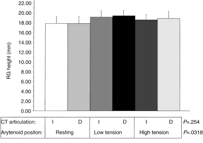
Mean (±SE) rima glottidis (RG) height (mm) for 9 cadaveric canine larynges before (resting) and after unilateral arytenoid lateralization with 2 different suture tensions (low tension, high tension) and with crycothyroid (CT) articulation intact (I) or disarticulated (D). P values are reported for each of the effects (CT disarticulation, suture tension).
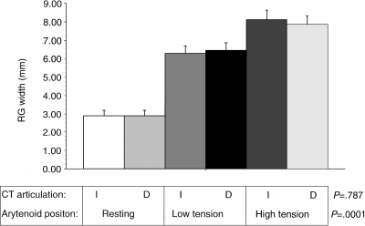
Mean (±SE) rima glottidis (RG) width (mm) for 9 cadaveric canine larynges before (resting) and after unilateral arytenoid lateralization with 2 different suture tensions (low tension, high tension) and with CT articulation intact (I) or disarticulated (D). P values are reported for each of the effects (CT disarticulation, suture tension).
Discussion
In interpreting our results, some limitations of a cadaveric study must be considered. Cadaver larynges have been used previously for evaluation of surgical treatment for laryngeal paralysis in dogs.19,21,22 The dynamic function of the larynx cannot be replicated, but cadaveric larynxes may provide information to explain some of the complications associated with arytenoid lateralization. In cadaveric larynges, the arytenoid cartilages are in a similar intermediate position as occurs in the paralyzed larynx in live dogs.19,21,22 We used only fresh larynges. The mobility of the arytenoid cartilages was maintained, and no problems were encountered abducting the arytenoid cartilages when performing the various procedures. Although motion of the epiglottis might not have been the same as in a live dog, the epiglottis could be mobilized without restriction, and epiglottis motion was consistent between low and high tension. Therefore, our conclusions related to suture tension should be valid.
Unilateral arytenoid lateralization with a low suture tension increased RG area with the epiglottis open without increasing the RG opening with the epiglottis closed. Suture tension did affect RG with the epiglottis closed and thus could increase the risk of aspiration pneumonia after unilateral lateralization. Aspiration pneumonia is a potential complication after surgical management of laryngeal paralysis and is the most commonly reported cause of death after surgery.1,12,20,21,23,24 Occasional coughing after drinking water can occur in as many as 89% of dogs after surgery.2,11,18,19,24,25 In a normal animal, aspiration pneumonia is prevented by adduction of the arytenoid cartilages and vocal folds and by epiglottic coverage of the RG during swallowing.20 Arytenoid cartilage lateralization makes normal arytenoid adduction during deglutition impossible, theoretically predisposing the animal to aspiration pneumonia.1,3,8,17,26–28 In our study, the aperture not covered by the epiglottis in a closed position was 467% larger with a high suture tension than with a low suture tension. Therefore, we can postulate that with a high suture tension, there is a greater risk for the development of aspiration pneumonia.
RG area increased by 82% with low suture tension and by 129% with high tension. Our results are similar to those previously reported.20,21 Degree of clinical improvement in animals with laryngeal paralysis has not been proven to be proportional to postoperative RG area.11,20 A significantly greater increase in RG area after unilateral cricoarytenoid lateralization compared with unilateral thyroarytenoid lateralization (207%v 140%) did not result in a different clinical outcome.29 The percentage increase in RG area required to relieve clinical signs of laryngeal paralysis is unknown, as is the percentage increase in RG during maximum inspiratory effort in normal dogs.20 More than 90% of dogs reportedly have good long-term respiratory function after unilateral lateralization despite increasing RG which was less than with a bilateral procedure.18,24 Jansson et al19 reported that a significant increase in airflow was possible with 80% abduction of one arytenoid cartilage in horses. We only measured the RG area and not the effect on airflow, which is the ultimate goal of any treatment for laryngeal paralysis. Airflow is dependent on the pressure gradient across the larynx and airway resistance. Because the area increased by 82% with low suture tension, the reduction in airway resistance should likely be sufficient to improve airflow through the larynx.
CT disarticulation has been recommended to increase surgical exposure during arytenoid lateralization.19,20 However, CT disarticulation could disrupt the lateral support of the larynx resulting in dorsoventral collapse of the RG because of flattening and overriding of the cricoid and thyroid cartilages.11,20 Decrease in RG height is minor with unilateral arytenoid lateralization, but it may become extreme when the procedure is performed bilaterally.11,20 Similar to Lussie et al,21 we did not find a difference in the RG area after CT disarticulation. Thus, we do not recommend CT disarticulation unless it is required for surgical exposure. The lateralization suture was placed between the cricoid cartilage and the arytenoid cartilage. It reproduces more closely the function of the dorsal cricoarytenoid muscle and provides a greater increase in the area of the RG.29
Another limitation of our study was that we did not measure actual suture tension. We attempted to reproduce the clinical situation and only used the limitation to abduction provided by resistance from the cranial part of the joint capsule to determine low suture tension. After we tightened the suture to high tension, it is possible that we stretched the joint capsule, which may have influenced arytenoid position after CT disarticulation and could potentially have influenced reproducibility of our experiment. However, RG area with low and high suture tension was similar with CT intact or disarticulated, which would suggest that we did not have significant variation in tension between similar parts of the experiment.
Use of cadaveric larynges does not allow evaluation of the effect that healing may have on RG area. In addition to the increase in RG area created surgically, further improvement may occur in clinical patients as inflammation and edema associated with the preexisting obstruction subside.22 Also, reported complications including fragmentation of the arytenoid cartilage, avulsion of the lateralization suture, and rupture of the suture5 could not be studied in this model Alternative methods of assessment such as measuring resistance to laryngeal airflow may yield a more accurate functional assessment of airway improvement after surgery.
We conclude that in the normal canine cadaver larynx, use of a low tension suture for unilateral lateralization significantly increases RG area with the epiglottis open, without significantly increasing the aperture not covered when the epiglottis is closed. Suture tension had an effect on the RG aperture not covered by the epiglottis when it was closed. Thus, we believe that high suture tension should be avoided to minimize the risk of aspiration pneumonia. CT disarticulation had no effect on the size of the RG and is not recommended unless additional surgical exposure is needed.


