Volume 52, Issue 4 pp. 313-317

Esophageal basaloid carcinoma with marked myoepithelial differentiation

Tomoyuki Hishida

Tomoyuki Hishida

Clinical Laboratory Division, National Cancer Center Hospital, Tokyo, Japan

Search for more papers by this author
Yukihiro Nakanishi

Yukihiro Nakanishi

Department of Surgery, National Cancer Center Hospital, Tokyo, Japan

Pathology Division, National Cancer Center Research Institute, Tokyo, Japan

Search for more papers by this author
Tadakazu Shimoda

Tadakazu Shimoda

Clinical Laboratory Division, National Cancer Center Hospital, Tokyo, Japan

Search for more papers by this author
Hiroyasu Igaki

Hiroyasu Igaki

Department of Surgery, National Cancer Center Hospital, Tokyo, Japan

Search for more papers by this author
Yuji Tachimori

Yuji Tachimori

Department of Surgery, National Cancer Center Hospital, Tokyo, Japan

Search for more papers by this author
Hoichi Kato

Hoichi Kato

Department of Surgery, National Cancer Center Hospital, Tokyo, Japan

Search for more papers by this author
Hajime Yamaguchi

Hajime Yamaguchi

Department of Internal Medicine, National Cancer Center Hospital, Tokyo, Japan

Search for more papers by this author
Gen Iinuma

Gen Iinuma

Diagnostic Radiology Division, National Cancer Center Hospital, Tokyo, Japan and

Search for more papers by this author
First published: 10 June 2002
Citations: 14
Yukihiro Nakanishi, MD, Pathology Division, National Cancer Center Research Institute, 5-1-1, Tsukiji, Chuo-ku, Tokyo 104-0045, Japan. Email: [email protected]

Abstract

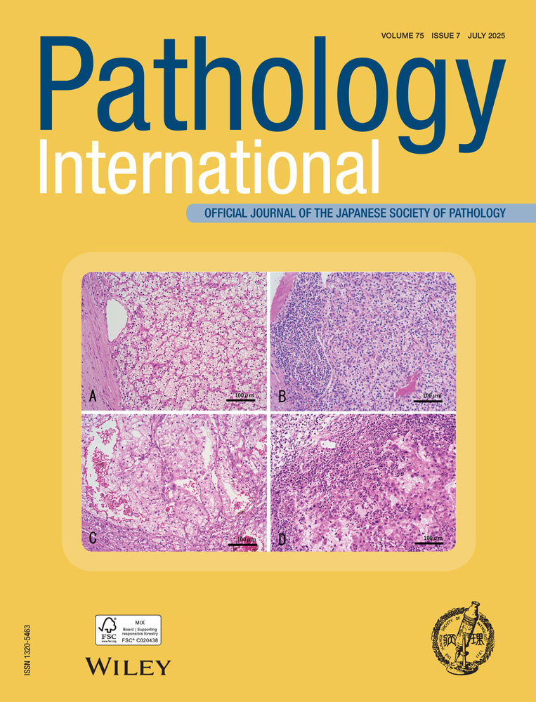
A case of esophageal basaloid carcinoma with marked myoepithelial differentiation in a 60-year-old man is reported. The tumor arose as an exophytic mass, measuring 65 × 60 mm, in the middle thoracic esophagus. Approximately two-thirds of the tumor surface was covered with non-cancerous esophageal epithelium. The depth of tumor invasion was limited to the submucosal layer. Histologically, about 70% of the tumor contained a typical basaloid carcinoma component and about 30% contained glandular and intercalated duct-like components with distinct epithelial and myoepithelial differentiation. The tumor presented no component of distinct squamous cell carcinoma, but a small portion of cribriform-like structure, which is typical of adenoid cystic carcinoma, was visible. The inner epithelium composing the intercalated duct-like structure showed immunohistochemical positivity for cytokeratin 14, and the outer epithelium lining adjacent to the stroma showed positivity for α-smooth muscle actin. These findings supported epithelial/myoepithelial differentiation. To our knowledge, our case is the first patient with an esophageal basaloid carcinoma showing marked myoepithelial differentiation.

The full text of this article hosted at iucr.org is unavailable due to technical difficulties.