Influence of arousal level for functional magnetic resonance imaging (fMRI) study: Simultaneous recording of fMRI and electroencephalogram
Abstract
Abstract Simultaneous recording of functional MRI (fMRI) and electroencephalogram (EEG) has been applied to several clinical fields, making it possible to monitor the arousal level of the subject during a cognitive task. The study confirmed that activated cerebral areas were different between high and low arousal levels during the smooth-pursuit eye movement task. When arousal level was high, activations in the parietal eye field, frontal eye field (FEF), supplementary eye field (SMA), visual fields (V1) and occipito–temporal junction (V5) were found. In contrast, when arousal level was low, activations were found only in V1 and FEF. The results indicate that the monitoring of the arousal level of subjects using fMRI and EEG recordings simultaneously is crucial for detecting cortical activations during a cognitive task.
INTRODUCTION
Electroencephalographic (EEG) monitoring during functional magnetic resonance imaging (fMRI) experiments has been increasingly applied to physiological1–3 and clinical4,5 studies. Intermittent acquisition of fMRI using EEG simultaneous recordings makes it possible to monitor the arousal level of the subject performing a cognitive task. We have developed a system that allows the simultaneous recording of fMRI and EEG,6 and have applied it to detect different cortical activation areas during smooth-pursuit eye movement (SPEM) tasks based on the simultaneous EEG pattern.
SUBJECTS AND METHOD
Three healthy right-handed volunteers provided written, informed consent to participate in the study, which was approved by the Local Ethics Committee of the Nihon University School of Medicine, Tokyo. The subjects were free from any neurological and psychiatric illnesses, and there were no abnormalities according to their structural MR images.
Functional MRI was performed with a 1.5 Tesla Magnetom Symphony (Siemens, Erlangen, Germany). The EEG was recorded inside the MRI scanner using our simultaneous EEG/fMRI recording system.
Ascending multislice gradient-echo Echo Planner Imaging (EPI) was used to produce 30 contiguous, 4-mm thick axial slices covering the whole brain [echo time (TE), 50 ms; repetition time (TR), 2948 ms; flip angle, 90°; field of view (FOV), 192 mm; 64 × 64 matrix; interval, 6 s]. Subjects were requested to perform six series, alternating between performing a SPEM task for 36 s and a control task for 36 s. The series consisted of 196 scans with a complete duration of 864 s. We also used the system of eye tracking in magnetic fields (Visible Eye; Avotec Inc., FL, USA), which can also present visual stimuli. The fMRI data were analysed using SPM99 software (SPM99; Wellcome Department of Cognitive Neurology, UK).
RESULTS
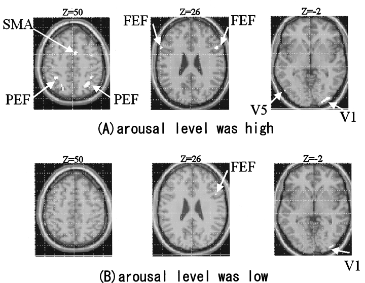
When beta waves on the EEG were observed during a SPEM task, we judged that the arousal level was high. Conversely, when alpha waves were found during a SPEM task with the subject’s eyes open, we evaluated that the arousal level was low. Figure 1 shows the activated areas of a subject’s brain during a SPEM task. When the arousal level was high, activations in the parietal eye fields (PEF), frontal eye field (FEF), supplementary eye field (SEF), primary visual fields (V1), and occipito–temporal junction (V5) were found (Fig. 1a). In contrast, when the arousal level was low, activations were found only in V1 and FEF (Fig. 1b). Similar results were obtained for the other two subjects.

. Activated areas during smooth pursuit eye movement in a subject when the arousal level was (a) high and (b) low. Statistical parametric maps were rendered onto a standard brain. Height threshold, which was corrected at P < 0.05, was used to extend each activated cluster.
DISCUSSION
The study’s results indicate that when the arousal level is high, the typical fronto-parietal network involved in the control of SPEM was activated. Supplementary Motor Area is thought to contribute to the programming of eye movements. Parietal eye fields are thought to be involved in visuo-spatial attention and V5 for the cognition of an object’s movements. Activations in these three regions decreased when the arousal level was low, indicating that the functions of accuracy for spatial cognition decreased when the arousal level was low. It could also be said that various cognitive tasks, the performances of which might be affected by the changes in arousal level, need to be conducted using the simultaneous fMRI/EEG monitoring techniques.


