Study of image findings in rapid eye movement sleep behavioural disorder
Abstract
Abstract To elucidate the cause of idiopathic rapid eye movement (REM) sleep behavior disorder (RBD), magnetic resonance imaging and single-photon emission computed tomography of the brain were conducted on 20 patients with RBD. Blood flow in the upper portion of both sides of the frontal lobe and pons was significantly lower in patients with RBD than in the normal elderly group. Among the patients with RBD, decreased blood flow in the frontal lobe showed no correlation with the extent of frontal lobe atrophy. Decreased blood flow in the upper portion of the frontal lobe and pons might be associated with the pathogenesis of idiopathic RBD.
INTRODUCTION
Rapid eye movement sleep behavioural disorder (RBD) refers to abnormal behaviour occurring during the night, which is based on pieces of dream experience. The disorder was first described in a paper by Schenck et al.1 in 1986, and was classified as a new sleep disorder in the International Classification of Sleep Disorders (ICSD)2 in 1990. Idiopathic RBD occurs in the elderly frequently, but its cause has yet to be identified. Under the circumstances, we studied the cause of idiopathic RBD by cranial magnetic resonance imaging (MRI) and cerebral single-photon emission computed tomography (SPECT) in patients diagnosed with RBD.
SUBJECTS
The subjects comprised 20 male patients who were diagnosed as having had idiopathic RBD according to the ICSD diagnostic standards based on clinical symptoms and polysomnographic findings (RBD group). As controls, seven healthy elderly men were included in the study (control group). The mean ages of the groups were 63.4 ± 8.5 years and 77.4 ± 4.2 years, respectively. Informed consent was obtained from all participants.
METHOD
Cerebral SPECT was performed at symptomatic intermittent intervals during the night in the RBD group. Essentially, the control group also underwent cerebral SPECT at the same times as those in the RBD group. The SPECT was performed on the upper portion of the frontal lobe, middle portion of the frontal lobe, parietal lobe, temporal lobe, occipital lobe, corpus striatum, thalamus, pons and cerebellum of the right hemisphere to establish the regions of interest, followed by the counting of I-123IMP accumulation. The count ratio based on the cerebellum of the same hemisphere was calculated for the cumulative count at each site. Moreover, a statistical comparative study was conducted on the count ratios at each site of the brain in the RBD and control groups, respectively, by using Student’s t-test.
RESULTS
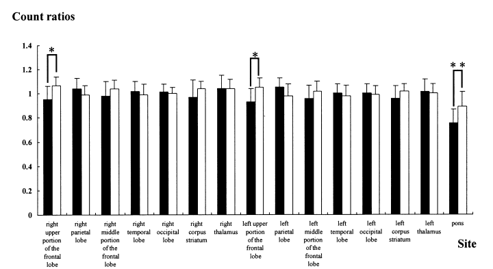
Cranial MRI findings revealed atrophy of the frontal lobe in nine patients of the RBD group and in five males of the control group. When compared with the control group, a statistically significant decrease in blood flow was observed in the upper portion of the frontal lobe on the left and right sides and the pons by cerebral SPECT in the RBD group (Fig. 1). In addition, there were no significant differences between the two groups at other sites. The patients in the RBD group were then divided into subgroups; that is, with and without atrophy of the frontal lobe. When the count ratios in the upper portion of the frontal lobe, which were obtained by cerebral SPECT, were compared between the subgroups, there was no significant difference in decreased blood flow between the two subgroups. The significant decrease in blood flow in the upper portion of the frontal lobe on the left and right sides were demonstrated in the RBD group, whereas there was no correlation between decreased blood flow in the frontal lobe and the extent of atrophy of the frontal lobe in the control group.

. Comparison of the ratios of cerebral blood flow volume between (▪) the group with rapid eye movement sleep behavior disorder and (□) the control group. *P < 0.05; **P < 0.0005.
DISCUSSION
Disorder of the dorsolateral tegmental portion of the pons is considered to be associated with the mechanism underlying the occurrence of RBD.3 The reduced blood flow in the pons observed in the present study supports this hypothesis. Alternatively, decreased blood flow in the frontal lobe was observed in the RBD group frequently, regardless of atrophy of the frontal lobe, and blood flow decreased markedly in the upper portion of the frontal lobe. Rapid eye movement sleep behavior disorder is considered a precursor symptom to Parkinson’s disease. As for SPECT findings of Parkinson’s disease, a report has shown that blood flow in the frontal lobe decreases, although the phenomenon is not observed constantly. Thus, the results of the present study indicate the close relationship between Parkinson’s disease and RBD, and suggest that decreased blood flow in the pons and frontal lobe is associated in some way or another with the pathology of idiopathic RBD.


