Hepatic cholangiocarcinoma and testicular mesothelioma in a wild-caught blue shark, Prionace glauca (L.)
Abstract
An adult male blue shark, Prionace glauca (L.), caught in July 2000 by a recreational fisherman off Long Island, New York, USA, had a retained fishing hook from a previous capture. The hook penetrated the gastric wall and lacerated the right liver lobe. Macroscopic lesions consisted of transmural gastritis and peritonitis. Alteromonas sp. and Vibrio alginolyticus were isolated from the peritoneal fluid. In addition, a well delineated, sessile mass was found on the otherwise normal serosa of the right testis. Histopathological findings included mesothelial hyperplasia and hypertrophy involving diffusely the gastric, hepatic and parietal serosae, and forming a discrete testicular capsular mass compatible with mesothelioma. In the liver an intrahepatic cholangiocarcinoma, chronic hepatitis, biliary hyperplasia and increased numbers of melanomacrophages were found. In addition organisms compatible with histozoic and coelozoic myxosporeans were found within the skeletal muscle of the abdomen and intrahepatic bile ducts, respectively. This is the third literature report of a liver tumour and the first report of a coelomic mesothelioma from a shark.
Introduction
Sharks, skates, rays and chimeras are gill breathing, jawed, finned, gas bladder-deficient fish with a calcified cartilaginous endoskeleton comprising the Class Chondrichthyes. All living members of this class are related monophyletically in a straight line of evolutionary descent. The term ‘shark’ generally refers to all chondrichthyans despite morphological divergences (Nelson, Crossman, Espinosa-Perez, Findley, Gilbert, Lea & Williams 2002). The blue shark, Prionace glauca (L.), is a large cosmopolitan carcharhinid species (Compagno 1984) that is one of the most far ranging and numerous of the large sharks (Lineaweaver & Backus 1970).
Since Deslongchamps described an enormous (8 × 30 cm), pedunculated fibroma on the tail of a thornback skate, Raja clavata L., in 1853 (Thomas 1931), 41 chondrichthyan neoplasms have been discovered (J. C. Harshbarger, unpublished data). These neoplasms originated in 18 (1.5%) of the 1168 total valid species of sharks (Campagno 1999). Approximately 250 species of bony fish with reported neoplasms represents 1.1% of the 23 450 total valid species comprising the Class Osteichthyes (Nelson 1994).
One neoplasm has been previously described in a blue shark (Schroeders 1908). That case was diagnosed as a hepatocellular adenoma. However, the liver was infiltrated with multiple tumours blending into the normal tissues which suggests that a diagnosis of hepatocellular carcinoma is warranted using current terminology. The only other chondrichthyan neoplasm arising in the hepatic parenchyma was a circumscribed hepatocellular adenoma in a red stingray, Dasyatis akajei (Muller & Henle) (Harshbarger 1980; Harshbarger, Charles & Spero 1981). The only genital tumour recorded in chondrichthyans was a seminoma in the testis of a thorny skate, Amblyraja radiata (Donovan) (Harshbarger et al. 1981).
This paper describes the first cholangiocarcinoma and the first testicular mesothelioma found in a shark. Both tumours arose in the same individual blue shark.
Materials and methods
An adult male blue shark was caught in June 2000 by a recreational angler in the northwestern Atlantic off Montauk, New York. The shark weighed 133 kg and had a fork length of 269 cm. The shark was necropsied immediately after landing. The peritoneal fluid was collected aseptically and inoculated onto two selective solid media plates tryptic soy agar (TSA) with 50% filtered ocean water (Micro Technologies, Inc., Richmond, ME, USA), and salt enriched blood agar (Binax-nel, Waterville, ME, USA). Plates were packaged on ice and shipped overnight to Micro Technologies, Inc. Laboratories for bacterial isolation.
Samples of the stomach, abdominal wall, liver and right testis were preserved in 10% buffered elasmobranch formalin (Prieur, Fenstermacher & Guarino 1976). After 48-h fixation these samples were processed routinely for paraffin embedding, embedded in paraffin, sectioned at 4–5 μm, stained with haematoxylin and eosin (H & E), and permanently mounted on glass slides using standard histological techniques. In addition, selected sections were stained with periodic acid-Schiff (PAS), Brown and Hopps (B & H), Ziehl-Neelsen (Z-N), trichrome (Sheehan & Hrapchak 1980), and an immunocytochemical method with a mouse cytokeratin squamous and non-squamous monoclonal pool (Immunon, Pittsburgh, PA, USA), according to the manufacturer's directions. Bright field microscopy was used to examine preparations.
Results
The shark appeared in good body condition and its body weight was within the normal range as reported for male blue sharks (Kohler, Casey & Turner 1996). The shark had a retained fishing hook from a previous capture. The hook (3 × 6 cm) penetrated the gastric wall and lacerated the right liver lobe. There was a localized but severe transmural necrotizing gastritis enclosing the fishing hook within fibronecrotic tissues (5 × 7 × 10 cm). The serosal surfaces of the stomach, right liver lobe and the associated parietal peritoneum appeared dull and granular due to short (0.1 × 0.3 cm) wart- and villous-like projections protruding diffusely from their surfaces. In addition a well delineated, firm, brown-red, sessile exophytic growth (0.5 × 3 × 3 cm) was present on the otherwise normal appearing serosa of the right testis (Fig. 1). There was a red-brown turbid, slightly granular peritoneal effusion, which yielded Alteromonas sp. and Vibrio alginolyticus on bacterial cultures.

Right testis of a blue shark with a brownish (0.5 × 3 × 3 cm), well delineated exophytic growth on the capsule (double arrow). Incidental cut in the capsule reveals epigonal tissue associated with the testis (single arrow).
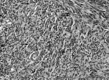
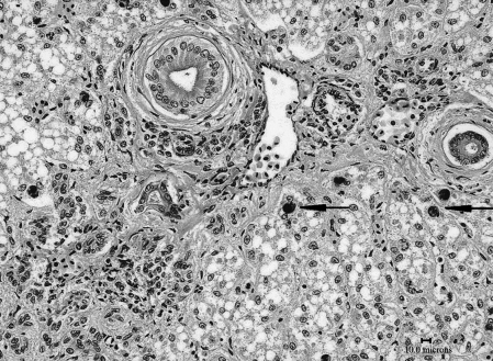
The histological lesions associated with the retained fishing hook in this shark are described in detail elsewhere (Borucinska, Kohler, Natanson & Skomal 2002). Briefly, they consisted of necrotizing, fibroproliferative gastritis and proliferative peritonitis with rare foci of mesothelial dysplasia. Herein we describe the lesions that were possibly not directly related to the fishing hook trauma. The parenchyma of the right liver lobe was effaced and expanded by chronic granulomatous hepatitis and fibrosis, which was bordering and overlapping with an infiltrating cholangiocarcinoma. The tumour cells were arranged into irregular clusters and into poorly to well defined ducts and rosettes separated by broad bands of fibroplasia (Fig. 2). The tumour cells were epithelioid to pleomorphic, had a moderate to scant lightly eosinophilic, slightly vacuolar cytoplasm, and large, vesicular, oval nuclei. One to two distinct nuclei were present, but mitotic figures were not conspicuous. Basement membranes and luminal mucin were seen in neoplastic rosettes with the PAS stain, and trichrome staining accentuated the marked desmoplasia within the tumour. Immunocytochemical staining with the cytokeratin polyclonal antibodies revealed variable immunoreactivity of normal epithelium in large bile ducts and no distinct staining of the neoplastic cells. Despite the lack of immunoreactivity, this tumour was classified as a cholangiocarcinoma based on cellular morphology and growth pattern. Within the remaining liver, marked biliary hyperplasia, peribiliary fibrosis and moderate portal lymphoplasmacytic infiltrates were present (Fig. 3). This liver also had an increased number of melanomacrophage cells (mean = 62 per high power field as compared with a mean = 12 found in four adult male blue sharks examined by JDB), and myxosporean organisms (sporogonic plasmodia) within some intrahepatic bile ducts. Special stains (Z-N and B & H) failed to detect bacteria within the hepatic parenchyma, but sparse colonies of Gram-negative coccoid to short rod bacteria were detected on the serosal surfaces of the stomach and liver.

Liver replaced by infiltrating cholangiocarcinoma with tumour cells forming distinct ducts → and accompanied by desmoplastic reaction  (H & E, bar = 10 μm).

Biliary hyperplasia, peribiliary fibrosis and moderate portal lymphoplasmacytic infiltrates in liver of shark with hepatic cholangiocarcinoma. Distinct melanomacrophages are present →(H & E, bar = 10 μm).
The serosa of the right liver lobe was thrown into papillomatous and cauliflower-like projections supported by a thin fibrovascular stroma extending from a thick fibrous layer separating the serosa from the hepatic parenchyma. Moderate lymphohistiocytic infiltrates were present within this stroma. The serosal cells were tall columnar (instead of cuboidal, which is normal in sharks) but retained their polarity. A similarly diffuse papillomatous proliferation involved the parietal serosa of the abdominal wall and the gastric serosa.
In contrast to these diffuse serosal lesions, a singular, well delineated, sessile, exophytic growth of the mesothelium was present on the capsule of the right testis. This growth was morphologically similar to the mesothelial proliferations described above, with the exception of sharp demarcation from normal mesothelium, lack of an inflammatory component in the lamina propria, the presence of abnormal mitotic figures, and cellular disorganization and loss of polarity (Fig. 4a,b). Based on the above, the testicular mass was classified as a papillary mesothelioma. Additional findings in this shark included myxosporean organisms within intracytoplasmic cysts in the skeletal muscle of the abdominal wall.

(a) Papillary mesothelioma of the testicular capsule in a shark with cholangiocarcinoma. Neoplastic mesothelium forms papillomatous projections supported by fibrovascular stroma. Tumour is sharply demarcated from normal testicular capsule →. Epigonal organ with haematopoietic tissue of the testis is present  (H & E, bar = 10 μm). (b) Higher magnification of testicular mesothelioma showing cellular disorganization, dysplasia, pleomorphism, loss of polarity and mitotic figures within the mesothelium → (H & E, bar = 10 μm).
Discussion
Approximately 40 tumours have been identified in elasmobranch fish (Schlumberger and Lucké 1948; Wellings 1969; Prieur et al. 1976; Wolke 1976; Stoskopf 1993; J. C. Harshbarger, unpublished data). The first reported tumour from a shark was a hepatic adenoma from a blue shark (Schroeders 1908) and the most recently reported tumour was a hepatic capsular fibroma in a captive Pacific swell shark, Cephaloscyllium ventriosum (Garman)(Berzins & Hovland 2000).
The tumours described in this report constitute the first cholangiocarcinoma and mesothelioma recorded from sharks. There were two pathological findings in this shark that could have been linked to the pathogenesis of these tumours. First, there was evidence of chronic exposure to environmental contaminants seen as increased numbers of hepatic melanomacrophage cells and biliary hyperplasia (Roberts 1975; Wolke, George & Blazer 1985; Ferguson 1989), and secondly, there was chronic granulomatous hepatitis probably initiated by the fishing hook injury and perpetuated by the bacterial peritonitis.
Epizootics of tumours occur in fish populations exposed to a wide range of environmental contaminants (Malins, McCain, Landahl, Myers, Krahn, Brown, Chan & Roubal 1988; Harshbarger and Clark 1990; Harshbarger, Spero & Wolcott 1993) and among these, piscine hepatic neoplasms are one of the most commonly described (Myers, Rhodes & McCain 1987; Myers, Landahl, Krahn & McCain 1991; Grizzle and Goodwin 1998). The tissue burdens of environmental contaminants were not examined in this shark, but sharks, like other top-predatory fishes, are known to bioaccumulate and bioconcentrate pollutants (Corsolini, Focardi, Kannan, Tanabe, Borrell & Tatsukawa 1995). High levels of anthropogenic toxins and carcinogens have been reported from shark tissues worldwide (Lyle 1986; Hornung, Krom, Cohen & Bernhard 1993) including the US coastal North Atlantic (Stevens & Brown 1974).
Chronic hepatitis as a result of toxic or infectious processes has been linked to hepatic neoplasms in vertebrates (Goodman 1997). This shark had a biliary infection with an organism morphologically compatible with Chloromyxum sp. (Myxosporea), but these are usually not associated with hepatic lesions (Borucinska & Frasca 2002). It is more likely that the hepatitis in this shark was secondary to bacterial peritonitis, which could have contributed to the development of the hepatic neoplasm.
The mesothelioma-like peritoneal proliferations and the testicular mesothelioma arose possibly from the persistent irritation by a foreign body (hook), and/or from bacterial infection leading to diffuse chronic proliferative peritonitis. The mesothelium of sharks is prone to hyperplastic and hypertrophic changes in response to injury (Borucinska, Martin & Skomal 2001) and has phagocytic properties. Iron particles in hyperplastic mesothelial cells were seen in two other sharks with proliferative peritonitis as a result of fishing hook injury (Borucinska et al. 2002). The role of chronic foreign body induced serositis progressing to neoplasia has been documented in man and other vertebrates exposed to asbestos (Barker 1993) and other inorganic compounds (Evans 1966). The possible progression of proliferative peritonitis to tumours of the peritoneum (mesotheliomas) in sharks with retained fishing hooks warrants further examination.
We believe that the low numbers of neoplastic lesions reported from elasmobranch fish result from the paucity of studies on this subject (Mawdesley-Thomas 1975). This lack of data facilitated the spread of a hypothesis that cartilaginous fishes are resistant to cancer. Indeed several studies examined the anti-cancer potential of products derived from shark tissues, mostly cartilage (Felzenszwalb, Pelielo de Mattos, Bernardo-Filho & Caldeira-de-Araujo 1998) and squalamine (Sills, Williams, Tyler, Epstein, Sipos, Davis, McLane, Pitchford, Cheshire, Gannon, Kinney, Chao, Donovitz, Laterra, Zasloff & Brem 1998), with rather discouraging results (Miller, Anderson, Stark, Granick & Richardson 1998). More studies, including case reports, on neoplasia in sharks are needed to fully understand their phylogenetic relationship with neoplastic disease.
Acknowledgements
We thank the fishermen, organizers and sponsors of shark fishing tournaments held on Long Island, New York and in Massachusetts who allowed us to examine and sample their catch, N. E. Kohler and L. J. Natanson (both with National Marine Fisheries Service) for facilitating our presence at these tournaments, Ione Jackman and Sallyann Gemme (both at the Veterinary Diagnostic Laboratory, University of Connecticut) for their excellent technical assistance, and the University of Hartford and University of Connecticut for logistic and laboratory support. This study was supported by the Coffin Grant from the University of Hartford.
References
Received: 17 June 2002 Accepted: 19 September 2002


