Histopathological study of feline eosinophilic dermatoses
Abstract
A retrospective study was conducted on skin specimens from 24 cats with eosinophilic granuloma complex. The specimens were stained with haematoxylin and eosin and Gallego’s trichrome stain. In all specimens, flame figures and/or large foci of so-called ‘collagen degeneration’ were detected and histopathological features were not predictive of the clinical picture. Use of the term eosinophilic dermatosis was advocated in diagnostic dermatopathology. On trichrome-stained sections, normally stained collagen fibres were identified in the middle of both flame figures and large foci of ‘collagen degeneration’ and the debris surrounding collagen bundles showed the same tinctorial properties as eosinophil granules. Eosinophil degranulation around collagen bundles seemed to represent the major pathogenetic event in these lesions, analogous with human flame figures. The term flame figures might therefore be more accurately used to designate those foci of eosinophilic to partly basophilic debris commonly referred to as ‘collagen degeneration’.
INTRODUCTION
The term eosinophilic dermatoses has been proposed to describe those feline diseases included in the eosinophilic granuloma complex (EGC),1 nevertheless, the term EGC is still commonly used.2 Eosinophilic plaque (EP), eosinophilic granuloma (EG) and indolent ulcer (IU) represent clinical entities classically included in EGC. Clinical and histopathological features distinctive of these three entities have been reported.3,4
EP occurs as pruritic, erythematous and erosive coalescing papules and plaques.4 Epidermal hyperplasia with spongiosis and prominent eosinophilic exocytosis with possible formation of intra-epidermal eosinophilic vesiculo-pustules and diffuse eosinophilic infiltration of the dermis are reported as common features.3,5
EG (synonyms: collagenolytic granuloma, linear granuloma) classically occurs as multiple nodules, variably pruritic, orientated linearly on the caudal thigh or, less commonly, as single nodular lesions located anywhere on the body, including the footpads, the lower lip and the oral cavity.4 Dermal foci of an amorphous to granular, eosinophilic to partly basophilic debris are reported as being distinctive histopathological features of EG. This debris is commonly referred to as, so-called, ‘collagen degeneration’, and is considered to represent a mix of degenerated collagen and degranulated eosinophils.3 Moreover, small foci of ‘collagen degeneration’, in which degenerated collagen fibres are surrounded by degranulated eosinophils, are commonly described and named flame figures.3,5 Dermal infiltrates in EG vary from predominantly eosinophilic to lymphocytic and histiocytic. In some cases epithelioid and multinucleated cells form a palisading granulomatous reaction around the eosinophilic debris.3,5 The epidermis is moderately hyperplastic and occasionally ulcerated.
IU (synonyms: rodent ulcer, eosinophilic ulcer, lip ulcer) refers to a painless, non-pruritic and non-bleeding ulcerated lesion most commonly located on the upper lip.4 Reported histopathological findings vary from an ulcerative dermatitis with a diffuse eosinophilic infiltrate and foci of ‘collagen degeneration’, although less prominent than in EG, to an ulcerative neutrophilic and fibrosing dermatitis.3,4
In spite of the report of a distinctive histopathological appearance, pathological features consistent with those of two or more of the recognized entities of the EGC have been observed simultaneously in feline cutaneous biopsies,6,7 although never reported in an original study.
The histopathogenesis of both flame figures and large foci of ‘collagen degeneration’ in the feline EG remains unclear. The primacy of ‘collagen degeneration’ remains the subject of debate. In a recent study of feline EG specimens,8 it was shown that Masson’s trichrome collagen staining abnormalities did not characterize collagen fibres entrapped in areas of ‘collagen degeneration’. Based on this finding, it was speculated that degeneration of collagen was not involved in the pathogenesis of the lesions. However, in this study, flame figures and large foci of ‘collagen degeneration’ were not differentiated even though it is still not known whether the two lesions have the same histopathogenesis.
The purposes of this study were to investigate the diagnostic specificity of the histopathological findings of the three entities of EGC and to study the histological features and staining properties of feline flame figures and large foci of ‘collagen degeneration’.
MATERIALS AND METHODS
A retrospective study was conducted on skin specimens from 24 cats with EGC lesions, diagnosed clinically by a veterinary dermatologist, and three normal skin specimens from one necropsied cat as the control. EGC lesions were represented by nine EP all located on the ventral abdomen, 11 EG with/without linear configuration and various cutaneous distribution, one intra-oral EG and three IU of the upper lip. One cutaneous biopsy from each case was examined. Specimens were processed in a routine fashion and stained with haematoxylin and eosin (H&E) and Gallego’s trichrome stain (IV variant).9
The H&E-stained specimens were examined in a blind fashion and the presence of flame figures and/or large foci of ‘collagen degeneration’ was recorded, as were other histopathological findings. Flame figures and/or large areas of ‘collagen degeneration’ were defined respectively as single collagen bundles surrounded by small amounts of amorphous to granular eosinophilic debris and as well-demarcated large areas of amorphous to granular, eosinophilic to partly basophilic, debris. With Gallego’s trichrome stain, collagen fibres stain blue, nuclei stain purple, and keratin, cytoplasm, muscle fibres and erythrocytes stain green. Gallego’s trichrome-stained sections were examined in a blind fashion and the presence of collagen bundles in the middle of flame figures and of large areas of ‘collagen degeneration’ was recorded. Moreover, collagen fibres were specifically scrutinized for abnormalities in size, outline and staining.
RESULTS
No abnormalities were detected in H&E-stained specimens in normal feline skin. The results of the histopathological findings in H&E-stained sections are summarized in Table 1. Histopathological findings are reported for each clinical entity.
| Case | Clinical diagnosis | Histopathological findings |
|---|---|---|
| 1 | EP | Flame figures |
| Ulcer, spongiosis, intraepidermal eosinophilic pustules | ||
| 2 | EP | Flame figures |
| Ulcer | ||
| 3 | EP | Flame figures |
| 4 | EP | Flame figures |
| Ulcer | ||
| 5 | EP | Flame figures |
| Ulcer | ||
| 6 | EP | Flame figures |
| Ulcer | ||
| 7 | EP | Flame figures |
| Ulcer, eosinophilic infiltrates in the subcutis | ||
| 8 | EP | Flame figures |
| Spongiosis, intraepidermal eosinophilic pustules | ||
| 9 | EP | Flame figures |
| Ulcer | ||
| 10 | EG | Flame figures and large foci of ‘collagen degeneration’ |
| Epithelioid macrophages and multinucleated cells | ||
| 11 | EG | Large foci of ‘collagen degeneration’ |
| Mild eosinophilic infiltrate, epithelioid macrophages and multinucleated cells | ||
| 12 | EG | Flame figures and large foci of ‘collagen degeneration’ |
| Ulcer, epithelioid macrophages and multinucleated cells | ||
| 13 | EG | Large foci of ‘collagen degeneration’ |
| Epithelioid macrophages and multinucleated cells | ||
| 14 | EG | Flame figures and large foci of ‘collagen degeneration’ |
| Spongiosis, transfollicular elimination of foci of ‘collagen degeneration’ | ||
| 15 | EG | Flame figures |
| Eosinophilic infiltrates in the subcutis | ||
| 16 | EG | Large foci of ‘collagen degeneration’ |
| Epithelioid macrophages and multinucleated cells | ||
| 17 | EG | Flame figures |
| Spongiosis | ||
| 18 | EG | Large foci of ‘collagen degeneration’ |
| Transepidermal elimination of foci of ‘collagen degeneration’ | ||
| 19 | EG | Flame figures |
| Ulcer | ||
| 20 | EG | Flame figures |
| Eosinophilic infiltrates in the subcutis | ||
| 21 | EG | Flame figures |
| Ulcer, epithelioid macrophages and multinucleated cells | ||
| 22 | IU | Flame figures |
| Ulcer, eosinophilic infiltrates in the muscle | ||
| 23 | IU | Large foci of ‘collagen degeneration’ |
| Mild eosinophilic infiltrate, epithelioid macrophages and multinucleated cells | ||
| 24 | IU | Flame figures |
| Ulcer |
In H&E-stained specimens, flame figures were detected in 19 cases (nine EP, eight EG and two IU) (Fig. 1) and large foci of ‘collagen degeneration’ were detected in eight cases (seven EG and one IU) (Fig. 2). Both flame figures and large areas of ‘collagen degeneration’ were recognized in three cases (all EG). The epidermis was variably ulcerated and/or spongiotic, and intraepidermal eosinophilic pustules were detected in two EP. A dermal, perivascular to diffuse, eosinophilic inflammatory infiltrate was observed in the entire dermis and extended to the subcutaneous tissue in one EP and one EG, and to the underlying muscle in one IU. Eosinophils were invariably present in the infiltrate and represented the predominant inflammatory cells in all but two specimens (one EG and one IU). Other inflammatory cells recognized were mast cells, usually perivascular and confined to the superficial dermis, lymphocytes and histiocytes. Neutrophils were present in the dermis underlying ulcers. Epithelioid macrophages and multinucleated cells were observed in seven specimens (six EG and one IU), surrounding both flame figures and large areas of ‘collagen degeneration’. In two biopsies (both EG), respectively, transfollicular (Fig. 3) and transepidermal elimination of foci of ‘collagen degeneration’ was detected.
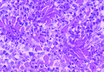
Granular eosinophilic debris accumulates along fragments of single collagen bundles (flame figures). Eosinophils and macrophages predominate in the inflammatory infiltrate (H&E, × 400).
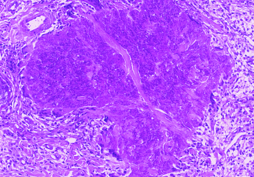
Large and irregularly shaped deposit of granular eosinophilic to partly basophilic debris (large area of ‘collagen degeneration’). Multinucleated cells surround the deposit (H&E, × 200).
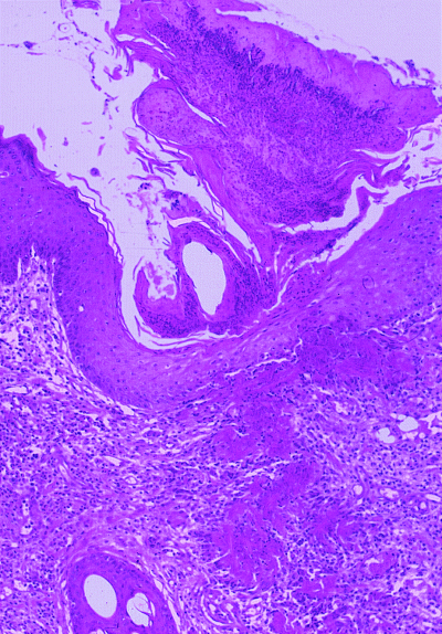
Multiple foci of ‘collagen degeneration’ are extruded from the dermis through the infundibular epithelium (transfollicular elimination of foci of ‘collagen degeneration’) (H&E, × 100).
In Gallego’s trichrome-stained sections, no abnormalities were observed in the skin specimens from the normal cat. In all EGC specimens examined, blue-stained collagen fibres were identified in the middle of both flame figures and large foci of ‘collagen degeneration’ (4, 5). Collagen fibres present in the middle of both flame figures and large areas of ‘collagen degeneration’ stained identically to collagen fibres observed elsewhere in the dermis. No size, surface contour or staining collagen abnormalities were detected in any of the specimens. In Gallego’s trichrome-stained sections, the eosinophilic debris observed in the H&E-stained sections appeared as a granular greenish material containing variable amounts of purple nuclear remnants, in both flame figures and large foci of ‘collagen degeneration’. In addition, green extracellular eosinophil granules were observed surrounding the greenish debris.
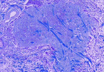
Same microscopic field as in Figure 2. In a large focus of ‘collagen degeneration’ blue collagen fibres are surrounded by granular greenish debris that contains purple nuclear remnants. Note that collagen bundles stain identically to those present in the surrounding dermis (Gallego’s stain, × 200).
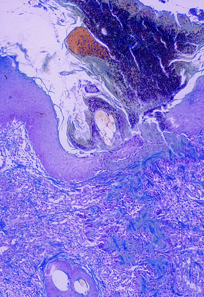
Same microscopic field as in Figure 3. Transfollicular elimination of amorphous deposits of greenish debris (large foci of ‘collagen degeneration’). Blue collagen fibres are recognizable in the middle of these deposits (Gallego’s stain, × 100).
DISCUSSION
In this study, flame figures and/or large foci of ‘collagen degeneration’ were observed in all the EGC specimens examined. These histopathological findings are considered typical of EG and IU in veterinary dermatopathology textbooks,3,5 whereas ‘collagen degeneration’ is not reported in EP.3 In our study, flame figures, but not large areas of ‘collagen degeneration’, were detected in all nine EP specimens examined. Moreover, in one IU, large foci of ‘collagen degeneration’ were observed and they appeared histologically indistinguishable from those classically described in EG. This finding does not agree with the statement that ‘collagen degeneration’ detected in IU is less prominent than in EG.1,3
Based on these observations, it appears that the histopathological features of the EGC are not always predictive of the clinical aspects of the disease complex and vice versa. The three entities of the EGC appear indistinguishable histopathologically. It may be suggested, therefore, that the terms EP, EG and IU should not be used in the dermatopathological diagnosis of feline EGC, because of the absence of a correlation between specific histopathological findings and clinical lesions. We propose that the terms EP, EG and IU, which correspond to clinically distinctive lesions, should be confined to clinical dermatology, whereas in diagnostic dermatopathology the term eosinophilic dermatoses should be used.
Feline eosinophilic dermatoses show histological similarities with human Wells’ syndrome, an uncommon chronic dermatitis, characterized clinically by well-demarcated, oedematous and erythematous plaques.10 Histopathological changes in Wells’ syndrome may evolve through three stages. The acute stage reveals dermal oedema and marked dermal eosinophilic infiltration, the subacute stage is characterized by the presence of flame figures and in the resolving stage histiocytes and multinucleated cells surround flame figures.11,12
Current opinion about the pathogenesis of flame figures in Wells’ syndrome is that eosinophil recruitment and degranulation represent the primary event.13 Collagen bundles in the middle of flame figures do not show staining abnormalities with Van Gieson stain14 and have a normal ultrastructure on transmission electron microscopy examination.11,13 This means that collagen is not primarily altered nor is it the primary target structure for damage in Wells’ syndrome, but rather it appears to be an innocent bystander entrapped in the middle of eosinophil granule products.11,13 When examined by immunofluorescence for major basic protein (MBP), flame figures show bright extracellular staining, suggesting that extensive eosinophil degranulation has occurred.15 Poor soluble eosinophil granule proteins, rather than degenerated collagen, are considered to provoke the granulomatous reaction around flame figures.14
In this study, in Gallego’s trichrome-stained sections, both flame figures and large foci of ‘collagen degeneration’, appeared to consist of blue collagen fibres surrounded by an amorphous to granular greenish debris and purple nuclear remnants. Feline flame figures and large areas of ‘collagen degeneration’ appear therefore to have the same structure, in spite of the different dimension. The debris surrounding collagen bundles showed the same trichrome tinctorial properties as eosinophil granules. In addition, the presence of purple nuclear fragments mixed with the debris likely indicates eosinophil destruction, which is a commonly reported mechanism of eosinophil granule content release.16 Based on these observations, we speculate that the greenish debris observed in Gallego’s trichrome-stained sections most likely represents eosinophil granule products, rather than degenerated collagen. In fact, it seems unlikely that, if the greenish material represented degenerated collagen, collagen fibres without any size, outline and staining abnormalities would be detected in the middle of this debris. Collagen fibres located in the centre of the lesions would probably be the first to undergo degeneration. The pathogenesis of both feline flame figures and large foci of ‘collagen degeneration’, which appear to have similar histogenesis, might therefore be analogous to that of human flame figures, i.e. eosinophil recruitment and degranulation around structurally normal collagen bundles.11 Consequently, we propose avoiding use of the term ‘collagen degeneration’ to designate the eosinophilic debris observed in H&E-stained sections in feline eosinophilic dermatoses. The term flame figures might be used, as proposed by Fernandez et al.,8 to designate both small and large foci of ‘collagen degeneration’, due to apparent similarity with the histopathogenesis of human flame figures. Nevertheless, findings in trichrome-stained sections do not confirm the eosinophil origin of the greenish debris or rule out the presence of collagen alterations not detectable with this stain. Immunological, as well as electron microscopy, studies are needed to confirm the hypotheses formulated, which are based mainly on histopathological studies and analogy with human medicine.
The similar structure of flame figures and large foci of ‘collagen degeneration’, as well as the presence of both lesions in the same biopsy specimens (three EG) support the hypothesis, suggested by Fairley,17 of a progression of histological lesions from flame figures to large areas of ‘collagen degeneration’. Similar to that reported in Wells’ syndrome,11 lesions might progress from an eosinophilic infiltrate to flame figures, to large foci of ‘collagen degeneration’, to granulomatous reactions around these structures. All these lesions, single or in combination, were recognized in our specimens in association with different clinical forms of EGC. A histopathological progression of the lesions implies that the disease stage in which the biopsy is taken is crucial to the histopathological picture and this might help to explain the difficulty in defining histopathological features specific for each clinical form of the EGC. However, progression of the lesions does not rule out that large foci of ‘collagen degeneration’ might form without being preceded by flame figures or that flame figures might provoke a granulomatous reaction without evolving to large foci of ‘collagen degeneration’, depending on the degree of eosinophil degranulation.
Transepidermal–transfollicular elimination of foci of ‘collagen degeneration’, detected in two EG, is a well-recognized phenomenon in a heterogeneous group of cutaneous human diseases, called ‘perforating dermatoses’. Degenerated collagen or elastic fibres, as well as keratin, may be extruded by means of transepidermal or transfollicular elimination.18 Transepidermal elimination of collagen has been reported previously in two cats and in one case it was interpreted as being the result of focal collagen degeneration due to collagen metabolic defects.19,20 However, electron microscopy studies did not reveal collagen structure abnormalities in the extruded collagen nor were systemic metabolic defects detected.19 Interestingly, both reported cases of feline transepidermal elimination of collagen histologically resembled an eosinophilic dermatosis. In our cases, the extruded material had the same microscopic and tinctorial characteristics, in both H&E- and Gallego’s trichrome-stained sections, as dermal foci of putative eosinophil degranulation around collagen bundles. The phenomenon was therefore interpreted, analogous to that reported in human ‘perforating dermatoses’, as a possible route of elimination of an irritating material acting as a foreign body, namely eosinophil granule products.18
In conclusion, from this study it appears that there is histopathological overlap among the three clinical forms of EGC and that eosinophil recruitment and degranulation probably represent the major pathogenetic events in these lesions.
REFERENCES
Résumé
Une étude rétrospective a été réalisée sur des prélèvements cutanés obtenus à partir de 24 chats présentant des lésions du complexe granulome éosinophilique. Les prélèvements ont été colorés à l’hématoxyline/éosine et par la coloration du trichrome de Gallego. Dans tous les prélèvements, des figures en flamme et/ou de larges zones de “dégénescence du collagène” ont été observés. Les signes histopathologiques ne permettaient pas de prédire le type de lésion clinique. Le terme de dermatose éosinophilique a été proposé comme diagnostic dermatopathologique. Sur les sections colorées par le trichrome, des fibres de collagène normalement colorées ont été identifiées au milieu des images de figures en flamme et des foyers de “dégénerescence du collagène”. Les débris entourant les fibres collagéniques présentaient les mêmes propriétés tinctoriales que celles des granulations éosinophiliques. Une dégranulation éosinophilique atour des fibres de collagène semble être l’élément pathogénique majeur dans ces lésions, comme pour les lésions de figure en flamme chez l’homme. Le terme de figure en flamme pourrait donc être plus adapté pour décrire les foyers de débris éosinophiles et parfois basophiles, couramment qualifiés de “dégénerescence collagénique”.
Resumen
Se realizó un estudio retrospectivo de muestras cutáneas de 24 gatos con complejo del granuloma eosinofílico. Las muestras fueron teñidas con hematoxilina y eosina y con tinción tricrómica de Gallego. En todas las muestras, se detectaron figuras en llama y/o grandes focos de la llamada “degeneración del colágeno”, y las características histopatológicas no predecían la presentación clínica. El uso del término dermatosis eosinofílica fue propuesto en dermatohistopatología diagnóstica. En las tinciones tricrómicas, se identificaron fibras de colágeno teñidas normalmente en el centro de las figuras en llama y en los grandes focos de “degeneración de colágeno’ y de detritus rodeando haces de colágeno mostraron las mismas características tintoriales que los gránulos de los eosinófilos. La degranulación de eosinófilos alrededor de los haces de colágeno parecía representar el hallazgo patogenético principal en estas lesiones, análogo a las figuras en llama humanas. El término figura en llama podría así ser más adecuado para designar estos focos de detritus eosinofílicos a parcialmente basofílicos referidos comúnmente como ‘degeneración de colágeno’.
Zusammenfassung
Die Hautproben von 24 Katzen mit eosinophilem Granulom-Komplex wurden in einer retrospektiven Studie untersucht. Die Proben wurden mit Hämatoxylin und Eosin sowie Gallego’s Trichromfärbung untersucht. In allen Proben wurden Flammenfiguren und/oder grosse Herde sogenannter ‘Kollagendegeneration” entdeckt und histopathologische Befunde gaben keine Hinweise auf das klinische Bild. Der Gebrauch des Ausdrucks eosinophile Dermatose wurde in der diagnostischen Dermatopathologie empfohlen. In den mit Trichrom gefärbten Schnitten wurden inmitten der Flammenfiguren und der grossen Kollagendegenerationsherde normale Kollagenfasern identifiziert und die die Gewebstrümmer umgebenden Kollagenbündel zeigten dieselben Färbeeigenschaften wie eosinophile Granula. Degranulation von Eosinophilen um die Kollagenbündel scheint pathogenetisch das bestimmende Ereignis in diesen Läsionen zu sein, genau wie bei Flammenfiguren in der Humanmedizin. Der Ausdruck Flammenfigur sollte daher akkurater zur Beschreibung von solchen Herden eosinophiler bis teilweise basophiler Gewebstrümmer verwendet werden, die gewöhnlich als Kollagendegeneration bezeichnet werden.


