Real time in-vivo miniature endoscopic surveillance system for imaging of nasopharynx
Abstract
Objective
To test the feasibility of a real time miniature endoscope system for imaging the nasopharynx.
Study design
Preclinical assessment on skull model and cadaver.
Methods
A 3.5 mm miniature endoscope was fabricated and the image capture of the nasopharynx was investigated by positioning the miniature camera system at the posterior free edge of the vomer bone. Wireless real time transmission of the images and quality was tested in a skull model. Next, three nasopharyngeal surveillance miniature camera system were developed for possible clinical translation. Two prototypes were anchored on the nasal septum and the last prototype was designed using a patient self-administered surveillance process. These prototypes were tested for feasibility on both the phantom skull and cadaveric model. Risk assessments were also performed to assess risk, safety and validate the reliability of the material utilized for clinical translation.
Results
Insertion and anchorage of the miniature surveillance endoscope prototypes at the vomer bone were feasible on all 3 prototypes. The quality of captured images was reasonable and miniaturized camera was responsive to pan at different angles so that the entire nasopharynx may be surveyed. Risk assessments on the material such as pull out test, breaking force analysis, finite element test and tensile strength test were reliable for possible clinical translation.
Conclusions
Real time miniature endoscope system for surveillance of nasopharyngeal cancer is feasible. Clinical translation of this technology was possible but requires further refinement in enhancing image quality and wireless transmission of the captured images.
Introduction
Nasopharyngeal cancer (NPC) is endemic in southern China and certain countries in southeast Asia including Singapore.1 Chemo-radiation is the primary treatment with favorable overall 5-year survival rate of 75%–83%.2,3 However, loco-regional recurrence occurs in up to 20% of patients and is a major cause of disease specific mortality.4-6 As with most strategies in cancer prevention, early detection of loco-regional recurrence in NPC will invariably improve survival. This is especially important since early detection of local recurrence will facilitate survival.4,6-9 Delayed diagnosis of local recurrence is often associated with involvement of critical structures surrounding the nasopharynx, which render surgical salvage nearly impossible.
Presently, the most straight means of detecting local recurrence is endoscopic examination of the nasopharynx. However, the early morphologic changes preceding the development of frank local recurrence are unknown. Most patients with local recurrence are picked up only when an obvious recurrent mass or ulcer is noted on routine follow up examination. Furthermore, radiological surveillance of the nasopharynx using either magnetic resonance imaging or computed tomography scan for local recurrence are not adequately sensitive or specific to diagnose early nasopharyngeal recurrence.10-14 Therefore, development of other strategies is much needed to detect early recurrent nasopharyngeal cancer. This study aims to investigate the feasibility of a miniaturized endoscope device in capturing real time images of the nasopharynx, so that application of this technology of cancer surveillance may be envisaged.
Materials and methods
Three prototypes of in-vivo-imaging were designed and tested in a cadaveric model. In brief, the heads were placed and the nasopharynx was inspected using the OLIVE Medical OVS1 Video System Portable nasal endoscope.
Miniature camera design, anchorage and imaging assessment
A 3.5 mm diameter miniature camera module encased by ObjetVeroclear™ (Stratasys) was fabricated through 3D printing technology. The ideal location for positioning the camera within the nasal cavity was tested on a phantom skull model. Real time wireless transmission of images was tested using a transmitter system and the image quality was evaluated.
Prototype design and anchorage
This miniature camera was subsequently incorporated onto 3 different miniature camera surveillance prototypes designed by our bioengineers from the Department of Biomedical Engineering, National University of Singapore (Fig. 1).15-17 Two of these prototypes function on the premise of anchoring the device onto the vomer bone; while the last prototype aims towards a patient self-administered surveillance system.

Preclinical assessment of prototypes for surveillance and safety
The feasibility of using the miniaturized endoscope prototypes was evaluated by the ability to insert and/or anchor this device in order to capture images of the nasopharynx for the purpose of cancer surveillance. Criteria for feasibility lies in the ability to insert and anchor the prototypes using existing endoscopic system and instruments (Karl Storz, Tuttlingen, Germany) for the 2 anchoring prototypes. Both phantom skull and cadaveric model were used to perform the feasibility test.
Results
Miniature endoscope system
Development of a novel camera module was the initial step towards long-term surveillance. Our design was based on a 3.5 mm diameter miniature camera module encased by the ObjetVeroclear™ material (Fig. 2). This material was fabricated through 3D printing technology (Stratasys). Next, wireless real-time transmission of the images was interfaced using 2 components with the inner components comprising of an illumination (light) source, lens and image sensor system. The outer components are the signal cable, battery and 2.5 GHz wireless emitter. These 2 components are integrated to function as a plug and play rule for ease of operation and replacement. Once the images were captured, the data was wirelessly transmitted to a display monitor in real time. Fig. 3 shows the image quality captured by the miniature camera.
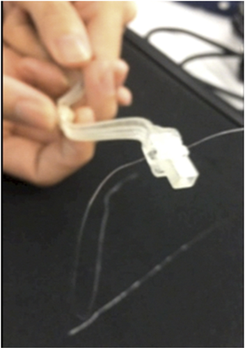
A 3D printed material encasing the miniature camera device.
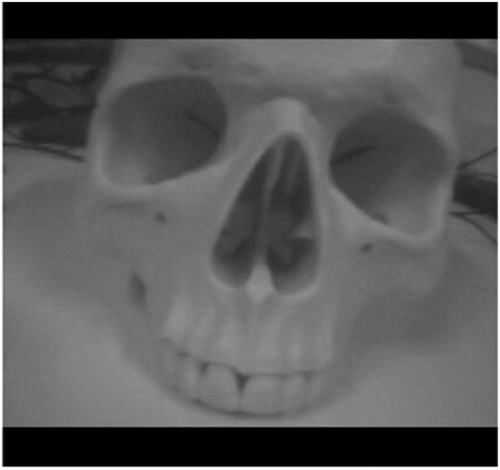
The quality of image captured and transmitted by the miniature camera in real time on skull model.
Anchorage
The vomer bone was deemed as the ideal site for image capture of the nasopharynx using our miniaturized endoscope system. Additionally, this location was amendable for anchoring the camera system, which presented a remote possibility for long-term nasopharyngeal surveillance. To that end, 2 of our initial prototypes were designed with an anchorage requirement. Our hypothesis was tested using 2 anchorage methodologies. The first method was exemplified by prototype 1 where there was a pre-fabricated latch to insert onto the free edge of the vomer bone. In the second prototype, a drilling system was designed to anchor onto the vomer bone. Moreover, the nasal vestibule may be another anchoring point, in addition to the vomer bone for improving the stability of the camera device as shown in Fig. 4. In the third prototype, long term anchoring of the camera system was not required. Nevertheless, the vomer bone remained an important site for securing the device for image capture (Fig. 5). In this manner, this device could potentially be self-administered for surveillance and image capture.
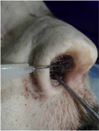
Cadaver with piercing of space near vomer bone and screw holes for fixation of screws from the 3 miniature camera prototypes.
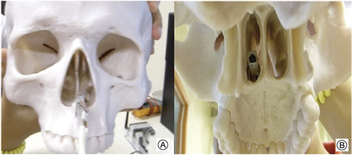
The position of miniature camera on the posterior edge of the vomer bone on skull model. A: Anterior posterior view from the camera insertion site; B: Posterior view from the camera insertion site.
Pre-clinical assessment and risk assessment of material tested
All 3 prototypes were tested on the cadaveric model and were found to be feasible for capturing stable images of the nasopharynx. Table 1 summarized the qualities and characteristics of these 3 prototypes. Overall, the third prototype represented the most ideal candidate for nasopharyngeal surveillance. Firstly, the L-shaped backing structure improved stability in the overall application of this device. Secondly, in the comparison of risk assessment among the 3 prototypes for breaking, pull out and tensile stress revealed the titanium alloy (Ti6Al4V) performed better under mechanical stress imposed on the prototype. The material does not break or bend until a maximum threshold of stress is imposed, suggesting that this material is durable and reliable for clinical application. Lastly, image distortion was greatly reduced in the third prototype due to improved stability from the L-shaped structure that increased stability and minimized motion of the camera during image capture. Lastly, positioning of the miniature camera system at the posterior end of the vomer bone allowed a full capture of the nasopharynx in all 3 prototypes. Table 2 shows the comparison of titanium alloy vs stainless steel as choice of metals for developing the prototypes.
| Testing standards | Prototype 1 | Prototype 2 | Prototype 3 |
|---|---|---|---|
| Dimensions (mm, L × W × H) | 70.0 × 5.5 × 6.0 | 90.0 × 6.2 × 1.5 | 35.0 × 7.5 × 5.0 |
| Weight (g) | 23.60 | 36.84 | 20.00–30.00 |
| Material | Titanium alloy (Ti–6Al–4V) | Medical grade stainless steel (316 L) | Titanium alloy (Ti–6Al–4V) |
| Anchorage | Vomer bone | N/A | Vomer bone |
| Angle (degree) | 180 (wide angle) | 90 (wide angle) | 180 (wide angle) |
| Factors | Titanium alloy | Stainless steel |
|---|---|---|
| Type & grade | Ti6 Al4V (grade 5) | Stainless steel medical grade 316 L |
| Tensile strength (Mpa) | 950 | 485 |
| Hardness (HB) | 334 | 217 |
| Elastic modulus (Gpa) | 113.8 | 193 |
| Property | Bio-compatible, corrosive and heat resistant | Biocompatible, corrosive and heat resistant |
| Risk | Easily contaminated, poor shear strength and surface wear property | Components may induce immune reaction and also interfere with MRI due to presence of ferrite |
| Application | Medical devices and implants | Medical devices and implants |
To verify the above standards, following risk assessments were performed on the prototypes. Pull out test provided the maximum force required to completely dislodge the device from its anchorage points. This information will help us determine the mechanical strength required so as to select the most appropriate material for this device. From the results obtained, the maximum force required to dislodge the device was 151 N with a stress pressure of more than 2.5 Mpa. However, these figures will hardly be achieved in reality and both titanium alloy and stainless steel will be suitable. Additionally, the pullout test for determining the breaking force for the 2 screws involved in anchorage was 34.9 N; which was unlikely to be breached for our clinical application.
Discussion
Our hypothesis of a real time nasopharyngeal surveillance device was validated in this preclinical study. Several key components were tested; namely, image capture using a miniaturized endoscope system, real time wireless transmission, design of the surveillance prototypes and material analysis.
In this first component, a 3.5 mm miniaturized endoscope system was deployed and navigated in the nasal cavity in order to capture images of the nasopharynx. Image quality was reasonable but lacked the high definition quality accorded in most clinical grade endoscopic system. When this camera was deployed in the nasopharynx, the image quality was compromised by the dim light source attached to the camera system. The dimness could be secondary of deflected light with the LED light source of the camera. With this present prototype, the images captured using the miniaturized camera system were not of sufficient quality for image recognition and processing. Further work is necessary to adapt a white light source for better optics visualization of the nasopharynx. Nevertheless, real time wireless transmission was possible with consistent images being captured seamlessly. This success underscores the possibility of a real time wireless transmission of these images for possible clinical translation. With these prospective images collection and analyses, machine learning using convolutional neural network (CNN) can be built up to optimize the accuracy of detecting NPC from these serial images.
Another key component was the feasibility of anchoring the miniaturized endoscope system. This aspect was tested on 2 of our prototypes. The free posterior edge of the vomer bone was found suitable as the site of anchorage due to the ability to insert screws for anchorage. However, a freshly designed 90-degree drill bit was necessary in order to create holes at the vomer bone from the nasal cavity. The cadaver model showed that the anchorage to be firm and sustained the camera in position after insertion. Additional anchorage at the nasal vestibule may be required for additional stability of this device. This may be necessary so that stable image may be captured seamlessly, although a major drawback is the potential discomfort at the nasal vestibule. The need for battery change also adds towards the complexity of using this anchored prototype into the system. This prototype will require period change (estimated 1–2 months) of the battery which may lead to obstacles in clinical translation of this device in clinical practice. Additionally, image quality may also be potentially affected by the mucus clouding the camera lens. These challenges will need to be overcome and the long-term durability of such a surveillance system needs to be systematically addressed before clinical translation is possible.
Given these limitations with an anchoring system, one of the prototypes was designed with the concept of a self-administration image capture system. In this prototype, the design was made to allow easy insertion of the camera by aligning the long axis of the shaft of the system with the floor of the nose. While this prototype was feasible, the possibility of inadvertent trauma to the nasal cavity structures may impede the insertion of this device trans-nasally. Further modification of this prototype is underway by incorporating a robotic steering capability to overcome this limitation.
Lastly, material analysis was addressed to identify the suitable material necessary for clinical translation. Given that a long-term placement of this device was ideal for long-term surveillance, durability and biocompatibility were the key factors in our consideration. Based on the information on the materials used in medical devices currently, we have focused our attention on titanium alloys and medical grade 316 L stainless steel in our material analysis. Titanium alloy Ti6 Al4V was our first choice due to its high tensile strength, lightweight, conformability and corrosion resistant nature. It also does not react with any of the nasal secretions and does not release toxic metal ion that precludes its clinical use. Furthermore, it has larger resistance and superior strength under repeated load of stress. Relative to stainless steel, the lower modulus of titanium makes it less rigid and hence, places less stress on bony structures. This feature was vital since our device was designed to be implanted in the posterior part of vomer bone.
Conclusions
Our study validates the possibility of a real time image capture of the entire nasopharynx using a miniaturized endoscope system. Image capture of the nasopharynx with real time wireless transmission is technically feasible using a 3.5 mm miniaturized endoscope system. Anchoring this system, while possible, requires a two-point anchoring process at the vomer and nasal vestibule for maximal stability. Material analysis showed that this concept is potentially translatable to the clinics but warrant further refinement in better image quality and optics development.
Declaration of Competing Interest
None.
Acknowledgement
Dr Lim Chwee Ming acknowledges the Pilot Grant from National University Health System for this work. Dr Ren Hongliang acknowledges NMRC Bedside & Bench under grant R-397-000-245-511, Singapore Academic Research Fund under Grant R-397-000-297-114 and the BME BN3101 Design module students for their contribution to this study. The author and co-authors have no conflict of interest to declare.


