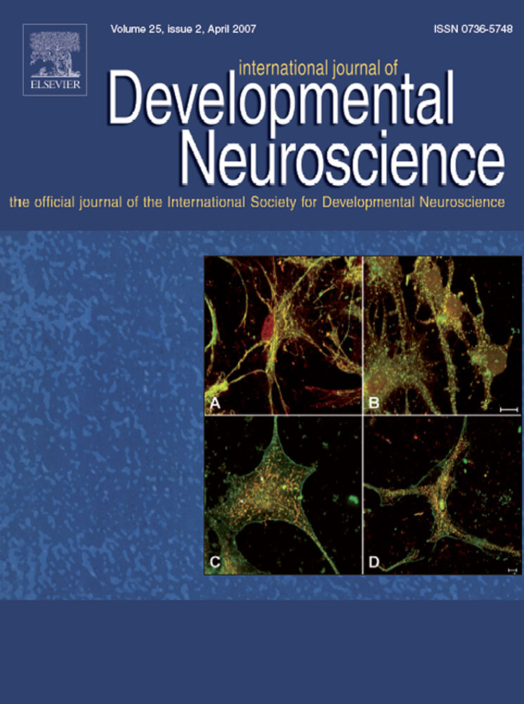
Changes in microtubule-associated protein-2 (MAP2) expression during development and after status epilepticus in the immature rat hippocampus
Niina S. Jalava
Department of Pharmacology, Drug Development, and Therapeutics, Institute of Biomedicine, University of Turku, Itäinen Pitkäkatu 4B, FIN-20014 Turku, Finland
Search for more papers by this authorFrancisco R. Lopez-Picon
Department of Pharmacology, Drug Development, and Therapeutics, Institute of Biomedicine, University of Turku, Itäinen Pitkäkatu 4B, FIN-20014 Turku, Finland
Search for more papers by this authorTiina-Kaisa Kukko-Lukjanov
Department of Pharmacology, Drug Development, and Therapeutics, Institute of Biomedicine, University of Turku, Itäinen Pitkäkatu 4B, FIN-20014 Turku, Finland
Search for more papers by this authorCorresponding Author
Irma E. Holopainen
Department of Pharmacology, Drug Development, and Therapeutics, Institute of Biomedicine, University of Turku, Itäinen Pitkäkatu 4B, FIN-20014 Turku, Finland
Medicity Research Laboratory, Tykistökatu 6A, 4th Floor, Institute of Biomedicine, University of Turku, FIN-20014 Turku, Finland
Corresponding author at: Medicity Research Laborotory, Tykistökatu 6A, 4th Floor, Institute of Biomedicine, University of Turku, FIN-20014 Turku, Finland. Tel.: +358 2 333 7018; fax: +358 2 333 7000.
E-mail address: [email protected] (I.E. Holopainen).
Search for more papers by this authorNiina S. Jalava
Department of Pharmacology, Drug Development, and Therapeutics, Institute of Biomedicine, University of Turku, Itäinen Pitkäkatu 4B, FIN-20014 Turku, Finland
Search for more papers by this authorFrancisco R. Lopez-Picon
Department of Pharmacology, Drug Development, and Therapeutics, Institute of Biomedicine, University of Turku, Itäinen Pitkäkatu 4B, FIN-20014 Turku, Finland
Search for more papers by this authorTiina-Kaisa Kukko-Lukjanov
Department of Pharmacology, Drug Development, and Therapeutics, Institute of Biomedicine, University of Turku, Itäinen Pitkäkatu 4B, FIN-20014 Turku, Finland
Search for more papers by this authorCorresponding Author
Irma E. Holopainen
Department of Pharmacology, Drug Development, and Therapeutics, Institute of Biomedicine, University of Turku, Itäinen Pitkäkatu 4B, FIN-20014 Turku, Finland
Medicity Research Laboratory, Tykistökatu 6A, 4th Floor, Institute of Biomedicine, University of Turku, FIN-20014 Turku, Finland
Corresponding author at: Medicity Research Laborotory, Tykistökatu 6A, 4th Floor, Institute of Biomedicine, University of Turku, FIN-20014 Turku, Finland. Tel.: +358 2 333 7018; fax: +358 2 333 7000.
E-mail address: [email protected] (I.E. Holopainen).
Search for more papers by this authorAbstract
In this study, we analyzed the spatiotemporal expression patterns of the high-molecular weight (MAP2a and b) and low-molecular weight (MAP2c and d) cytoskeletal microtubule-associated protein-2 (MAP2) isoforms with Western blotting, and the cellular localization of the high-molecular weight MAP2 isoforms with immunocytochemistry in the hippocampi of 1- to 21-day-old rats. Moreover, the temporal profile (from 30 min to 1 week) of MAP2 isoform reactivity to kainic acid-induced status epilepticus was studied in P9 rats. During development, the expression of the high-molecular weight MAP2 isoforms significantly increased, while the low-molecular weight isoforms decreased, the most prominent changes occurring during the second postnatal week. This developmental increase in the high-molecular weight MAP2 expression was also confirmed with immunocytochemistry, which showed increased immunoreactivity, particularly in the molecular layers of the dentate gyrus, and in CA1 and CA3 stratum radiatum. In 9-day-old rats, status epilepticus resulted in a rapid transient increase (about 210%) in the high-molecular weight MAP2 expression, without any effect on the low-molecular weight MAP2. Moreover, disturbed dendritic structure in the CA1 and CA3 stratum radiatum was manifested as formation of varicosities 3 h after the kainic acid treatment. The strictly developmentally regulated MAP2 isoform expression suggests different functional roles for these proteins during the postnatal development in the rat hippocampus. Moreover, high-molecular weight MAP2s may play a role in nerve cell survival during cell stress.
References
- C. Arias, I. Arrieta, L. Massieu, R. Tapia. Neuronal damage and MAP2 changes induced by the glutamate transport inhibitor dihydrokainate and by kainate in rat hippocampus in vivo. Exp. Brain Res. 116: 1997; 467–476
- G.P. Ballough, L.J. Martin, F.J. Cann, J.S. Graham, C.D. Smith, C.E. Kling, J.S. Forster, S. Phann, M.G. Filbert. Microtubule-associated protein-2 (MAP-2): a sensitive marker of seizure-related brain damage. J. Neurosci. Methods. 61: 1995; 23–32
- S.A. Bayer. Development of the hippocampal region in the rat. I. Neurogenesis examined with 3H-thymidine autoradiography. J. Comp. Neurol. 190: 1980; 87–114
- L.I. Binder, A. Frankfurter, H. Kim, A. Caceres, M.R. Payne, L.I. Rebhun. Heterogeneity of microtubule-associated protein-2 during rat brain development. Proc. Natl. Acad. Sci. U.S.A. 81: 1984; 5613–5617
- A. Caceres, G.A. Banker, L. Binder. Immunocytochemical localization of tubulin and microtubule-associated protein-2 during the development of hippocampal neurons in culture. J. Neurosci. 6: 1986; 714–722
- A. Caceres, L.I. Binder, M.R. Payne, P. Bender, L. Rebhun, O. Steward. Differential subcellular localization of tubulin and the microtubule-associated protein MAP2 in brain tissue as revealed by immunocytochemistry with monoclonal hybridoma antibodies. J. Neurosci. 4: 1984; 394–410
- C. Charriere-Bertrand, C. Garner, M. Tardy, J. Nunez. Expression of various microtubule-associated protein-2 forms in the developing mouse brain and in cultured neurons and astrocytes. J. Neurochem. 56: 1991; 385–391
- M.E. Chicurel, K.M. Harris. Three-dimensional analysis of the structure and composition of CA3 branched dendritic spines and their synaptic relationships with mossy fiber boutons in the rat hippocampus. J. Comp. Neurol. 325: 1992; 169–182
- W.J. Chung, S. Kindler, C. Seidenbecher, C.C. Garner. MAP2a, an alternatively spliced variant of microtubule-associated protein-2. J. Neurochem. 66: 1996; 1273–1281
- D.A. Dawson, J.M. Hallenbeck. Acute focal ischemia-induced alterations in MAP2 immunostaining: description of temporal changes and utilization as a marker for volumetric assessment of acute brain injury. J. Cereb. Blood Flow Metab. 16: 1996; 170–174
- P. De Camilli, P.E. Miller, F. Navone, W.E. Theurkauf, R.B. Vallee. Distribution of microtubule-associated protein-2 in the nervous system of the rat studied by immunofluorescence. Neuroscience. 11: 1984; 817–846
- L. Dehmelt, F.M. Smart, R.S. Ozer, S. Halpain. The role of microtubule-associated protein-2c in the reorganization of microtubules and lamellipodia during neurite initiation. J. Neurosci. 23: 2003; 9479–9490
- B.T. Faddis, M.J. Hasbani, M.P. Goldberg. Calpain activation contributes to dendritic remodeling after brief excitotoxic injury in vitro. J. Neurosci. 17: 1997; 951–959
- L. Ferhat, A. Represa, W. Ferhat, Y. Ben-Ari, M. Khrestchatisky. MAP2d mRNA is expressed in identified neuronal populations in the developing and adult rat brain and its subcellular distribution differs from that of MAP2b in hippocampal neurones. Eur. J. Neurosci. 10: 1998; 161–171
- J.M. Fritschy, O. Weinmann, A. Wenzel, D. Benke. Synapse-specific localization of NMDA and GABA(A) receptor subunits revealed by antigen-retrieval immunohistochemistry. J. Comp. Neurol. 390: 1998; 194–210
10.1002/(SICI)1096-9861(19980112)390:2<194::AID-CNE3>3.0.CO;2-X CAS PubMed Web of Science® Google Scholar
- C.C. Garner, A. Matus. Different forms of microtubule-associated protein-2 are encoded by separate mRNA transcripts. J. Cell Biol. 106: 1988; 779–783
- R. Hartel, A. Matus. Cytoskeletal maturation in cultured hippocampal slices. Neuroscience. 78: 1997; 1–5
- Y. Ikegaya, J.A. Kim, M. Baba, T. Iwatsubo, N. Nishiyama, N. Matsuki. Rapid and reversible changes in dendrite morphology and synaptic efficacy following NMDA receptor activation: implication for a cellular defense against excitotoxicity. J. Cell Sci. 114: 2001; 4083–4093
- C. Ikonomidou, M.T. Price, J.L. Mosinger, G. Frierdich, J. Labruyere, K.S. Salles, J.W. Olney. Hypobaric–ischemic conditions produce glutamate-like cytopathology in infant rat brain. J. Neurosci. 9: 1989; 1693–1700
- S. Kaech, B. Ludin, A. Matus. Cytoskeletal plasticity in cells expressing neuronal microtubule-associated proteins. Neuron. 17: 1996; 1189–1199
- S. Kaech, H. Parmar, M. Roelandse, C. Bornmann, A. Matus. Cytoskeletal microdifferentiation: a mechanism for organizing morphological plasticity in dendrites. Proc. Natl. Acad. Sci. U.S.A. 98: 2001; 7086–7092
- J.A. Kim, K. Mitsukawa, M.K. Yamada, N. Nishiyama, N. Matsuki, Y. Ikegaya. Cytoskeleton disruption causes apoptotic degeneration of dentate granule cells in hippocampal slice cultures. Neuropharmacology. 42: 2002; 1109–1118
- Y. Li, N. Jiang, C. Powers, M. Chopp. Neuronal damage and plasticity identified by microtubule-associated protein-2, growth-associated protein 43, and cyclin D1 immunoreactivity after focal cerebral ischemia in rats. Stroke. 29: 1998; 1972–1980
- F.R. Lopez-Picon, M. Uusi-Oukari, I.E. Holopainen. Differential expression and localization of the phosphorylated and non-phosphorylated neurofilaments during the early postnatal development of rat hippocampus. Hippocampus. 13: 2003; 767–779
- F. Lopez-Picon, N. Puustinen, T.K. Kukko-Lukjanov, I.E. Holopainen. Resistance of neurofilaments to degradation, and lack of neuronal death and mossy fiber sprouting after kainic acid-induced status epilepticus in the developing rat hippocampus. Neurobiol. Dis. 17: 2004; 415–426
- F. Lopez-Picon, T.-K. Kukko-Lukjanov, I.E. Holopainen. Calpain inhibitor MDL-28170 and AMPA/KA receptor antagonist CNQX inhibit neurofilament degradation and enhance neuronal survival in kainic acid-treated hippocampal slice cultures. Eur. J. Neurosci. 23: 2006; 2686–2694
- D.F. Matesic, R.C. Lin. Microtubule-associated protein-2 as an early indicator of ischemia-induced neurodegeneration in the gerbil forebrain. J. Neurochem. 63: 1994; 1012–1020
- W. Matsunaga, S. Miyata, Y. Hashimoto, S.H. Lin, T. Nakashima, T. Kiyohara, T. Matsumoto. Microtubule-associated protein-2 in the hypothalamo-neurohypophysial system: low-molecular-weight microtubule-associated protein-2 in pituitary astrocytes. Neuroscience. 88: 1999; 1289–1297
- A. Matus, R. Bernhardt, T. Hugh-Jones. High molecular weight microtubule-associated proteins are preferentially associated with dendritic microtubules in brain. Proc. Natl. Acad. Sci. U.S.A. 78: 1981; 3010–3014
- M. Morales, E. Fifkova. Distribution of MAP2 in dendritic spines and its colocalization with actin. An immunogold electron-microscope study. Cell Tissue Res. 256: 1989; 447–456
- J. Noraberg, B.W. Kristensen, J. Zimmer. Markers for neuronal degeneration in organotypic slice cultures. Brain Res. Brain Res. Protoc. 3: 1999; 278–290
- R.S. Ozer, S. Halpain. Phosphorylation-dependent localization of microtubule-associated protein MAP2c to the actin cytoskeleton. Mol. Biol. Cell. 11: 2000; 3573–3587
- Z. Pang, G.H. Umberger, J.W. Geddes. Neuronal loss and cytoskeletal disruption following intrahippocampal administration of the metabolic inhibitor malonate: lack of protection by MK-801. J. Neurochem. 66: 1996; 474–484
- J.S. Park, M.C. Bateman, M.P. Goldberg. Rapid alterations in dendrite morphology during sublethal hypoxia or glutamate receptor activation. Neurobiol. Dis. 3: 1996; 215–227
- E.M. Quinlan, S. Halpain. Emergence of activity-dependent, bidirectional control of microtubule-associated protein MAP2 phosphorylation during postnatal development. J. Neurosci. 16: 1996; 7627–7637
- A. Rami. Ischemic neuronal death in the rat hippocampus: the calpain–calpastatin–caspase hypothesis. Neurobiol. Dis. 13: 2003; 75–88
- C. Sanchez, J. Diaz-Nido, J. Avila. Phosphorylation of microtubule-associated protein-2 (MAP2) and its relevance for the regulation of the neuronal cytoskeleton function. Prog. Neurobiol. 61: 2000; 133–168
- B. Shafit-Zagardo, N. Kalcheva. Making sense of the multiple MAP-2 transcripts and their role in the neuron. Mol. Neurobiol. 16: 1998; 149–162
- R. Siman, J.C. Noszek. Excitatory amino acids activate calpain I and induce structural protein breakdown in vivo. Neuron. 1: 1988; 279–287
- B. Stein-Behrens, M.P. Mattson, I. Chang, M. Yeh, R. Sapolsky. Stress exacerbates neuron loss and cytoskeletal pathology in the hippocampus. J. Neurosci. 14: 1994; 5373–5380
- R.P. Tucker. The roles of microtubule-associated proteins in brain morphogenesis: a review. Brain Res. Brain Res. Rev. 15: 1990; 101–120
- R.P. Tucker, L.I. Binder, C. Viereck, B.A. Hemmings, A.I. Matus. The sequential appearance of low- and high-molecular-weight forms of MAP2 in the developing cerebellum. J. Neurosci. 8: 1988; 4503–4512
- L.R. Vega, F. Solomon. Microtubule function in morphological differentiation: growth zones and growth cones. Cell. 89: 1997; 825–828
- C. Viereck, R.P. Tucker, A. Matus. The adult rat olfactory system expresses microtubule-associated proteins found in the developing brain. J. Neurosci. 9: 1989; 3547–3557


