Sclerite assembly, articulation and protective system of Lower Devonian machaeridians
Abstract
Machaeridians are a unique group of Palaeozoic annelids that secreted a calcitic armour consisting of dorsally interlocking sclerites running along the entire length of the animal. Preserved remains are most commonly found as disarticulated sclerites, although rare articulated specimens have also been preserved as moulds, recrystallized calcite and even pyrite replacement. These preservational modes unfortunately have obscured direct observation of the dorsal interlocking hinge, thus impeding our understanding of the functional protection offered by this biological armour. Herein, we document and describe articulated silicified lepidocoleids (Lepidocoleus caliburnus sp. nov., Lepidocoleus shurikenus sp. nov. and Lepidocoleus sp.) in association with isolated sclerites of Lepidocoleus cf. kuangguoduni from the Lower Devonian (Pragian) Garra Formation of New South Wales, Australia. Articulated specimens include straight and enrolled scleritome configurations possessing opposing and alternate dorsal hinge articulation, respectively. Silicified scleritomes were analysed using x-ray microtomography to investigate the three-dimensional morphology, cross-sectional geometry and dorsal hinge articulation and configuration of individual sclerites in the armour assembly. Virtual dissection of articulated specimens alongside reconstructions of isolated sclerites reveal distinctions in sclerite morphology across opposing and alternate articulation mechanisms, including a novel system not previously observed in lepidocoleids. The combination of heterogeneous sclerite cross-sectional geometry with sclerite overlap also serves to maintain a uniform thickness of the armour assembly and presumably improve resistance to predatory attack.
Machaeridians were once an enigmatic group subject to placement across the animal kingdom but have recently been constrained within crown group phyllodocids (Parry et al. 2019), a placement that groups them with modern aphroditiform annelids including scaleworms. Unlike their closest relatives, let alone any other representatives of the annelid phylum, machaeridians are unique in their construction of a biomineralized armour composed of two or four longitudinal series of variably articulating dorsal sclerites (Vinther et al. 2008). The machaeridians are separated into three families (Plumulitidae Jell, 1979; Turrilepadidae Clarke, 1896; and Lepidocoleidae Clarke, 1896) based on the morphology, articulation and arrangement of their sclerites (Withers 1926; Parry et al. 2014). In the plumulitids, the inner and outer sclerites articulate on the dorsal surface; in turrilepadids and lepidocoleids, modified inner sclerites in addition to laterally displaced outer sclerites enclose most or all of the body. The lepidocoleid body plan is considered the most derived, with the inner sclerites aligned along the sagittal length of the body and articulated along a dorsal hinge (Parry et al. 2014). Two articulation mechanisms exist in lepidocoleids, opposing and alternate (Högström & Taylor 2001a). In the opposing form, sclerite series abut each other directly (Högström 1997, 2000), whereas in the alternate form the sclerites are offset from each other (Högström & Taylor 2001a, figs 2C, 3B). These scleritome configurations presumably provided an efficient means of protection, although the mechanisms involved in the articulation of the armour assembly and the potential relationships with the ecology of these organisms have largely eluded close study due to preservational bias (Gügel et al. 2017).
Three-dimensionally preserved articulated machaeridians have predominantly been represented by internal or external moulds (Högström & Taylor 2001b), recrystallized calcite (Högström & Taylor 2001a) and, in rare cases, replacement by pyrite (Klug et al. 2008; Högström et al. 2009). Documentation of disarticulated silicified sclerites helped lay the foundation for machaeridian sclerite terminology and revealed morphological features unknown from mouldic preservation, such as a tongue and groove mechanism for opposing articulation (Adrain 1992). However, silicification remains an atypical preservational mode in machaeridians and has not previously been associated with any fully articulated specimens. Although silicification can be a relatively coarse mode of preservation, possibly removing fine-scale ornament and ultrastructural details, it does provide a unique opportunity to investigate the three-dimensional structure, geometry, articulation and functional morphology of the sclerites in an articulated scleritome.
Here, we provide the first report of articulated lepidocoleids from Australia, including two new and one indeterminate species, based on remarkably well-preserved silicified specimens from the Lower Devonian (Lochkovian–Pragian) Garra Formation of New South Wales, Australia. X-ray tomographic microscopy (μCT) was used to create three-dimensional reconstructions of both isolated sclerites of Lepidocoleus cf. kuangguoduni and articulated machaeridian scleritomes. Selected specimens were virtually dissected in order to: (1) examine and measure scleritome thickness relative to cross-sectional geometry; (2) measure the amount of sclerite-to-sclerite overlap; and (3) reveal the dorsal hinge morphology and mechanisms of articulation between opposing and alternate forms. This study provides novel insights into the complex functional armour of this extinct group and a basis for comparative functional morphological studies with other extant multi-element scleritomes.
Geological setting
Silicified machaeridian material is derived from two stratigraphic sections (‘Golf Course’ and ‘Golf Course Revisited’) measured through the Lower Devonian Garra Formation near Wellington Caves Reserve, approximately 8 km south of Wellington, NSW, Australia (Fig. 1). This thick carbonate sequence was deposited on the Mumbil Shelf as part of a shallow marine carbonate platform that developed from the late early Silurian to late Early Devonian (Hoare & Farrell 2004). Conodont biostratigraphy indicates a late Lochkovian to early Pragian age for the Garra Formation in this region, which hosts the Pedavis pesavis – Eognathodus sulcatus transition (c. 410.8 Ma) (Wilson 1989). All specimens reported here occur in the E. sulcatus zone (Mawson et al. 1988; Wilson 1989).
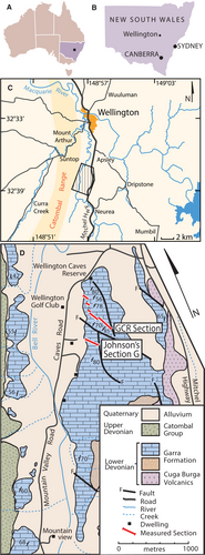
In the Wellington Caves region, the Garra Formation conformably overlies the Cuga Burga Volcanics, a suite of andesitic lavas, tuffs and detrital volcanic sediments (Johnson 1975). After an initial transgressive phase involving reworked volcanics, conglomerates, siltstones and shales, sediments of the Garra Formation transition into a predominately limestone sequence with the onset of carbonate deposition. The lower half of the formation consists of thinly bedded limestones deposited in subtidal–tidal settings on a shallow platform. The silicified horizons are found in the uppermost part of the lower half of the formation, commonly associated with gradual shallowing recorded in the upper 340 m (Chatterton et al. 1979). In the upper half of the formation, further shallowing led to deposition of massive, marl-rich and dolomitized sequences representative of a sabkha-style environment (Johnson 1975; Chatterton et al. 1979). The contact between the Garra Formation and overlying clastics of the Catombal Group is clearly marked by an erosional angular unconformity (Conolly 1963).
The Garra Formation is renowned for its diverse silicified faunal assemblages, including corals (Etheridge 1903, 1907; Hill & Jones 1940; Jones & Hill 1940; Packham 1953; Strusz 1965, 1966, 1967; Strusz & Jell 1970; Wright 2000), sponges (Pickett & Rigby 1983; Plusquellec & Wright 2018), brachiopods (Savage 1969), ectoprocts (Ross 1961; Wass & Byrnes 1969), trilobites (Chatterton et al. 1979), molluscs (Hoare & Farrell 2004; Frýda & Farrell 2005), tentaculitids (Sherrard 1967), crinoids (Jell et al. 1988), foraminiferal linings (Winchester-Seeto & Bell 1994) and chitinozoans (Winchester-Seeto 1993). Specimens documented herein were acid-processed in dilute (10%) hydrochloric acid at the Macquarie University Acid Leaching Facility by Brian D. Johnson in 1981 for his PhD dissertation research from two measured sections (sections G and GC in reference to the ‘Golf Course’), and additional material later recovered via resampling by students at the Macquarie University Centre for Ecostratigraphy and Palaeobiology (MUCEP) at the ‘Golf Course Revisited’ section (GCR in Fig. 1). The GCR section measures 588 m, with the top of Johnson’s original section G approximately equivalent to 257.6 m along the section (see Mawson et al. 1988). The P. pesavis – E. sulcatus conodont transition, constraining the late Lochkovian to early Pragian age, occurs at GCR/35, 34.9 m above the base of the section (Mawson et al. 1988; Wilson 1989). The silicified fauna previously recovered from Johnson’s section G are all E. sulcatus in age or younger, placing these silicified faunas in the early Pragian (Wilson 1989).
Material and method
Sampled material and terminology
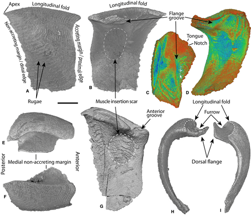
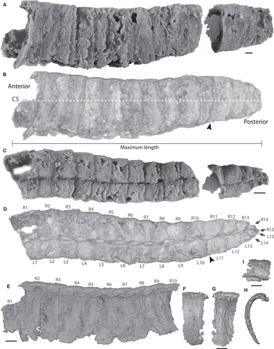
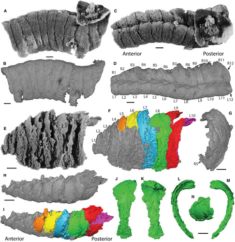
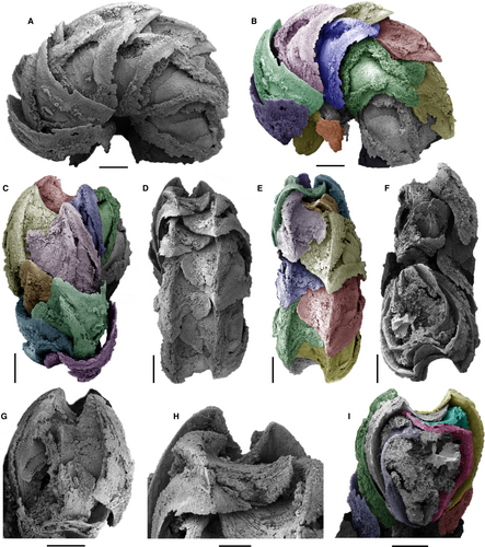
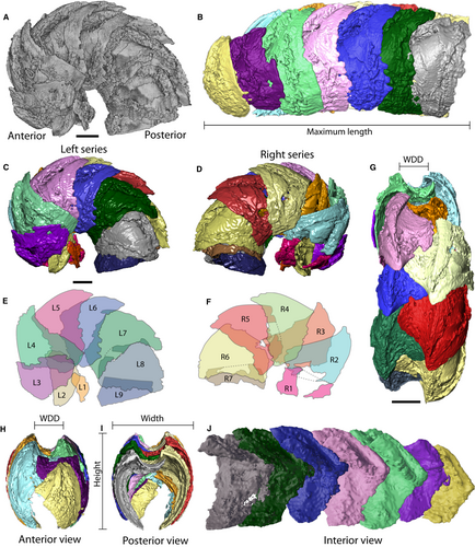
As noted above, specimens documented herein were derived from archived PhD material at Macquarie University (Johnson 1981) and student-collected processed samples reposited at the Londonderry Drillcore Library, NSW, Australia. From these collections, 127 loose sclerites and four articulated specimens were examined. Two isolated sclerites of Lepidocoleus cf. kuangguoduni from opposing sides of the scleritome (AM F.1465879 and AM F.146580; Fig. 2), plus the four articulated specimens were scanned using µCT. Articulated specimens represent two new and one indeterminate species that exhibit different degrees of lateral curvature and forms of articulation. Lepidocoleus caliburnus (AM F.146581, AM F.146582; Figs 3, 4A–D) and Lepidocoleus sp. (AM F.146808; Fig. 4E–N) are relatively straight and display opposing articulation. Lepidocoleus shurikenus (AM F.146809; Figures 5, 6) is partially enrolled and shows alternate articulation. The terminology of the major characteristics used throughout this paper follows that previously established in the literature (Adrain 1992; Högström 1997; Högström & Taylor 2001a, b; Högström et al. 2009) and is summarized in Figures 2 and 6. Given that all articulated forms are incomplete, individual sclerites are numbered sequentially from the anterior-most sclerite in each respective series (left or right) towards the posterior.
Imaging and data visualization
Gigapixel-resolution photomosaics of specimens were acquired using a GIGAmacro Magnify2 Robotic Imaging System with Canon EOS Rebel T6i DSLR and Nikon T1 ×1. Additional high-resolution micrographs were collected on a JEOL 6480LV analytical scanning electron microscope (SEM) at Macquarie University. X-ray microscopy was conducted using a Zeiss Xradia MicroXCT-400 at the Australian Centre for Microscopy and Microanalysis (ACMM), Sydney University, and a Zeiss Xradia 510 Versa at the X-ray Microanalysis Core (MizzoµX), University of Missouri. Parameters used for scanning, the number of projections used and voxel size for each sample can be accessed for each specimen in the associated MorphoSource Project (Jacquet 2021; Appendix S1). Three-dimensional rendering and segmentation were conducted by importing tomograph stacks into AVIZO version 9.4. and Dragonfly version 4.1 for Windows (Object Research Systems (ORS) Inc., 2018; http://www.theobjects.com/dragonfly). Articulated material was manually segmented to manipulate individual sclerites and remove unwanted preservational artefacts (i.e. silicified infill). Sclerite size metrics were measured using the public imaging package Fiji (Schindelin et al. 2012; http://imagej.net).
Cross-sectional geometry and scleritome thickness
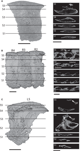
In individual sclerites, including those segmented from articulated scleritomes, external morphology was observed using tomographic reconstructions of the µCT data. In addition, two-dimensional cross-section slices were captured at 20% (S1), 40% (S2), 60% (S3) and 80% (S4) of the sclerite height and across the dorsal flange (S5) (Fig. 7) to map dorsoventral changes in geometry, thickness and structure.
In the most complete and straightest specimen, AM F.146581, cross-sectional morphology, overlap and individual sclerite thickness were measured and imaged in the frontal plane via an adjusted slice module in AVIZO (Figs 3B, 8A). The adjusted slice was set to intersect the entire specimen at the point of maximum scleritome width. Sclerite-to-sclerite overlap, expressed as a percentage, was measured using maximum sclerite length (Ls) and the combined length of overlapping regions (Fig. 8B). Spatial distribution of thickness was measured using the maximum thickness of the individual sclerite (Ts), as well as the maximum and minimum thicknesses of overlapping sclerite regions (Fig. 8C).
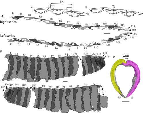
Lateral sclerite overlap area and dorsal hinge morphology
In articulated specimens, sclerite-to-sclerite overlap was measured using left and right sagittal projections (lateral view; see Figs 6E, F, 8D). Projections were applied with a transparency filter so each individual sclerite could be visualized within the scleritome composite. Outlines of each sclerite in a composite scleritome were traced from these projections in Fiji. The total individual sclerite area and the sum of the two overlapping regions were measured, and total overlap was calculated as a percentage of the overall sclerite area.
To determine the mechanism of sclerite articulation in the dorsal hinge area, a transparency filter was applied to the scleritome in both articulated species. Sclerites were observed both in their articulated position and as segmented individuals to display morphological features of the dorsal flange in detail. Three-dimensional renderings of isolated sclerites were exported as stereolithography (.stl) files into free online software Autodesk Meshmixer (http://www.meshmixer.com/) and Blender (https://www.blender.org/) and reconfigured to the most parsimonious agreement of the scleritome or articulation of sclerites.
Results
Three-dimensional structure and geometry of sclerites
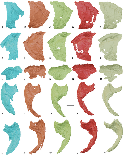
Consistent across all specimens, the longitudinal fold dips towards the anterior (lateral view) to articulate with the adjacent sclerite (Figs 2A, G, 3E). The magnitude of the dipping angle varies between specimens, with the greatest angle measured for Lepidocoleus shurikenus at c. 30°, and that for Lepidocoleus caliburnus at c. 18°. Consequently, the proximal edge is always shorter in height than the distal edge with respect to the height of the sclerite. In anterior profile, the lateral sclerite surface is evenly curved with the ventral edge roughly in alignment with the longitudinal fold (Fig. 2E, F). In interior view, muscle insertion scars are evident in isolated sclerites (Fig. 2B, G), and those of L. shurikenus, positioned sub-centrally below the dorsal flange (Fig. 6J). Such depressions are not apparent in L. caliburnus, probably as a consequence of the quality of preservation.
Two-dimensional cross-sectional slices of sclerites at 20% intervals across the dorsoventral sclerite height reveal a non-uniform geometry (Fig. 7). Generally, sclerites are sub-centrally thickened, with a convex bulge on the interior sclerite wall that tapers to a short blunt point along the proximal edge, and a sharper point along the distal edge. The exterior edge exhibits a generally straight cross-sectional profile that becomes increasingly curved dorsally (Fig. 7). Isolated sclerites exhibit a gradual ventral to dorsal thickening, with a convex bulge appearing at c. 70% of the sclerite height and concentrated around one-third of the shell sclerite length (S3 in Fig. 7A). Sclerites R2–R4 of L. caliburnus show thickening of the wall from <20% shell height (Fig. 7B). In slice S2, the bulge becomes visibly more flattened on the interior surface (Fig. 7B). This geometry is less apparent in sclerites of L. shurikenus owing to the lack of preserved exterior sclerite surface, but they still possess a comparable general morphology (Fig. 7C).
Cross-sectional scleritome thickness
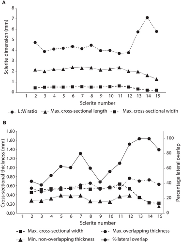
Lepidocoleus caliburnus was the only complete scleritome preserved in a straight configuration with the exterior surface of the sclerites largely intact. Lateral sclerite-to-sclerite overlap and spatial change in thickness were measured from a frontal slice of the articulated scleritome (CS in Fig. 3B) at the point of maximum width (Fig. 8A). Overall, the left and right series exhibit little discrepancy in average maximum sclerite thickness and minimum and maximum overlapping thickness (Table 1). The mean maximum sclerite thickness (0.45 mm ± 0.13, n = 28) of the entire scleritome is slightly less than the mean maximum thickness of overlap between two sclerites (0.59 mm ±0.11, n = 28). The central trunk portion (sclerites 1–10; Fig. 8A) exhibits minimal variation in the mean sclerite length : width (L:W) ratios (Fig. 9A), although gentle sinuosity of the scleritome increases both cross-sectional thickness and lateral sclerite-to-sclerite overlap in the left compared with the right series (Table 1). This is similarly responsible for the variation seen in the total percentage overlap for the trunk portion of the scleritome (Fig. 9B). In contrast, the distal portion of the scleritome (sclerites 11–15) displays a notable increase in sclerite length : width (L:W) ratio in sclerite pairs 13–15 (Fig. 9A). Lateral sclerite overlap steadily increases (Fig. 9B), with sclerites pairs 13 and 14 in the right series, and 13–15 in the left series completely overlapping with adjacent sclerites (Table 1). For the combined left and right series, lateral sclerite overlap ranges from 22.6% to 154.6% with an overall mean of 65.8%. The combination of lateral sclerite-to-sclerite overlap and variable cross-sectional sclerite thickness functions to maintain a relatively constant overall thickness of the scleritome assembly. In sclerite pairs 11–15, the cross-sectional thickness of individual sclerites gradually decreases along with cross-sectional length (Fig. 9A). However, the integrity of the distal scleritome is maintained by increased lateral overlap (Fig. 9B).
| Max. cross-sectional length (mm) | Max. thickness of individual sclerite (mm) | Max. thickness of overlap between two sclerites (mm) | Min. non-overlapping thickness of sclerite (mm) | Lateral overlap of sclerite tail (mm) | Total lateral overlap | % lateral overlap | |
|---|---|---|---|---|---|---|---|
| Right | |||||||
| Average | 2.092 | 0.489 | 0.611 | 0.322 | 0.709 | 1.323 | 67.5 |
| Geometric mean | 2.064 | 0.473 | 0.598 | 0.305 | 0.634 | 1.170 | 61.4 |
| SD | 0.328 | 0.115 | 0.134 | 0.104 | 0.293 | 0.577 | 27.7 |
| Min | 1.280 | 0.255 | 0.410 | 0.152 | 0.196 | 0.378 | 22.6 |
| Max | 2.609 | 0.639 | 0.875 | 0.508 | 1.172 | 2.064 | 122.1 |
| Left | |||||||
| Average | 2.139 | 0.478 | 0.597 | 0.323 | 0.811 | 1.513 | 75.8 |
| Geometric mean | 2.110 | 0.451 | 0.591 | 0.313 | 0.732 | 1.433 | 70.5 |
| SD | 0.331 | 0.141 | 0.083 | 0.078 | 0.304 | 0.503 | 31.3 |
| Minimum | 1.290 | 0.191 | 0.454 | 0.162 | 0.142 | 0.657 | 31.7 |
| Maximum | 2.567 | 0.643 | 0.752 | 0.411 | 1.318 | 2.568 | 154.6 |
| Scleritome | |||||||
| Average | 2.115 | 0.483 | 0.604 | 0.322 | 0.760 | 1.418 | 71.6 |
| Geometric mean | 2.087 | 0.462 | 0.594 | 0.309 | 0.681 | 1.295 | 65.8 |
| SD | 0.325 | 0.127 | 0.110 | 0.090 | 0.297 | 0.541 | 29.3 |
| Minimum | 1.280 | 0.191 | 0.410 | 0.152 | 0.142 | 0.378 | 22.6 |
| Maximum | 2.609 | 0.643 | 0.875 | 0.508 | 1.318 | 2.568 | 154.6 |
Sclerite-to-sclerite overlap area
Lepidocoleus caliburnus sp. nov
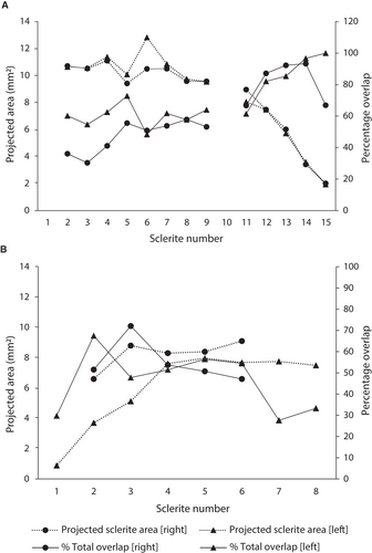
Estimates of the projected area and extent of sclerite overlap area are commensurate with the quality of silicification of each sclerite (Fig. 3A, C). Where silicification was incomplete, the extent of the overlapping area was inferred based on the preserved morphology of each sclerite. Notably, sclerite pairs 1 and 10 were excluded from analysis due to partial damage, but preserved overlap is highlighted in Figure 8D.
In the straight central trunk of the scleritome (sclerite pairs 1–10), sclerites overlap only with those immediately adjacent, with the proximal and dorsal edge of the successive sclerites running approximately parallel or displaying a slight taper in overlap area dorsally (Fig. 8D). Conversely, sclerite pairs 13–15 at the terminus of the scleritome partially overlap with up to 3 adjacent sclerites. The average total sclerite overlap for the respective series, including both the central trunk and distal portion, is 68.2% (n = 13) on the left, 57.2% (n = 13) on the right, and 62.5% for the overall scleritome. Generally, sclerite pairs possess similar projected sclerite areas (excluding L6, Fig. 10A), although sclerite pairs 11–15 exhibit a decrease in area in connection to a reduction in sclerite size (Fig. 10A). Simultaneously, pairs 11–15 increase in percentage sclerite overlap, in some cases overlapping entirely with adjacent sclerites (Fig. 10A). Overhang of the distal edge of the longitudinal fold extends on average 38% in sclerite pairs 1–9 over subsequent sclerites. At the terminus, the anteriorly directed dip of sclerite pairs 13–15 exposes less of the longitudinal fold superficially, but dorsal overlap with adjacent sclerites increases up to c. 76%.
Lepidocoleus shurikenus sp. nov
Lateral views of the left and right sclerite series in L. shurikenus are shown in Figure 6E and F, respectively. In this enrolled position the ventral portion of a sclerite can partly overlap up to four adjacent sclerites (Fig. 6E, F). The mean sclerite overlap for the respective series is 44% (n = 5) on the left and 54.4% (n = 8) on the right, averaging 47.7% for the overall scleritome. The highest percentage overlap is found towards the anterior in L2 and R3, and subsequently drops to c. 50% in sclerites 4–6 of either side (Fig. 10B). In lateral view, the distal edge of the longitudinal fold almost always projects over the proximal edge of the successive sclerite (Fig. 6E, F), with the exception of L1 and L2. This overhang ranges from c. 10% of the length of the longitudinal fold to up to c. 50%.
Dorsal hinge mechanism and articulation
Isolated lepidocoleid sclerites
Two distinct morphologies in the left and right series, respectively, were identified in L. cf. kuangguoduni (Figs 2, 11A, B). In dorsal view, the dorsal flange of the sclerite from the left series is sub-rectangular in outline, with a straight medial non-accreting margin (Fig. 2E). In anterior view, the dorsal flange is strongly concave such that the medial non-accreting margin recurves up to a height just below that of the longitudinal fold (Fig. 2I). Interior views show that the medial margin of the flange follows the same profile of the longitudinal fold (Fig. 12A, B). At the anterior-most portion of the longitudinal fold, directly in front of the dorsal flange, there is a small hemispherical groove, here termed the ‘anterior groove’ (Fig. 2G).
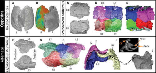
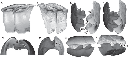
Alternatively, in the right series the dorsal flange exhibits a sub-triangular outline in dorsal view (Fig. 2F). The dorsal flange is shorter and flares up to a point below the height of the longitudinal fold (Fig. 2H). A narrow elongate ‘flange groove’ is located immediately below the flange on the inner surface of the sclerite and runs approximately parallel to the longitudinal fold (Fig. 2B–D). Additionally, in a comparable position to the left series, the anterior portion of the dorsal flange exhibits a small ‘tongue’ distinguished by an adjacent notch along the medial non-accreting margin (Fig. 2G).
Three-dimensional reconstructions of these sclerites in paired configurations reveal a complex mechanism for articulation. In order to interlock effectively, the longitudinal fold and dorsal flange are angled downward towards the anterior in both series (Fig. 12A, B). The site of the hinge articulation is directed anteriorly while the posterior part of the sclerite is broadly concave, enabling overlap with the subsequent opposing pair. The anterior groove of the right series and tongue of the left interlock, while simultaneously the straight medial margin of the dorsal flange in the left series articulates with the flange groove of the right series (Fig. 12C). This is clearly demonstrated in oblique ventral view (Fig. 12E), and together produces the hinge and locking mechanism for the opposing series (Fig. 12F). Overlap of the dorsal flanges in opposing series produces a distinct zigzag suture along the longitudinal length of the dorsal groove in dorsal view (Fig. 12G), while the full extent of the overlap is revealed by the ventral interior view (Fig. 12H).
Lepidocoleus caliburnus sp. nov
This species has opposing articulation with a narrow dorsal depression wherein the dorsal flanges of the right series overlie those on the left (Figs 3B, C, 4C, D). The silicification of the dorsal flanges in the left series of specimen AM F.146581 is coarse, significantly obscuring morphological details. Sclerite pairs 7 and 8 are the most complete and exhibit a wider dorsal flange that extends medially under the longitudinal fold of the opposing right sclerite (Fig. 11C–E). In dorsal view, the dorsal flange of the left series (L8) expands towards its widest point at approximately mid-length, then tapers towards the posterior (Fig. 11C). Sclerites in the right series possess an anteriorly developed dorsal flange that tapers towards the posterior in dorsal view (Fig. 11C). The margin of the dorsal flange extends from approximately the proximal edge to two-thirds of the length of the longitudinal fold, terminating before the distal edge. In the right series, the anterior-most margin of the dorsal flange articulates under both the immediately previous left and right sclerites (Fig. 11D). While the dorsal view reveals relatively minimal overlap (Fig. 11D), internal views show the extent of suturing between the opposing series (Fig. 11E). In the preserved configuration of specimen AM F.146581, the dorsal flange of a single sclerite along the main trunk of the scleritome will generally only overlap partially with that of two (or more) adjacent sclerites, the opposing and previous (Fig. 11D, E).
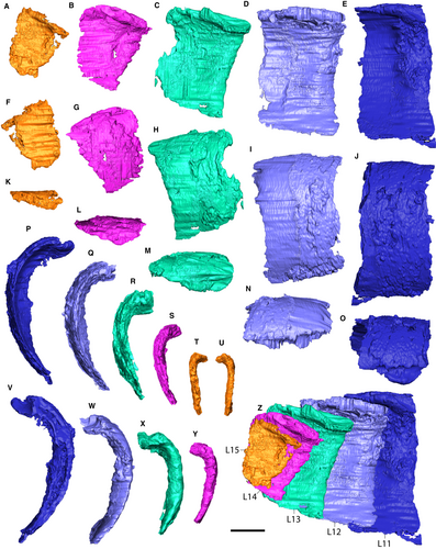
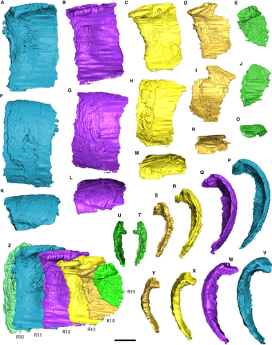
The distal portion of the scleritome exhibits slightly offset sclerite pairs, and the dorsal depression becomes very narrow (Fig. 3C, D). Sclerite pairs 11–15 in specimen AM F.146581 possess near straight longitudinal folds with minimal dip towards the anterior and a comparatively narrow dorsal flange (Figs 13, 14).
Lepidocoleus shurikenus sp. nov
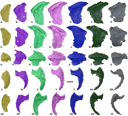
This specimen exhibits alternate articulation with the right series displaced posteriorly in relation to the left one (Figs 5, 6, 11G). Virtual disarticulation of the individual sclerites reveals no evidence of a tongue and groove mechanism locking the sclerites of the opposing series. Instead, the dorsal flange of each offset sclerite overlaps the subsequent one from anterior to posterior (Fig. 11G). The structural integrity of each series is reliant on the continuous interlocking of flanges with the opposite series. This is achieved by the specialized mirrored morphology of the dorsal flange in each respective series, as seen in dorsal view (Figs 11F, 15, 16). The longitudinal fold is gently curved and the anterior and posterior edges of the dorsal flange are concave, rapidly becoming convex towards the midpoint (Fig. 11H). The alternate configuration of the sclerites results in increased overlap; in an enrolled position, the dorsal flange of a single sclerite can partially overlap with up to three adjacent sclerites (Fig. 11G), whereas in an extrapolated straight position it overlaps with four. Interlocking of sclerites is achieved via a distinct void under the apical end of the longitudinal fold, which is large enough to accommodate both the longitudinal fold of the subsequent sclerite and the flared medial non-accreting margin of the opposite dorsal flange (Fig. 11H–J).
Discussion
The effectiveness of the exoskeleton in achieving protection and biomechanical function in metazoans is dependent not only on the composition and integrity of the armour but also on the morphology and structure of the individual armour ‘sub-units’ (Connors et al. 2012). In the case of the lepidocoleid annelids, although the composition is assumed to have been calcareous, the specific microstructural geometry and multilayering of the sclerites have thus far precluded examination due to recrystallization. With no extant analogue, the structural and functional morphology documented in other modern natural protective systems (Song et al. 2010; Connors et al. 2012) provides our only reference for assessing the defensive efficacy of the machaeridian armour sub-units (i.e. individual sclerites). Moreover, this offers insight into the evolutionary trajectory of the machaeridian body plan and potential selective pressures driving these anatomical and behavioural adaptations. This reiterates the importance of conducting biomechanical studies of the scleritome itself, such as presented here.
The armour of lepidocoleids
At the sub-unit level, tomographic slices in the frontal plane show a heterogeneous dorsoventral cross-sectional geometry (Fig. 7). Each sclerite has a sub-central sclerite thickening bracketed by differential degrees of tapering along the accreting and non-accreting sclerite margins. The thicker, more mature accreting margin is overlain by the thinner juvenile non-accreting margin of the preceding sclerite (Högström 2000; Figs 8, 7). Although these features are obscured by coarse silicification in Lepidocoleus shurikenus, they are well-preserved in Lepidocoleus caliburnus and isolated sclerites of Lepidocoleus cf. kuangguoduni. At the scleritome level, this cross-sectional sclerite geometry combined with sclerite-to-sclerite overlap contributes to a relatively constant thickness in the armour, as exhibited in the straight body configuration of L. caliburnus. Such geometric shapes are closely comparable to modern armour-bearing organisms, such as the tapered anterior and posterior margins of overlapping lateral sclerites in the three-spine stickleback (fish), Gasterosteus aculeatus (Song et al. 2010), and the calcareous plates of the polyplacophoran (chiton), Tonicella marmorea (Connors et al. 2012). Notably, the respective life modes of these taxa differ significantly, with G. aculeatus adopting an actively motile pelagic habit compared with the motile benthic habit of T. marmorea. The presumably motile infaunal to semi-infaunal habit of the extinct lepidocoleids is also distinct. Hence, maintaining a constant thickness of the protective assembly appears to provide an effective enclosure to protect the soft tissues, and is not indicative of, or constrained by, a specific life mode.
The extent of sclerite area overlap is influenced by sclerite configuration and the degree of scleritome curvature. Although it is important to consider intraspecies-level variation, the enrolled scleritome configuration in L. shurikenus has a reduced mean percentage of sclerite-to-sclerite area overlap of 47.7%, compared with the straight scleritome configuration in L. caliburnus with 62.5%. Although both specimens are incomplete, missing the posterior and anterior portions of the scleritome, respectively, these estimates are remarkably close to the percentage overlap measured in enrolled (48%) and straight (62%) configurations of shell plates in the modern chiton T. marmorea (Connors et al. 2012). This may imply underlying biomechanical constraints on the flexibility and mobility of the armour in exoskeletal scleritomes.
The reduced overlap area exhibited in the enrolled specimen of L. shurikenus is most pronounced towards the dorsal surface. To combat this reduction in protection, the longitudinal fold extends over the succeeding sclerite by 10–50% of the overall length of the sclerite. In the dorsal depression, the mirrored morphology of the dorsal flange in the opposing series maximizes overlap in both enrolled and reconstructed straight positions. In L. caliburnus, percentage overlap remains relatively consistent until the posterior, where the overall size of the sclerites decreases in conjunction with an increase in sclerite-to-sclerite overlap (up to 100%, Figs 9B, 10A). In addition, the width of the dorsal flange is greatly reduced and the last two sclerite pairs (14–15; AM F.146581) lose their transverse curvature in order to close the posterior opening by effectively sandwiching adjacent sclerites together. This telescoped configuration of the sclerites in the terminal portion of the scleritome maintains the integrity and coverage of the armour but may also reduce or completely prevent dorsal rotation in this region of the exoskeleton.
The functional hinge of lepidocoleids
Opposing articulation
Although the virtual dissection of L. caliburnus offers some insight into the interlocking tongue and groove articulation mechanism and anteriorly located hinge as documented by Adrain (1992), the dorsal hinge is heavily silicified and it is difficult to distinguish morphological details. The isolated sclerites of Lepidocoleus cf. kuangguoduni herein reveal a hinge structure located on the interior surface, positioned under the dorsal flange, which has not been previously documented. There are two distinct morphotypes among the isolated sclerites: a right-grooved sclerite and a left sclerite possessing a strongly curled, elongate dorsal flange. Functionally, the elongated medial non-accreting margin in the left sclerite slots into the groove on the opposing right sclerite (Fig. 8E). Notably, the groove and muscle attachment observed in the current material differs slightly to that of other descriptions. For instance, Lepidocoleus sclerites from the Silurian (upper Wenlock) Halla Beds of Gotland, Sweden, exhibit an inner groove extending diagonally from the apex to approximately three quarters of the total sclerite height, and the muscle scar is located parallel to the posterior margin (Högström 1997, figure 2B, D). The inner groove in this case seems less likely to have been involved in the hinge mechanism of the sclerites.
The anteriorly located tongue and groove described by Adrain (1992) is considerably less conspicuous in this material, mainly because the tongue and notch on the right series sclerite are not particularly well-pronounced. Nevertheless, the accommodating groove on the left series is well-defined (Fig. 2), and virtual reconstructions of the cumulative articulation demonstrate the complexity of the interlocking mechanism utilizing both hinge loci (Fig. 12E, F). Although these combined structures strengthen the overall dorsal hinge by locking the opposing sclerites into position, a drawback may be the possible limitation on flexibility in certain directions. This arrangement allows for some degree of linear motion in the armour sub-units achieved by sliding opposing sclerite pairs over each other in the longitudinal axis. This linear displacement of sclerites has been suggested as a key mechanism behind machaeridian locomotion, specifically with an emphasis on the Turrilepadids (Vinther & Briggs 2009), but the extent to which this overlap could be achieved in the Lepidocoleus scleritome requires additional investigation. Limited lateral motion is also possible, most notably demonstrated in slightly sinusoidal articulated specimens of Lepidocoleus sigmoideus Withers, 1926 (sclerite 1, fig. 1), L. strictus Withers, 1926 (sclerite 2, fig. 14), L. elongatus Sieverts, 1935 (fig. 1) and also herein with L. caliburnus exhibiting a slight lateral offset (Figs 3C, D, 4C, D). Traces preserved in association with L. hohensteini Högström et al., 2009, from the Lower Devonian (lower Emsian) Hunsrück Shale of Germany suggest the potential for a high degree of lateral flexibility in some species (Högström et al. 2009; Vinther & Briggs 2009). However, the extent of lateral movement is not necessarily representative of normal movement behaviour, given that the ichnofossil in question was interpreted as an escape trace (Vinther & Briggs 2009).
Alternate articulation
Alternate articulation is a comparatively rare morphotype in Lepidocoleus, insofar as that it is known only in L. sarlei Clarke, 1896, from the Silurian of New York State (see also Högström & Taylor 2001a) and in the new taxon proposed herein, L. shurikenus. The articulation mechanism for L. sarlei has been described as having the sclerites simply overlap with the underlying musculature system primarily responsible for maintaining the scleritome assembly in place (Högström & Taylor 2001a). Virtual dissection reveals no evidence of a tongue and groove hinge in L. shurikenus; instead, each sclerite articulates by means of a flared dorsal flange tucking below the apical fold under the adjacent sclerite on the opposite series. Plausibly, the void between the sclerites was also occupied by epithelial tissue similar to that beneath the scales of polyplacophorans (Checa et al. 2017). Alternatively, the void remained open, akin to modern elytra-bearing scale worms in which the elytra are attached via elytrophores that protrude from the dorsal surface of the notopodia (Aneli et al. 2017). A steep anteriorly directed downward angle of both the longitudinal fold and the dorsal flange functioned to keep the sclerites articulated without a physically interconnected hinge joint. This configuration also permits a higher degree of sclerite rotation in the sagittal direction, given that there is no physical hinge constraint on sclerite motion on either side of the longitudinal body axis. Comparable degrees of scleritome enrolment are seen in an articulated specimen of L. sarlei (Högström & Taylor 2001a, pl. 1, figs 8, 9). In terms of lateral flexibility of the scleritome, alternate articulation is suggested to produce a stiffer scleritome due to the lateral connection between segments (Högström & Taylor 2001a). In L. shurikenus, lateral motion is mainly constrained by the rotation afforded by the dorsal flange that articulates with the associated space (and any connective soft tissue) under the alternating sclerite apex, in conjunction with reduced sclerite-to-sclerite overlap.
Dorsal rotation of the sclerites sufficient to enable ventral opening and closing of the scleritome depends largely on the width of the dorsal depression (WDD). In opposing scleritome assemblies such as L. caliburnus, the general trend is that as the WDD increases, the higher the degree of rotation without the opposing longitudinal folds abutting each other. In specimens with very narrow dorsal depressions, there may be minimal to no room for dorsal rotation. Lepidocoleus shurikenus possesses a remarkably wide dorsal depression due to the width of the dorsal flange. Three-dimensional reconstruction of L. shurikenus in a straight configuration (Fig. 6B, H, I) suggests that the ventral portion of the scleritome remains open by at least c. 49% of the body width. Consequently, the scleritome appears unable to fully close due to the medial non-accreting margin of the dorsal flange directly abutting the longitudinal fold of the alternating sclerite (Fig. 11H). In the enrolled configuration, however, the sclerites at the posterior portion of the preserved scleritome almost entirely close the ventral opening due to displacement of the sclerites in the sagittal direction.
Enrolment or conglobation is a defensive behaviour that has evolved convergently in numerous taxa spanning the marine and terrestrial realm throughout the Phanerozoic. Among those adopting such a strategy are several living mammals (armadillos, echidnas, hedgehogs, pangolins), arthropods (isopods, millipedes, trilobites, shrimp larvae) and polyplacophoran molluscs (chitons). This defensive response is a basic strategy to protect delicate appendages or soft tissues by withdrawing them within a rolled-up or coiled posture under an external dorsal armour of varying nature (e.g. mineralized plates, spines, scales or tergites) (Tuf et al. 2016). Conglobation principally acts to dissuade skeleton-crushing or gape-limited predators from manipulating the scleritome or swallowing the organism whole. Perhaps the strongly enrolled behaviour exhibited in L. sarlei and L. shurikenus is a similar strategy that developed to protect the broader ventral visceral field. It is worth noting, however, that the response elicited by an organism capable of conglobation may be influenced by variable circumstances and behaviours. For instance, chitons may adopt a fully enrolled position, but this response is not primarily a defensive mechanism, rather it is a method by which to right itself and reattach to the substratum (Sigwart et al. 2019). Millipedes such as Phyllogonostreptus nigrolabi will tightly coil (commencing at the head) in response to external stimuli, although the intensity of the response will differ depending on the activity that the organism is engaged in at the time (i.e. feeding, resting or walking) and relative body inclination in its environment (Srinivasa & Mohanraju 2011). In pill millipedes, both the type and frequency of the stimulus (e.g. touching, squeezing or dropping) will affect the length of time enrolled (Tuf et al. 2016). The response is also often tailored to a specific predatory cue, wherein enrolment may not only assist in protection against direct contact with a predator, but it will also increase the mobility of the organism (Tenaza 1975) by enabling it to roll away and escape the danger, or by lowering the potential of detection (Tuf et al. 2016). Besides a defensive behaviour against predatory attack, other suggestions for enrolment in lepidocoleids include a voluntary contraction of the scleritome in response to prolonged anaerobic conditions (Högström 2001), or alternatively, it may result from contraction of the muscular tissues post mortem.
Adaptation and selective pressures
The evolution of the machaeridian body plan displays a clear trajectory towards increasingly well-armoured protective systems in conjunction with behavioural adaptations. The plumulitids, interpreted as the most ancestral lineage, were most likely to have been epibenthic organisms using a ‘passive’ dorsal armour consisting of relatively thin sclerites (Jell 1979; Vinther & Briggs 2009). By contrast, the more derived turrilepadids and lepidocoleids, inferred to have been infaunal to semi-infaunal, enclosed their bodies within laterally compressed, thickened external calcareous sclerites (Vinther & Briggs 2009; Parry et al. 2014). This transition to infaunalization prompted Parry et al. (2014) to separate the burrowing machaeridian groups into their own clade, the Cuniculepadia. This leads to a discussion of whether the defensive traits are principally predator induced, insofar as that predatory guilds are the principal drivers for diversification of different defensive morphologies and behaviours, or alternatively habitat mediated, whereby habitat divergence or exploitation of novel environments enhance predation risk from a specific predator guild (Broeckhoven et al. 2018a). It is probable that these drivers work in synergy, although the extent to which each has played a role in the development of machaeridian armour and behaviour has not been considered.
Lepidocoleid remains have a wide environmental distribution (Hints et al. 2004), typically recovered from shales, mudstones and claystones associated with offshore to deep subtidal ramp environments (Withers 1922, 1926; Högström 1997; McCobb & Bassett 2008; Gügel et al. 2017); shallower subtidal mid-ramp settings with intercalated bioturbated marls and limestones with occasional skeletal wackestones (Högström 1997, 2000, 2001; Klug et al. 2008; Vinther & Briggs 2009; De Baets et al. 2010); bioclastic limestones formed adjacent to shallow water carbonate banks (Withers 1922, Adrain et al. 1995; herein); and even nearshore environments with brackish lagoons and evaporites (Ruedemann 1925; Plotnick 1999). Given that these are normally low energy environments only intermittently influenced by high energy events (e.g. storms or turbidites), the selective pressure imposed to protect against environmental factors alone is difficult to reconcile. However, many of these environments are considerably more open habitats lacking shelter compared with topographically complex reefs environments. With the substratum providing the primary local refuge against predators, adopting an infaunal habit or dorsal protective scleritome would be advantageous for living in such diverse open benthic environments.
For modern organisms possessing segmented exoskeletal systems, several have been shown to provide effective resistance to different types of predatory attack. For instance, a fundamental adaptation to resist piercing, pinching and bending attacks is the combination of sclerite cross-sectional geometry, lateral articulation and sclerite overlap to provide a spatially uniform thickness in the armour assembly (Song et al. 2010; Connors et al. 2012). Altogether, these structural attributes disperse multiaxial stresses produced by external loading (i.e. biting) and assist to alleviate weakness at the sub-unit interconnections. This adaptive morphospace conforms to the geometric and morphometric traits of the lepidocoleid armour assembly, particularly given the homogenous scleritome thickness of L. caliburnus. A rigid scleritome, however, has notable trade-offs including reduced manoeuvrability (particularly lateral movement) and speed (Manton 1954). This has consequences for increasing predation risk, especially in habitats with variable predator guilds. For example, experiments using three-spine sticklebacks tested the influence of fish predation on lateral plate number in two environments, unsheltered pelagic and sheltered benthic habitats (Leinonen et al. 2011). Stickleback survival probability in the unsheltered environments corresponded to an increasing number of bony lateral plates, given that these offer effective protection against gape-limited, toothed predators such as fish. Conversely, those in the sheltered environments where predatory fishes are mostly absent possessed fewer plates, favouring swimming performance and potentially also avoidance of more non-gape-limited predators such as arthropods (Leinonen et al. 2011; Broeckhoven et al. 2018a). Lateral plate phenotypes show a clear transition across open marine and freshwater environments, implying that both predation pressure and habitat differentiation play an important role in the adaptation of protective body armour for this species (Leinonen et al. 2011). Mounting evidence suggests that defensive trait differentiation may not always evolve solely in response to predation pressures, but instead in concert with habitat-induced factors (Stankowich & Campbell 2016; Broeckhoven et al. 2018a, b).
Currently, there are no reported instances of durophagous shell repair or drill holes in disarticulated or articulated fossil specimens of lepidocoleids. The functional integrity of the armour described here suggests that the scleritome was an efficient deterrent against crushing, biting and drilling predatory attack. We predict that such an armour assembly in conjunction with enrolment would be most effective against gape-limited predators, such as bony fish.
Adaptation of dorsal biomineralized armour does, however, raise the question of why such a defensive trait would be retained in conjunction with an infaunal lifestyle. From an evolutionary and metabolic standpoint, the investment required to construct a biomineralized external skeleton seems counterintuitive with an organism inferred to have led a predominantly active infaunal mode of life. Although many taxa with hard skeletons are infaunal (e.g. bivalves, echinoderms and several arthropod species), they typically occupy semi-permanent or permanent domiciles within the substrate. Vinther & Briggs (2009) note that the use of biomineralized sclerites in burrowing is a unique functional transition for annelids when considering that the clade is inferred to have been ancestrally epifaunal (Parry et al. 2014). Consequently, to have retained the dorsal scleritome probably implies some underlying ecological or functional benefit for such a life mode. Vinther & Briggs (2009) inferred that the burrowing machaeridians, including the turrilepadids and lepidocoleids, must have used a form of peristaltic locomotion aided by the pre-existing external rugose sculpture of the sclerites to assist in forward movement of the scleritome through the sediment. The enclosed form and interlocking dorsal articulation of the lepidocoleid scleritome (especially in opposing forms) probably meant that locomotion was slow with limited lateral flexibility. The implications for such are difficult to assess given that, unfortunately, no modern analogues truly exist for lepidocoleids. Moreover, although several taxa use a similar passive defensive strategy, the life modes and phylogenetic disparity between the groups prevents direct comparison.
Conclusion
This study reveals some of the key structural adaptations contributing to the protective assembly and biological function of the lepidocoleid body plan. The high-fidelity preservation combined with high-resolution x-ray tomographic microscopy enables virtual dissection of both articulated and disarticulated specimens of silicified machaeridians, as well as digital manipulation of individual sclerites. These three-dimensional reconstructions help to reveal previously unknown morphological features as well as differences between opposing and alternate lepidocoleid sclerite assemblies and their respective dorsal hinge mechanisms. With a greater understanding of sclerite morphology, geometry and interlocking structures, future investigations can focus on biomechanical constraints of linear, lateral and sagittal motion of scleritomes belonging to these remarkable extinct worms.
Institutional abbreviations
AM, Australian Museum, Sydney, NSW, Australia; PIMUZ, Paläontologisches Institut und Museum der Universität Zürich, Switzerland.
Systematic palaeontology
A compilation of the known species attributed to the genus Lepidocoleus and associated dimensions and features is provided in Appendix S1.
Phylum ANNELIDA Lamarck, 1809
Class MACHAERIDIA Withers, 1926
Order LEPIDOCOLEOMORPHA Schallreuter, 1985
Family LEPIDOCOLEIDAE Clarke, 1896
Genus LEPIDOCOLEUS Faber, 1886
Type species
Lepidocoleus jamesi (Hall & Whitfield, 1875); from the Cincinnati Group (Upper Ordovician), Ohio River at Cincinnati, Ohio, USA.
Lepidocoleus caliburnus sp. nov.
Figures 3, 4A–D, 13, 14
LSID
urn:lsid:zoobank.org:act:5A5AD1B4-7B7B-4F4D-9656-A91E2215AADC
Derivation of name
Derived from the Latin name of the famous sword ‘Excalibur’ from the Arthurian legend, The Sword in the Stone. Metaphorically, this is akin to extracting these straight, intact silicified machaeridian specimens from the rock via acid digestion.
Type specimen
Holotype AM F.146581 (Figs 3, 11, 12), paratype AM F.146582 (Fig. 4A–D).
Type locality & horizon
Lower Devonian (lower Pragian) Garra Formation, 8 km south of Wellington, NSW, Australia; horizon GCR/354.
Material
2 silicified articulated specimens. Holotype (AM F.146581) broken into two pieces, representing the trunk and posterior terminus of the scleritome from a horizon along strike ‘North of GCR/354’. Paratype (AM F.146582) representing a partial trunk of the scleritome from horizon GC/0.0.
Diagnosis
Lepidocoleus with more than 30 sclerites (>15 pairs), c. 0.6–1 exposed sclerites/mm along mid-length. Dorsal hinge with opposing articulation; dorsal depression narrow, only 9–14% of the entire width of the scleritome with repeating diamond-shaped furrows. Sclerites dorsoventrally elongated with longitudinal fold dipping gently anteriorly; sub-rectangular in outline along mid-length, posterior-most become short and square. Accreting margin and non-accreting margin almost parallel and similar in length. Left and right sclerites morphologically distinct. In dorsal view, left series sclerites have straight longitudinal fold and dorsal flange broadest towards posterior; right series sclerites have gently sinusoidal longitudinal fold and dorsal flange broadest towards anterior. In articulated pairs the dorsal flange of the right series sclerites rests on top of those in the left series.
Description
The holotype (AM F.146581) is incomplete and broken into two pieces, including the straight central and posterior portion of the scleritome (Fig. 3A, C). A total of 31 sclerites are preserved with opposing articulation. Specimen measures a maximum of 21.2 mm in length, 4.3 mm wide and 5.7 mm high. Dorsal depression is narrow (9–14% of the overall scleritome width) and shallow; WDD ranges from virtually closed (<100 µm) towards the posterior up to 540 µm along the trunk. In paired sclerites the longitudinal fold tapers inward towards the dorsal depression to produce a series of diamond-shaped furrows. Hence the furrow of the dorsal depression exhibits a zigzagged edge (Figs 3C, D, 4C, D). External rugae not preserved; apical or marginal spines on sclerites lacking.
Sclerites in the central trunk portion of AM F.146581 range between 1.6 and 2.7 mm in length and between 4.2 and 5.7 mm in height. The height : length (H:L) ratio of exposed sclerites surfaces is c. 3.4. In lateral view, sclerites are sub-rectangular in outline; those centrally located are more dorsoventrally elongated compared with those at the posterior end. In both series, the longitudinal fold and dorsal flange are angled downward (c. 22° on the right and c. 14° on the left) towards the anterior, facilitating articulation between adjacent sclerites. Morphology of the longitudinal fold and dorsal flange differ between right and left series sclerites.
In the right series, the longitudinal fold is gently sinusoidal in dorsal view, being slightly concave near the anterior and more convex near the posterior with respect to the scleritome midline (Figs 3I, 11C). The dorsal flange is short, narrow and rests directly on top of the opposing dorsal flange of the paired left sclerite. In dorsal view, the dorsal flange is broadest close to the anterior and tapers to meet the convex part of the longitudinal fold. The medial non-accreting margin of the dorsal flange ends approximately two-thirds along the sagittal length of the sclerite. The medial non-accreting margin of the dorsal flange is approximately parallel with the external surface of the sclerite. In anterior view, the dorsal flange is concave, producing a shallow furrow along the length of the flange (Fig. 3H).
In the left series, the longitudinal fold is virtually straight in dorsal view (L8 in Fig. 11C). Silicification obscures many of the details of the dorsal flange, although segmented sclerites reveal a convex outline, broadest approximately midway along the sagittal length of sclerite. In anterior view, the furrow is narrow and the median accreting margin of the dorsal flange curls upward more than the right side (Fig. 8E). Left and right series sclerites have a consistent curvature, producing a heart-shaped cross-section in anterior view.
Sclerites in the posterior portion of specimen AM F.146581 (pairs 10–15) are morphologically distinct from those in the main trunk of the scleritome. Sclerites measure 1.2–2.5 mm in length and 1.7–4.0 mm in height, and dorsal flanges gradually decrease in width (<0.5 mm). In lateral view, sclerites gradually shorten dorsoventrally to become sub-trapezoidal in outline (Figs 13A, B, 14D, E) and increasingly dip anteriorly, with the longitudinal fold dipping up to 30°. In dorsal view, the longitudinal fold becomes straighter closer to the terminus. In anterior view, the curvature of the sclerite exterior surfaces gradually reduces to a flat surface in sclerite pair 15 (Figs 13P–Y, 14P–Y).
Specimen AM F.146582 preserves an incomplete central portion of the scleritome trunk with 24 sclerites (Fig. 4A–D), and measures c. 15.6 mm long, 5.7 mm high and 4.5 mm in width. Sclerites in the left series are better preserved, with 8 extending to the ventral margin, and an H:L ratio of exposed sclerites surfaces of 4.1. Those on the right preserve mainly the dorsal portion, including the longitudinal fold and hinge section. The dorsal depression is narrow (c. 10% of overall scleritome width) and has the characteristic repetitive diamond-shaped furrows resultant from the longitudinal fold curving inward (Fig. 4C, D).
Remarks
The opposing articulation of L. caliburnus clearly distinguishes it from taxa possessing alternate articulation such as Silurian species L. sarlei and the new taxon L. shurikenus documented herein. Likewise exhibiting some degree of offset in sclerite pairs along the trunk portion of their scleritomes are L. illinoiensis Savage, 1913, from the Lower Devonian, Clear Creek Chert, Illinois, USA; L. polypetalus Clarke, 1896, from the Lower Devonian, Lower Helderberg Group, New York, USA; and L. rugatus Klug et al., 2008, from the Lower Devonian (lower Emsian) of Ouidane Chebbi and Mdâour-El-Kbîr (Dra region), Anti-Atlas, Morocco.
Among examples of opposing articulated lepidocoleids, L. caliburnus possesses a higher number of sclerite pairs (>15), although the exact number is difficult to determine with only incomplete trunk and terminal portions. This distinguishes the new taxon from other complete (or near complete) specimens, including L. hohensteini Högström et al., 2009 from the Lower Devonian, Hunsrück Slate, Germany, with 15 pairs; L. gleidorfense (Wolburg, 1938) from Middle Devonian (Eifelian) Fredeburger Schiefe, Germany with 14/15 pairs; L. jamesi Hall & Whitfield, 1875, Upper Ordovician Cincinnati Group, Ohio, USA with 14 pairs; and L. kuangguoduni Gügel et al., 2017, with c. 11 pairs.
With respect to gross morphology, L. caliburnus is wider than the markedly slender scleritomes of L. grayae Withers, 1926, from the Upper Ordovician Drummuck Group, UK; and L. britannicus Withers, 1926, from the middle Silurian ‘Wenlock Beds’, Worcestershire, UK, which also exhibit narrow dorsal depressions (the WDD approximately making up c. 9% and c. 8% of the entire width of the scleritome, respectively). With regard to the structure of the dorsal depression as compared with other species, L. caliburnus lacks the near-parallel longitudinal folds of opposing series that commonly create a straight-edged dorsal depression. This feature appears to be present in L. britannicus (Withers 1926, pl. 2, fig. 11; see also L. cf. britannicus in Högström 2000, fig. 3C–D); L. grayae (Withers 1926, pl. 1, fig. 8); L. sigmoideus (Withers 1926, pl. 1, fig. 1), from the Middle Ordovician Trenton Limestone; and L. strictus (Withers 1926, pl. 2, fig. 14) from the middle Silurian Richmond Group, Indiana, USA. In a few cases, such as L. polypetalus (Withers 1926, pl. 4, fig. 6); L. latus (Withers 1926, pl. 4, fig. 11) from the Middle Devonian Czech Republic; and possibly also L. eifelianus Sieverts, 1935, from the Middle Devonian (Eifelian) Rhenish Massif, Germany (Seilacher & Gishlick 2014, pl. 21.5h), the articulation of opposing series leaves no room for a dorsal depression and instead the longitudinal folds of each pair appear to directly abut each other, although this may be preservational.
Dorsoventral elongation of sclerites is notable in many taxa, typically manifesting as a sub-rectangular outline (see Gügel et al. 2017, fig. 9, for inferred sclerite profiles). Lepidocoleus caliburnus on average has a higher H:L ratio of the exposed surface (on average 3.8) than that of species with similar morphologies (e.g. c. 2.8 in L. grayae; c. 2.3 in L. strictus; c. 2.5 in L. ketleyanus; and c. 2.9 in L. illinoiensis).
Lepidocoleus caliburnus is perhaps most similar in gross morphology to the Late Ordovician species L. ulrichi Withers, 1926, from the Prosser Member (Clitambonites bed), Galena Limestone, Minnesota, USA. The holotype and only specimen of L. ulrichi is an incomplete anterior portion of a scleritome (Withers 1926, pl. 1, figs 2–3). A closely comparable feature in L. ulrichi is the presence of a narrow dorsal fold with diamond-shaped furrows produced by the morphology of the longitudinal fold in paired sclerites (Withers 1926, pl. 1, fig. 3). The WDD of L. ulrichi is c. 11% of the entire width of the scleritome, which falls within the range of L. caliburnus (9–14%). The respective species differ only marginally in the density of sclerites along the length of the scleritome; in L. caliburnus there are 0.6–1.0 exposed sclerites per 1 mm along the mid-length, whereas in L. ulrichi there are 1.1 per mm. An articulated specimen referred to L. cf. ulrichi from the Upper Ordovician (lower Katian) Trenton Group, Verulam Formation, Ontario, Canada, has a more complete scleritome, with 40 pairs of sclerites (Högström & Taylor 2001b, fig. 1). In both L. ulrichi and L. cf. ulrichi (Högström & Taylor 2001b) there is no information on the hinge structure, and much of the sclerite morphology is obscured by articulation. A subtle difference between the sclerites in the respective species is the outline of their non-accreting margins and relative position of the apex. In L. ulrichi and L. cf. ulrichi the apex appears to extend slightly further posteriorly beyond the rest of the distal edge, whereas in L. caliburnus the apex and the non-accreting margin meet at almost 90° (Figs 3F, G, 13C–E, H, I, 14A–C, F–H), producing a distinct rectangular outline in lateral view. In their assessment of L. cf. ulrichi, Högström & Taylor (2001b) noted the difficulties in differentiating lepidocoleid species with dorsoventrally elongated sclerites in the absence of ontogenetic variants. The conservative morphology of lepidocoleids may play havoc with the characterization of taxonomically informative traits, but it is a testament to its success as a specialized scleritome morphology suited to the infaunal lifestyle of the genus that has endured across palaeobiogeographical terranes and throughout their biostratigraphical range.
Stratigraphical & geographical range
Type locality only.
Lepidocoleus shurikenus sp. nov.
Figures 5, 6, 15, 16
LSID
urn:lsid:zoobank.org:act:2395948F-4E0D-42AB-B635-D7CC3DA7CB86
Derivation of name
Named for its resemblance (in its enrolled form) to the outline of some shuriken (手裏剣), the Japanese noun for ‘throwing stars’.
Type specimen
Holotype AM F.146809
Type locality and horizon
Lower Devonian (lower Pragian) Garra Formation, 8 km south of Wellington, NSW, Australia; horizon G/9.0.
Material
1 incomplete, enrolled specimen.
Diagnosis
Enclosed scleritome with two series of longitudinal imbricating sclerites, dorsal hinge with alternate articulation producing a zigzag overlap. Dorsal depression wide, 30–45% of total scleritome width. Sclerites dorsoventrally elongated, accreting margin about two-thirds the length of the non-accreting margin. Longitudinal fold arched and non-accreting margin concave below apex. Distinct and broad dorsal flange that mirrors those on the opposing series of the scleritome.
Description
Single partially enrolled articulated scleritome consisting of two longitudinal series of imbricating sclerites, 16 in total. Posterior portion incomplete (Fig. 5A, F, I). Scleritome length enrolled is 8.3 mm (estimated true linear length >13 mm), height 3.8 mm and width 3.4 mm. Most fine-scale ornament, such as rugae and striae, destroyed by silicification; exterior surface either smoothed or absent so the total number of rugae per sclerite is imperceptible. Dorsal hinge with overlapping alternate imbrication of the respective series produces a zigzagged, uniformly concave dorsal depression (Figs 5C–E, 6G, H, I). Dorsal depression is between 1 and 1.6 mm wide, c. 30–45% of total width of scleritome and 0.5 mm deep. Imbrication of the dorsal flanges produces a heart-shaped cross-section (Fig. 5I), and the flange overlap between successive sclerites extends to the longitudinal fold of the opposing sclerite in dorsal view (Figs 5C–E, 6G).
The most complete sclerites (L2–L8 and R2–R6, Figs 15, 16) measures 1.5–3.3 mm in length, 2.9–4.2 mm in height, and 1.5–2.7 mm in width, with L2 representing the smallest and R5 the largest of the measured sclerites. Dorsal flange measures 0.5–1.6 mm. In lateral view, all sclerites are sub-triangular in outline; height of sclerites gradually increases posteriorly (L1–L9, R1–R7) to produce dorsoventrally elongate forms (Figs 15A–G, 16A–E). Longitudinal fold arches towards sclerite apex, corresponding laterally with anterior dip of the dorsal flange (Figs 15H–N, 16F–J). In dorsal view, the longitudinal fold forms a gently curved convex ridge, thickened at the anterior and tapering towards the apex (Figs 15O–U, 16K–O). In both right and left series, the median non-accreting margin is anteriorly and posteriorly concave, rapidly becoming convex towards the midpoint. In anterior view, the dorsal flange is concave with highest point on the medial non-accreting margin just below the level of the longitudinal fold (Figs 15V–AB, 16P–T). The resultant furrow is broader at the anterior end (compare Figs 15V–AB, 16P–T and 15AC–AI, 16U–Y). Apical and marginal spines on sclerites lacking.
Internal surface features of sclerites are difficult to discern due to coarse silicification. A few sclerites have a shallow depression directly below the dorsal flange that abuts a thickened ridge running almost parallel with the posterior margin (Figs 15I, L, 16G, H, J). This depression, inferred as a muscle insertion scar, occupies 10–16% of the total interior surface area of the sclerite.
Remarks
The scleritome of Lepidocoleus shurikenus is incomplete and has been sheared off at the posterior. At least 9 sclerite pairs are preserved, but the full complement of sclerites remains unknown. The partially enrolled nature of the scleritome makes it difficult to determine scleritome length, but three-dimensional reconstruction provides an estimate of >13 mm. Silicification has obscured much of the exterior sclerite surface, and with it important taxonomic features, such as the number of rugae. Nevertheless, the alternate articulation and distinct morphology of the individual sclerites separates this taxon from other Lepidocoleus species.
Lepidocoleids reported to have alternate placement of sclerites along the length of the body include L. gleidorfense (Wolburg, 1938), L. polypetalus, L. sarlei Clarke, 1896, L. rugatus and L. kuangguoduni.
The single specimen of L. gleidorfense from the Middle Devonian of the Fredeburger Schiefer, Germany is an incomplete and semi-articulated mould (Wolburg 1938, fig. 1). The displacement of the left and right series of posterior sclerites appears to be the result of taphonomic processes rather than true anatomical alternation. Sclerites of L. gleidorfense are weakly rhomboidal in outline, expanding transversely, but there is minimal information regarding the morphology of the dorsal flange (Wolburg 1938, fig. 2). In the original diagnosis for the genus Aulakolepos Wolburg, 1938, under which L. gleidorfense was originally placed, there is mention of morphological differences between the left and right sides. Wolburg (1938) noted that the right hinge lobes are shorter and articulate onto the broader left ones. If so, L. gleidorfense differs from the dorsal flanges in L. shurikenus, which have approximately the same shape and size in both sclerite series.
Lepidocoleus sarlei Clarke, 1896, from the Silurian (Wenlock, Sheinwoodian) Rochester Shale, Lewiston Member, New York (Högström & Taylor 2001a) can be discriminated from L. shurikenus, which has a slightly wider WDD, up to 45% of the total scleritome width, compared with the c. 30% in L. sarlei. The depth of the WDD was not recorded for L. sarlei but there seems to be a steep transition (c. 90°) from the longitudinal fold to the dorsal flange in anterior views (Högström & Taylor 2001a, fig. 3B). This differs from the gentle curvature of the flange in L. shurikenus (Fig. 5I). The morphology of the dorsal flange and degree of sclerite overlap in L. sarlei bear some similarity to the new species (Högström & Taylor 2001a). The main difference between the two species is the curvature exhibited in the longitudinal fold in dorsal view. In L. sarlei it is relatively straight or gently curved, with the fold curving inward toward the midline (Högström & Taylor 2001a, text-fig. 3B; pl. 1, fig. 12; pl. 2, fig.1). In the new species, the longitudinal fold is gently curved, although the curvature is approximately parallel to that of the exterior surface (Fig. 15O–U), in contrast to that in L. sarlei.
Lepidocoleus rugatus bears little resemblance to the new species (De Baets et al. 2010). The dorsal depression is very narrow: only 17% of the width based on the three documented specimens. The offset between alternating articulated sclerites is minimal and might suggest taphonomic alteration of the sclerite position (De Baets et al. 2010, fig. 6C–E). If it is truly alternating morphology then the degree of posterior displacement of the left row is c. 27% in specimen PIMUZ 28388. To a similar extent, the displacement exhibited by L. polypetalus (Withers 1926, pl. 4, fig. 6) and L. kuangguoduni (Gügel et al. 2017, fig. 6B) is also minimal. Unlike L. shurikenus, the morphology of the left and right dorsal flanges is clearly distinct in these species.
Stratigraphical & geographical range
Type locality and horizon only.
Lepidocoleus sp.
Figure 4E–N.
Material
AM F.146808. One incomplete articulated specimen, left series of anterior-most scleritome preserved from horizon GC/17, Garra Formation, 8 km south of Wellington NSW, Australia; Lower Devonian (lower Pragian).
Description
Single specimen of an incomplete portion of the anterior scleritome with 10 sclerites preserved in the left series (Fig. 4E–I), measuring 10.6 mm in length and 6.3 mm in height. Sclerites increase in size rapidly from the anterior, becoming dorsoventrally elongate and ranging from 1.0 to 6.3 mm in height. In lateral view (Fig. 4E, F, J, K), sclerites are sub-rectangular in outline, with the accreting margin and exterior surface twisted slightly inward in anterior direction (Fig. 4G). Longitudinal fold displays strong curvature in lateral view, most pronounced in anterior-most sclerites and dipping between 20° and 40°. In dorsal view (Fig. 4H, I), ridge of longitudinal fold straight to slightly convex with respect to scleritome midline. Dorsal flange is sub-triangular in outline (Fig. 4H, I, N), broadest midway along longitudinal fold and terminating at the apex. External rugae not preserved; apical or marginal spines on sclerites lacking.
Remarks
The single specimen of Lepidocoleus sp. is coarsely silicified and details of individual sclerite morphology could not be seen on three-dimensional segmentation to the same extent as other articulated specimens described here. Although lacking an opposing row of sclerites, partial preservation of the ventral portion of sclerites on the right side (directly adjacent to those in the left series, Fig. 4G) suggests opposing articulation of the scleritome.
Preservation of the anterior portion of the scleritome is rare in machaeridians. Among lepidocoleids, only four species are recorded with the anterior, including Lepidocoleus jamesi (Hall & Whitfield 1875; fig. 1A–C, H–J), L. sarlei (Högström & Taylor 2001a; pl. 2, figs 1–3, 5–10, text-fig. 4A, B), L. hohensteini (Högström et al. 2009, fig. 2a, c, k, l) and L. shurikenus herein. Lepidocoleus sp. can easily be distinguished from each of these species based on sclerite morphology and the density of sclerites in the preserved series. In Lepidocoleus sp. the sclerites are closely packed, with significant overlap, and have a gradual dorsoventral elongation in at least the first 6 sclerites (L1–L6 in Fig. 4E, F). Although segmentation of the anterior-most sclerites was not possible, their outline remains sub-rectangular, with no clear evidence of lateral extension. By comparison, the aforementioned species exhibits a decreased size in only the first 2–3 sclerites compared with sclerites along the mid-length of their respective scleritomes. In these species, the first sclerites in each row have a clear sub-triangular lateral outline. Although the anterior-most sclerites of Lepidocoleus sp. are not completely preserved, the gradual dorsoventral elongation of the sclerites and pointed anterior profile inferred for the new species is distinct from the short and blunt profile of the anterior portion in other species.
Lepidocoleus cf. kuangguoduni Gügel et al., 2017
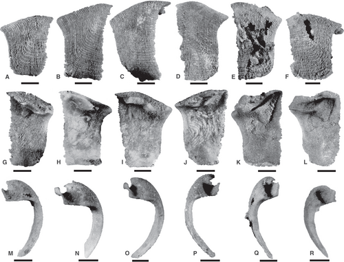
Figures 2, 12, 17
Material
127 loose sclerites in total; 71 from the left series and 56 from the right series. Two sclerites derived from the Johnson (1981) collections marked as derived from horizon ‘WG/1-5’ (AM F.1465879) and ‘WG/3-5’ (AM F.146580), respectively (Figs 2, 12). 125 specimens from horizon GC/15; figured specimens include AM F.152505–AM F.1525014 (Fig. 17). All material collected from the Garra Formation, 8 km south of Wellington NSW, Australia; Lower Devonian (lower Pragian).
Description
Sclerites from left and right series, dorsoventrally elongated with rectangular to subtrapezoidal outline. Commencing from the longitudinal fold, the accreting margin follows a concave trajectory before running sub-parallel to the also slightly concave non-accreting margin, then curves evenly before running straight anteroposteriorly (Fig. 17A–F). The longitudinal fold is arched in lateral profile, reaching a maximum height approximately two-thirds along its length. Length of longitudinal fold ranges between 1.6 and 3.8 mm (n = 79; geometric mean, 2.8 mm), is gently sloping in smaller specimens (Fig. 17A, F) and well-expressed in larger specimens (Fig. 17B–E). Sclerite height ranges between 1.9 and 5.5 mm (n = 71; geometric mean, 3.2 mm) and H:L ratio ranges from 0.7 to 1.6. Left and right series sclerites have even curvature in anterior view. External ornament sometimes clearly preserved as raised evenly spaced rugae on the outer surface but is not apparent on the dorsal flange, probably due to silicification. Inflections I3 and I4 are present (see Högström et al. 2009, fig. 1, for reference), where I3 is pronounced throughout ontogenetic variants and I4 approximates 90° in all specimens with ornament preserved (Fig. 17A–F). Number of rugae ranges from c. 30 to 50. Density of dorsoventrally aligned rugae 7–17 per mm, anteroposteriorly aligned rugae 5–9 per mm. Apical and marginal spines on sclerites lacking.
Sclerites in each series have a unique dorsal flange morphology. In the left series, the medial margin runs parallel to the profile of the longitudinal fold both in dorsal and interior views (Figs 2E, 12A, B, 17G–I). A small hemispherical anterior groove positioned in front of the dorsal flange is present in well-preserved specimens (Fig. 2G). In anterior view, the dorsal flange is strongly concave with a narrow furrow, and a medial non-accreting margin recurved almost to the height of the longitudinal fold (Figs 2I, 17M–O). Dorsal furrow in left series c. 0.6–0.9 mm wide and 0.5–0.6 mm deep. In the right series, the dorsal flange is short and narrow (Figs 2, 11A, B, 17J–L) with a sub-triangular outline in dorsal view (Fig. 2F). The medial non-accreting margin flares up to a point at a height just below the longitudinal fold in anterior view (Figs 2H, 17J–L). Towards the anterior, the dorsal flange exhibits a small tongue distinguished by an adjacent notch along the medial non-accreting margin, although it is not frequently preserved due to coarse silicification (Figs 2G, 17K). Dorsal furrow in right series c. 0.8–0.9 mm wide and 0.3–0.5 mm deep.
Internal surface features characterized by a shallow distinct depression below dorsal flange approximately midway along height of sclerite (Fig. 2). Sclerites from the left series possess a distinct narrow groove that runs from midway below the dorsal flange to the apex under the longitudinal fold (Figs 2B–D, 17H, I).
Remarks
Isolated sclerites from the Garra Formation are better preserved than articulated specimens, retaining finer details including flange grooves and external rugae. As discussed above, they can be readily distinguished from L. shurikenus in possessing left and right morphotypes with respect to dorsal flange morphology. Moreover, isolated sclerites exhibit a sub-rectangular outline with approximately parallel accreting and non-accreting margins, and proximal and distal distinct concavity (corresponding to inflection I3) causing the longitudinal fold to project forward in front of the accreting margin. Conversely, in L. shurikenus the respective distal and proximal edges taper straight towards the ventral margin with no curvature along the accreting margin. The sclerites in L. caliburnus are similarly dorsoventrally elongate and rectangular in outline, however, they also lack the concave margins below the longitudinal fold. The lateral profile of the longitudinal fold in the isolated material exhibits strong dorsal arching compared with the gentle slope of that seen in L. caliburnus (compare Fig. 17A–F with Fig. 3F, G).
The possession of this anterior and posterior concavity along the proximal and dorsal edges in conjunction with the pronounced dorsal arching distinguishes these loose sclerites from most other species and warrants comparison with the Middle Devonian species L. kuangguoduni from China (Gügel et al. 2017, fig. 4). Volume renderings of L. kuangguoduni share several diagnostic features with the Australian material, including the shape of the longitudinal fold. However, the respective series are reversed, which supports Adrain’s (1992) observations that mirror-image enantiomorphs can exist between and perhaps also within species. As seen in dorsal view, the type material follows a sinusoidal trajectory in the left series (Gügel et al. 2017, fig. 4A3–C3) whereas the Australian material displays the same feature but only in the right series (Fig. 2F). Similarly, the straight to slightly bent trajectory of the longitudinal fold, parallel to the medial non-accreting margin, in the right series of the type material (Gügel et al. 2017, fig. 4D3–G3) is present in the opposing series described herein (Fig. 2E). Both species exhibit an overlap in sclerite dimensions, with the specimens herein documenting slightly larger forms, probably because of a larger sample size. The number and density of rugae is also similar, wherein the type material has c. 9–11 per mm with up to 40 rugae per sclerite and the Australian material (depending on whether measured dorsoventrally or anteroposteriorly) has 5–17 per mm and c. 30–50 rugae.
Acknowledgements
The authors acknowledge: Dr Brian Johnson and past members of the Macquarie University Centre for Ecostratigraphy and Palaeobiology (MUCEP) who originally collected the material described herein; Dr Yong-Yi Zhen for access to reposited material at the Londonderry Drillcore Library, NSW, Australia; Dr Matthew Foley for technical assistance with the Zeiss Xradia MicroXCT-400 and AVIZO at the Australian Centre for Microscopy and Microanalysis (ACMM), Sydney University; and Kim Costello and Brock Anderson for assistance with picking and segmentation of CT data. Thanks to Anette Högström (Norwegian Arctic University Museum) and Kenneth De Baets (Geozentrum Nordbayern, Germany) for their constructive reviews. JDS is supported by the National Science Foundation EAR CAREER-1652351; JDS and TS are supported by EAR/IF-1636643; GAB was provided with internal Macquarie University Research Development grants for this research.
Author contributions
SMJ was responsible for conceptualization, interpretation and project administration/supervision, and carried out visualization using the μCT data and prepared figures. TS performed the μCT investigation at MizzoµX. JDS and GAB assisted with project administration/ supervision and conceptualization. SMJ wrote the manuscript and supplement, with significant input from all of the authors.
Open Research
Data archiving statement
This published work and the nomenclatural acts it contains have been registered in ZooBank: http://zoobank.org/References/42FB71F7-6623-4CD5-8622-E4AA5010C33C. All figured specimens are reposited in the Australian Museum (AM), Sydney, NSW, Australia. CT scans and select .stl meshes of the material documented herein are available in the MorphoSource Repository: https://www.morphosource.org/projects/000363753.


