A 27-year-old female presented with diffuse alveolar haemorrhage due to breast angiosarcoma with lung metastasis
Abstract
Angiosarcomas are rare soft-tissue sarcomas of endothelial cell origin and have a poor prognosis. Diffuse alveolar haemorrhage (DAH) is a syndrome characterized by the accumulation of intra-alveolar red blood cells. However, neoplastic diseases are not commonly considered in the differential diagnosis of DAH. We report a case of a 27-year-old female with the diagnosis of primary breast angiosarcoma developing DAH. This raised our concern that angiosarcoma with lung metastasis may present with DAH and should be considered as an important fatal differential diagnosis of DAH.
Introduction
Angiosarcomas are rare soft-tissue sarcoma and have a high rate of local recurrence, metastasis, and poor outcome 1. Primary angiosarcoma of the breast is not common and represents only 0.04% of all breast tumours and about 8% of breast sarcomas 2. The prognosis of primary breast angiosarcoma is unfavourable with a 5-year survival between 8% and 50%. Unlike angiosarcoma elsewhere, primary breast angiosarcoma tends to affect younger females 2. Such sarcomas principally spread haematogenously, with the lungs the most common site for metastases 1.
Diffuse alveolar haemorrhage (DAH) is a distinct clinicopathological syndrome characterized by the accumulation of intra-alveolar red blood cells originating from the alveolar capillaries 3. DAH is associated with pulmonary capillaritis, bland pulmonary haemorrhage, diffuse alveolar damage, and miscellaneous histology 3. However, neoplastic diseases are not commonly considered in the differential diagnosis of DAH. There are few reports of primary breast angiosarcoma with lung metastasis presenting with DAH and acute respiratory failure.
Here we report a 27-year-old female who presented with massive haemoptysis and deteriorated rapidly into acute respiratory failure and death. She was finally diagnosed with primary angiosarcoma of the breast with DAH secondary to lung metastasis.
Case Report
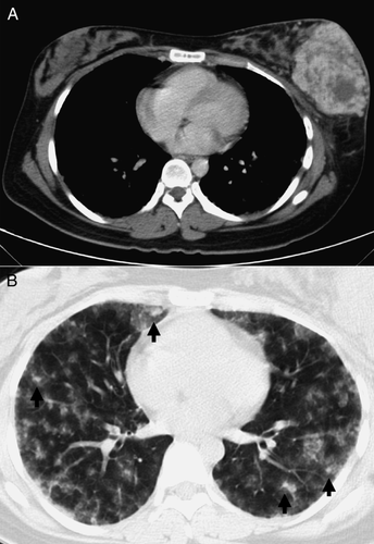
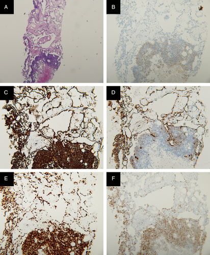
A 27-year-old female with no significant systemic disease presented with cough and blood-tinged sputum for three weeks. The symptoms worsened and shortness of breath developed. Her medical history revealed that she gave birth to a healthy infant 10 months prior to admission and breastfed the infant until admission. However, she reported noticing a lump in the left breast half a year ago. She did not seek medical advice for the mass and continued breastfeeding. Her dyspnoea worsened one week before this admission, so the patient went to a regional hospital to seek advice. Pneumonia was suspected by chest X-ray findings and antibiotics were administered. In addition, aspiration of the left breast mass was performed and 600 mL bloody content was drained. Computed tomography (CT) scan revealed a hypervascular mass in the left breast (Fig. 1A) and multiple nodular lesions with diffuse ground-glass patches in both lung fields (Fig. 1B). Due to poor response to the initial treatment for one week, the patient was transferred to our hospital. Core needle biopsy of the left breast was performed after admission. Haemoptysis soon deteriorated and the patient was intubated due to acute respiratory failure on the third day after admission. On bronchoscopy, diffuse oozing of blood from bilateral segmental bronchi was seen on the fourth day. The serial laboratory examinations included anti-glomerular basement membrane antibodies, anti-neutrophil cytoplasmic antibodies, antinuclear antibodies, and galactomannan test for aspergillus, but the results were all negative. No tubercle bacillus or fungi were found in the sputum and the sputum bacterial culture yielded negative result. The pathological result of breast biopsy revealed a high-grade angiosarcoma. The results of immune histochemical staining for AE1/AE3, CD31, CD34, ERG, and c-MYC were positive (Fig. 2). The patient rapidly deteriorated and prone positioning was applied, but she died eight days after admission.


Discussion
Angiosarcomas are malignant vascular tumours with a poor prognosis that arise from the endothelium. The cutaneous form is the most common type of angiosarcoma and the head and neck are the most frequently involved regions 2. Such sarcomas principally spread haematogenously, with the lungs the most common site for metastases 2. Metastatic pulmonary angiosarcoma displays a variety of radiographic appearances. The most common features of angiosarcoma in chest CT scan are multiple solid nodular lesions and the second most common, multiple thin-walled cysts 4. Tateishi et al. 5 suggested that ground-glass attenuation surrounding solid nodules might correspond to alveolar haemorrhage. Despite the lack of direct pathological evidence of lung metastasis in our case, the CT findings of multiple solid nodular lesions accompanied by ground-glass attenuation surrounding solid nodules could be an important hint of metastasis angiosarcoma in lung with alveolar haemorrhage. In addition, the pathological finding of breast mass is consistent with high-grade angiosarcoma due to the positive results of immunohistochemical stains for AE1/AE3, CD31, CD34, ERG, and c-MYC.
Wang et al. 3 conducted the only retrospective study that indicated angiosarcoma with pulmonary metastasis often presents with multiple pulmonary nodules and DAH. The common initial misdiagnoses included tuberculosis, auto-immune-related vasculitis, non-tuberculous infectious disease, and constrictive pericarditis 3. The most common findings of patients with haemoptysis in CT scan were nodules that were bilateral, randomly distributed, variably shaped and sized, as well as ground-glass opacities (GGO) 3-5. However, CT-guided needle biopsy was difficult to perform in most patients due to the nodules being small in size 3. The median overall survival was very poor, only 5.0 months, for the nine followed up patients and only one patient with pulmonary symptoms had the chance to receive anti-tumour therapy 3.
Angiosarcoma with pulmonary metastases is a rare disease that should be considered in patients with haemoptysis, especially accompanied with GGO and nodules on chest CT scan. This is, to our knowledge, the first case of breast angiosarcoma with lung metastasis reported to cause DAH. Physician in dealing with such fatal DAH in young patients should consider the possibility of angiosarcoma with lung metastasis.
Disclosure Statement
Appropriate written informed consent was obtained for publication of this case report and accompanying images.


