Interpreting mortuary treatment from histological bone diagenesis: A case study from Neolithic Çatalhöyük
Abstract
This study examines the evidence for differential mortuary practices at the Neolithic site of Çatalhöyük, in Anatolia, Turkey, using the histological examination of bacterial microbioerosion in archaeological human bone. In order to analyse bacterial microbioerosion, thin sections were prepared from the midshaft of thoracic ribs 6–8, of n = 162 individuals (adults and juveniles) and analysed using qualitative analysis of microscopic focal destructive changes with polarized light microscopy. The extent of destructive change was assessed, and the degree of preservation was examined using the Oxford Histological Index. Individual, in situ interment was assessed using burial and skeletal data. Results show no differences in histological bacterial microbioerosion between male and female burials, but histological preservation was seen to differ between juveniles and adults, findings that are interpreted as the result of differential mortuary practices at the time of death. The challenges and potential of examining recurring taphonomic signatures in bone histology and their relationship to differential burial practices are discussed.
1 INTRODUCTION
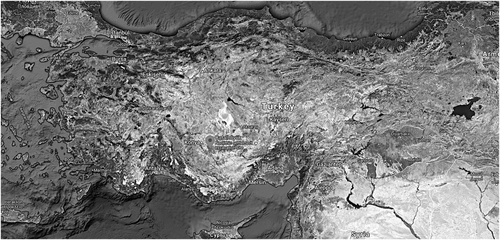
Çatalhöyük is a Neolithic settlement located in south-central Turkey (Figure 1) dating from roughly 7100 to 6000 cal BC (Bayliss et al., 2015). The site was first excavated by James Mellaart in the 1960s, then followed by a large excavation project under the direction of Ian Hodder from 1993 to 2017. Çatalhöyük is located in south-central Anatolia, approximately 50 km from the modern city of Konya. The site is known for its architecture of stacked buildings that demonstrate meticulous dedication to repetition of reconstructing of the same building layout, over multiple sequences in time (Farid, 2014; Matthews, 2005; Russell et al., 2014). This dedication to tradition or social control is also observed in the communal funerary practices, the location of paintings within houses and the repetitive replastering and repainting of reliefs during the lifecycle of a building (Czeszewska, 2014).
Paleoenvironmental soil analysis suggests that Neolithic Çatalhöyük was located on alluvial wetlands and during its occupation coincided with a period of active river alluviation, surrounding most of the area in floodwater each spring (Roberts & Rosen, 2009). Analysis of soil cores by Ayala et al. (2017) reveals four stratigraphic phases from the early Holocene onwards in which humid conditions shifted to increasingly dryer climate. The basilar layer of sediments is primarily composed of marl and sands with gravel. The lower and upper layers of sediment were dominantly silty-clays or silts and clay.
Over 700 skeletons have been excavated from the site and represent complex mortuary practices, including the retrieval and reuse of skulls and dismemberment of individuals (Hodder, 2014). Haddow et al. (2020) suggest that there are a number of instances where the crania and mandibles of individuals have been removed then eventually reburied as headless primary burials at Çatalhöyük. The approximate number of primary burials where the cranium and mandible have been intentionally removed is predicted to be 15 (Pilloud, Haddow, Knüsel, & Larsen, 2016). In addition, according to Pearson and Meskell (2015), individuals were occasionally found to be interred with plaster encasing the skeletonized remains and or with red paint on the skull. In a recent analysis of the interment categories at Çatalhöyük (Haddow et al., 2020), six burial categories have been defined, with four main depositional interment: secondary, tertiary, primary undisturbed and primary disturbed burials. Secondary burials are if a partial or complete skeleton has been moved by Neolithic people from a previous location and deposited in a final interment context. Tertiary burials are disarticulated, partially articulated, or isolated skeletal elements found in noninterment contexts (e.g., midden). Primary undisturbed burials are articulated or nearly complete individuals found in their original place of interment. Primary disturbed burials are in situ skeletal remains found in their original burial location and subsequently disturbed by Neolithic people. Secondary burials account for 13% of the total number of burials, tertiary burials are for 23%, the primary undisturbed burials are 39% and primary disturbed burials are 25% of the interment observed at Çatalhöyük. Individuals are primarily buried within houses under platforms and floors but have been also interred within building foundations, infill, benches and middens (Boz & Hager, 2013, 2014). These intramural burials placed the living both physically and symbolically with the dead (Nakamura & Meskell, 2009).
Over the years, the study of the human remains has been a collaborative effort that has contributed much to our understanding of mortuary practice, social structure, health, diet and lifestyle at Çatalhöyük. There have been numerous analyses applied to the human remains with the objective to interpret significance between variations in mortuary treatments (Agarwal et al., 2017; Boz & Hager, 2013, 2014; Haddow & Knüsel, 2017; Haddow, Sadvari, Knüsel, & Hadad, 2016; Hillson, Boz, & Hager, 2013; Larsen et al., 2015; Pilloud et al., 2016; Pilloud & Larsen, 2011). It remains unclear why particular individuals received differential mortuary treatments at Çatalhöyük; however the large number of isolated and partially articulated skeletal elements may be interpreted as a communal relation with the dead. This might suggest that delayed burial practice was a part of the social structure of the community.
Cut marks from manual excarnation, the removal of flesh using tools, such as sharp obsidian has been observed in 2% of the sample population. These individuals display evidence of linear markings on the diaphysis of the bone. It has been suggested by Russell, Twiss, Orton, and Demirergi (2013) that application of obsidian tools might have lessened the need for deep cuts that could potentially leave incisions on the bone. Furthermore, paintings of vultures surrounding headless bodies have been observed at Çatalhöyük during the Mellaart (1967) excavations. Pilloud et al. (2016) investigated preinterment vulture excarnation as a mortuary treatment and found the scavengers to be skilled at the removal of soft tissue from the bone, often leaving surrounding ligaments and tendons fully intact. Based on the manner and consistency of skeletal element articulation at the site, it is possible that vulture excarnation was utilized at Çatalhöyük (Pilloud et al., 2016).
It is well understood that the macroscopic appearance of whole skeletal elements may differ from the microstructural appearance of its tissues. Therefore, it is valuable to incorporate histological findings when assessing pathological, traumatic and postmortem diagenetic changes to the microstructure of skeletal tissues (Bell, Skinner, & Jones, 1996; Hollund et al., 2012; Jans, Nielsen-Marsh, Smith, Collins, & Kars, 2004; Nielsen-Marsh et al., 2007; Parker Pearson et al., 2005; Smith, Nielsen-Marsh, Jans, & Collins, 2007; Turner-Walker & Jans, 2008). There are three primary diagenetic pathways that cause macroscopic and microscopic alteration to the bone including hydrolysis; the chemical deterioration of the bone organic component, dissolution; the chemical deterioration of the inorganic component of the bone and microbial attack; the bacterial deterioration of the tissue composite (Child, 1995; Collins et al., 2002; Hedges, 2002; Hedges & Millard, 1995; Hedges, Millard, & Pike, 1995; Henderson, 1987; Millard, Brothwell, & Pollard, 2001; Trueman & Martill, 2002; Turner-Walker, 2008). The most common of the three primary destructive changes in archaeological bone is deterioration by microbial attack (Brönnimann et al., 2018; Collins et al., 2002; Hedges, 2002; Jans et al., 2004; Nielsen-Marsh et al., 2007; Nielsen-Marsh & Hedges, 2000; Turner-Walker, Nielsen-Marsh, Syversen, Kars, & Collins, 2002). Diagenetic change is visible in the bone at a microscopic level and consists of five primary categories identifiable with normal and polarized transmitted light microscopy: type of destruction, the presence of inclusive and/or infiltrated materials, the presence of microfissures and the intensity of birefringence (Jans, 2005). Microbial destruction attacks both the collagen and mineral phases of the bone, impacts the pore structure and is most often indicated by areas of remineralization originating from within the Haversian canals as they proceed to fill osteons.
Hackett (1981) characterized microbial attack to the bone by its distinguishing features of size, shape, presence of a hypermineralized rim (ranging between 10 and 60 μm in diameter), reorganization of the bone microstructure and the presence of tunnel like structures. Microbial attack leaves a hypermineralized rim through the dissolution and redistribution of hydroxyapatite, creating a higher density area with respect to the surrounding bone (Dal Sasso, Maritan, Usai, Angelini, & Artioli, 2014). Hackett (1981) characterized four microfocal destruction tunnel types: lamellate, budded, linear longitudinal and Wedl tunnels. Recent research by Jans et al. (2004), Dal Sasso et al. (2014) and Turner-Walker (2012) indicate that while using high magnification light microscopy, the tunnels are a series of interconnected pores, often surrounded by a hypermineralized border and might suggest the vascular canalicular network of the bone assists in the transportation of bacteria.
Microbial diagenesis to the bone tissue microstructure has been observed in both terrestrial (Baud & Lacotte, 1984; Hackett, 1981; Jackes, Sherburne, Lubell, Barker, & Wayman, 2001) and aquatic environments (Bell, Boyde, & Jones, 1991; Bell et al., 1996; Davis, 1997; Pesquero, Ascaso, Alcalá, & Fernández-Jalvo, 2010) and is sometimes attributed to attack by fungi (Carlile, Watkinson, & Gooday, 2001; Marchiafava, Bonucci, & Ascenzi, 1974; Morgenthaler & Baud, 1956; Roux, 1887; Schaffer, 1894; Wedl, 1864). In terrestrial interment, it is undetermined whether the microorganisms that demineralize bone are primarily endogenous and exogenous, but it is most likely that the two are intertwined as the severity and type of diagenetic change are often controlled by the external environment (Grupe & Harbeck, 2019).
A number of histological methods have been developed to semiquantify the extent of preservation of the bone microstructure (Garland, 1989; Hackett, 1981; Hedges et al., 1995; Stout, 1978) and may contribute to better interpretation of an individual's burial history. This study is the first to histologically quantify microbioerosional change in relation with secondary burials; those displaying manipulation by human action, to investigate differential diagenetic expressions in the bone microstructure between biological age and sex groups and those with pathologies as seen on the gross skeleton. Individuals with possible alteration by anthropogenic manipulation might indicate a different diagenetic expression than primary undisturbed burials, if microbial attack is endogenous in origin. Findings of the study will advance our understanding of the process of diagenetic change in archaeological human skeletal tissue and potentially further our understanding of the social conditions that determined specific mortuary treatments to particular individuals at Çatalhöyük.
2 MATERIALS
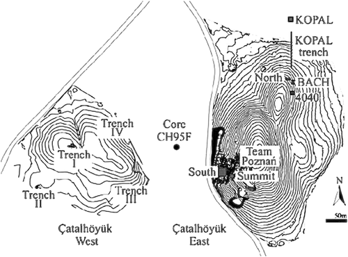
Rib sections were sampled from juvenile and adult skeletonized individuals (n = 162) who were excavated between field seasons 1997 to 2010. Individuals included in this study were sampled from the following excavation areas: South Shelter (S), North Shelter (N), BACH Area, 4040 Area (4040) and the Team Poznan (TP) Area (Figure 2). The sample is comprised of 90 juveniles (aged neonate to 12 years old), six adolescents (12–20 years old) and 66 adult (20–50+ years old) individuals. Age groups included in the sample were neonates (n = 25), infants (n = 41), child (n = 24), adolescents (n = 6), young adult (n = 12), mature adult (n = 15), old adult (n = 10) and adult (n = 29).
The sample included 32 males, 31 females, 85 individuals recorded as too young to determine sex (O) and 14 individuals recorded as indeterminate (I/?). The biological sex and age of the individuals were estimated by a number of osteological team members as outlined in Hillson et al. (2013), and all age and sex estimated were reassessed and/or confirmed at the time of sample collection by authors Agarwal and/or Beauchesne. Biological sex was estimated using standardized morphological features of the pelvis and skull (Buikstra & Ubelaker, 1994). In some cases, the remains did not display diagnostic elements for biological sex estimation. Sex estimation of the skull included assessment of the mastoid process, supramastoid crest, temporal line, glabella, supraorbital ridge, nuchal line and external occipital protuberance. The mandible was assessed for robusticity of the coronoid process, ramus, gonial angle and mental eminence. The os-coxa was evaluated using the shape of the greater sciatic notch, pubis, ventral arc, subpubic concavity and inferior ramus ridge. Age at death in adults was estimated using the Brooks and Suchey (1990) pubic symphysis stages and the Smith (1984) occlusal tooth wear (attrition) scores. Wear in the dentition was used to divide the adults into young, middle and old age groups. The age of subadults was estimated using developmental indicators including measurements defined by Fazekas and Kósa (1978) of the long bones and skull and dental development using both the Schour and Massler (1941) and Hillson (2005) methods.
Interment data such as burial location, in situ interment position, the association of grave goods and indications of pathology or body parts moved were evaluated using published online Çatalhöyük archive reports (http://www.catalhoyuk.com/research). Most interment were found within domestic buildings beneath the north and east platforms of the main room, with the exception of infants who were sometimes interred in floors of the side rooms, hearths or ovens (Andrews, Molleson, & Boz, 2005; Boz & Hager, 2013). Burial preference was for individuals to be interred in the flexed position, with minor variations of extended legs or arms (Boz & Hager, 2013). Most interment lacked burial goods, and when burial goods were present, they were limited in number (Boz &Hager, 2013). Burial goods were investigated on a presence or absence basis. Fibre analysis revealed that adults were interred with sedge fibres, and juveniles were consistently interred with a variety of plant fibres including those for funerary baskets (Rosen, 2005). Different kinds of mineral pigments have been found associated with interment including red and yellow ochre, and green and blue cinnabar in adult burials (Boz & Hager, 2013). Red ochre has been found on the skulls of adults, juveniles and infants.
As noted above, postmortem manipulation of bodies is a characteristic feature of burial at Çatalhöyük (Boz & Hager, 2014; Hodder, 2005). In a study of human remains from field seasons 1996 to 2008, Boz and Hager (2013) observed that decapitation seemed to occur in all age categories. Individuals in the sample were assessed for indicators of dismemberment (appendages dismembered or disarticulated), decapitation (head removed prior to excavation) (Table 1) and widely assessed for pathological conditions including evidence of metabolic stress and degenerative diseases (Table 2). No individuals in the sample had evidence of infectious disease. It is unclear if disarticulated individuals had their heads and limbs intentionally moved or if it is an artefact of an addition of a new individual to a multi-burial grave. Due to this discrepancy, individuals in the sample whom were documented in the archive report as disarticulated or with their heads moved are included in assessment of postmortem manipulation as it signifies the movement of body parts postmortem, regardless of intentionality.
| Skeletal number | Area | Age | Burial position | Sex | Dismemberment & decapitation | Burial deposition | OHI score |
|---|---|---|---|---|---|---|---|
| 2362 | S | Neonate | Flexed | Too young to determine | Disarticulated | Primary disturbed/secondary | 0 |
| Left arm and leg detached | |||||||
| Skull smashed | |||||||
| 2017 | S | Neonate | NA | Too young to determine | Disarticulated | NA | 0 |
| Only parts of burial present | |||||||
| 11657 | S | Child | Flexed | Too young to determine | Headless | Primary disturbed | 0 |
| 16697 | 4040 | Adult | Flexed | Female | Cranium removed | Secondary | 0 |
| 14139 | 4040 | Adult | Flexed | Male | Skull removed | Secondary | 0 |
| 14108 | 4040 | Adolescent | Flexed | Male | Skull removed | Secondary | 0 |
| 4593 | S | Young adult | On back | Male | Skull removed | Primary disturbed | 0 |
| Evidence of cut marks on atlas | |||||||
| Wooden plank placed on body | |||||||
| 12863 | S | Young adult | NA | Female | Incomplete, disarticulated | Secondary | 1 |
| 10033 | 4040 | Infant | Crouched | Too young to determine | Disarticulation | NA | 1 |
| No long bones present | |||||||
| 15640 | 4040 | Adult | Flexed | Female | Headless | Primary disturbed | 1 |
| Missing vertebra and upper limbs | |||||||
| 17412 | 4040 | Adult | On back | Male | Headless | Secondary | 1 |
| Limbless torso | |||||||
| 11659 | S | Mature adult | Crouched | Female | Skull removed | Primary disturbed | 1 |
| 11982 | 4040 | Adolescent | Flexed | Indeterminate | Head moved 5 cm above rest of body | Primary disturbed | 1 |
| 2125 | N | Infant | Crouched | Too young to determine | Headless | Secondary | 1 |
| 17698 | TP | Adult | Flexed | Female | Skull removed | Primary disturbed | 1 |
| 13162 | 4040 | Adult | Flexed | Female | Missing skull | NA | 1 |
| Displaced cervical vertebrae | |||||||
| Interred with full term fetus in abdominal and pelvic regions. | |||||||
| 1466 | N | Mature adult | Flexed on back | Male | Missing skull and atlas, axis missing dens | Undisturbed | 1 |
| Tarsal bones of right foot ossified, including pathological modification of medial phalanx | |||||||
| 2772 | S | Neonate | Extended | Too young to determine | Partially disarticulated | Primary | 1 |
| 13609 | 4040 | Old adult | On side | Male | All limbs including scapula and clavicle removed | NA | 2 |
| 16304 | 4040 | Old adult | On side | Female | Skull, mandible and cervical vertebra removed | Secondary | 2 |
| 17536 | 4040 | Adult | Sitting | Female | Head missing | Primary disturbed | 2 |
| 10814 | S | Adult | Flexed | Female | Head removed and recovered in postretrieval pit | Primary disturbed | 4 |
- Note: Burial data of individuals sampled for histological assessment collected from the Çatalhöyük archive reports including indication of dismemberment, decapitation (skull indicates both cranium and mandible), pathology, biological age and sex, burial position and deposition, and excavation area. Recording of NA for burial position and burial deposition indicates when this information was not documented in the Çatalhöyük archive report or project database
| Skeletal number | Area | Age | Burial position | Sex | Pathology | Burial deposition | OHI score |
|---|---|---|---|---|---|---|---|
| 2033 | S | Child | Flexed | Too young to determine | Cribra orbitalia on left orbit | Primary | 0 |
| Remodelling on inner right parietal | |||||||
| 11494 | S | Mature adult | Flexed | Male | Degenerative joint disease | Primary disturbed | 0 |
| Periodontal disease | |||||||
| 18457 | S | Adult | Flexed | Female | Tunnel like lesion through right scaphoid with a smoothed out, remodelled surface | Primary | 0 |
| 8113 | BACH | Young adult | Indeterminate | Spondylolysis | Primary disturbed | 0 | |
| 6303 | BACH | Adult | Flexed | Male | Advanced osteoporosis | Primary | 1 |
| Roots penetrate inside of most bones | |||||||
| Fragmentary cranium, mandible and ribs | |||||||
| Burnt/carbonized wooden plank over feet | |||||||
| 1884 | S | Child | Flexed | Too young to determine | Cribra femora | Primary | 1 |
| 1885 | S | Child | Flexed | Too young to determine | Cribra orbitalia | Primary | 1 |
| Spina bifida occulta | |||||||
| 8425/8410 | BACH | Adult | Flexed | Male | Degenerative joint disease | Primary | 0 |
| 2115 | N | Old age | Female | Head Lesion | Primary | 0 | |
| 2728 | S | Infant | Flexed | Too young to determine | Pathology at thorax: deflation and spacing of ribs suggest shallow breathing | NA | 2 |
| Cribra orbitalia | |||||||
| 3368 | S | Young adult | Flexed | Male | Systemic bone pathology | Primary | 2 |
| 8410 | BACH | Mature adult | Flexed | Male | Degenerative joint disease at spinal column, pelvis and hands | Primary | 4 |
| 2056 | S | Mature adult | Flexed | Male | Possible disarticulation: left leg | Primary/secondary | 4 |
| Mid cervical vertebrae: osteophyte growth, eburnation and degeneration | |||||||
| Degenerative change to hamate and metacarpals indicating hand injury. | |||||||
| 2886 | S | Young adult | Crouched | Male | Fracture of lateral incisor, clavicle first rib, manubrium and chest. Scheuermann's asymmetry of dorsal vertebrae | Primary | 4 |
| 10813 | S | Adult | On back flexed | Male | Bony growth at bregma | NA | 5 |
3 METHODOLOGY
Thin sections were prepared from the midshaft of thoracic ribs (midshaft rib #6–8). Sections were removed using a Buehler Isomet 1000™ precision saw and embedded in Buehler's Epothin resin. Thin sections, approximately 70–100 microns were made using a Bueller Petro ThinTM sectioning system (Agarwal, Glencross, & Beauchesne, 2013). After mounting on a glass slide, sections were examined with polarized light microscopy using a Leica MX6 dissection upright microscope, with a plain magnification factor of 0.8× and an eyepiece magnification of 10×. The degree of destructive change and preservation was assessed using the Oxford Histological Index (OHI), a classification system developed by Hedges et al. (1995), so the extent of microbioerosion could be quantified. The OHI method was developed on thick sections of the bone (Neolithic and Paleolithic) but may be applied to thin sections of tissue for detailed analysis. The OHI method has been used in both experimental forensic and biological archaeological studies (Booth, 2015; Booth & Madgwick, 2016; Brönnimann et al., 2018; Kontopoulos, Nystrom, & White, 2016; Madgwick, 2008; Redfern, 2008; White & Booth, 2014). Each histological section was assigned a score based on the amount of microstructurally recognizable preserved bone and given a numerical value to semiquantitate diagenetic alteration. A bone section was evaluated on a scale of zero to five to summarize the degree of postmortem microstructural change, with zero, being less than approximately 5% of the bone intact, to five, where greater than 95% is unaffected (Table 3).
| Index | Approx % of intact bone | Description |
|---|---|---|
| 0 | %3C5 | No original features identifiable, other than Haversian canals |
| 1 | %3C15 | Small areas of well-preserved bone present or some lamellar structure preserved by pattern of destructive foci |
| 2 | %3C33 | Clear lamellate structure preserved between destructive foci |
| 3 | %3E67 | Clear preservation of some osteocyte lacunae |
| 4 | %3E85 | Only minor amounts of destructive foci, otherwise generally well preserved |
| 5 | %3E95 | Very well preserved, virtually indistinguishable from fresh bone |
To validate statistical significance of preservation between biological sex and age groups, chi-square tests of independence were applied using R statistical analysis software (R Core Team, 2019). Comparison groups investigation included preservational differences between juvenile and adult age categories, juveniles without the presence of adolescents for comparison with the adult age categories, differences of preservation between biological males and females, differences of preservation between old age and the young adult age categories and preservational differences between old age males and old age females. Comparative analysis was also conducted between the amount of preservation and individuals with indications of pathology or postmortem manipulation.
4 RESULTS
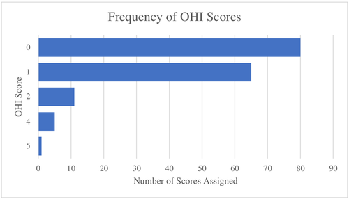
Results of analysis show that 90% of the sample was assigned a score of 0 and 1 for containing less than 15% of the preserved bone microstructure (Figure 3). Eleven individuals were assigned a score of two, indicating less than 33% of the bone intact. Six individuals were given a score of four or five who had 85% or more bone intact.
To evaluate for differences between subadult and adult age groups, all subadult age categories—neonates, infants, children and adolescents—were combined then compared with the combined adult categories—adult, young adult, mature adult and old age (Figure 4). Results showed significant differences in preservation between adult and subadult age categories (p = 0.002241) indicating subadults to be less well preserved than adults. Only adults received OHI scores four and above (%3E85% of bone intact).
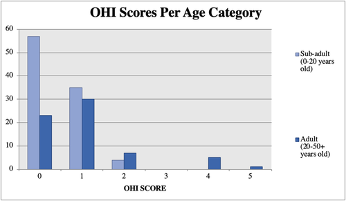
Adolescence (12–20 years) is the generalized biological start of adulthood and the period when the human skeleton goes through a series of developmental changes. At birth all, primary ossification centres of the rib are present, but it is not until 12–14 years that secondary ossification centres of the rib begin to develop at the sites of articulation (Scheuer & Black, 2004). The rib is not considered fully adult until after the rib head and tubercle epiphyses develop, between the ages 17 and 25 (Scheuer & Black, 2004). As such, the subadult group was analysed without the adolescent individuals. Statistically significant differences of preservation between adult and subadult age groups were maintained when the adolescent group was removed (p = 0.001674). Additionally, juveniles were observed to be dismembered or disarticulated less than their adult counterparts when reviewing the archive reports.
To check if there were differences in preservation within the adult age categories, a chi-square test of independence was conducted between the old age adult group and the young adult category but was not statistically significant (p = 0.3258). The adult age group was excluded from this analysis as it was not possible to estimate which broad age category they should be included.
To examine possible differences in microstructural preservation and burial treatments between male and female groups, chi-square tests of independence evaluated differences been males and females of all adult age groups. No statistically significant differences were observed in preservation between males and females (p = 0.6912). Although no statistically significant differences were validated between males and females, four of the six individuals assigned an OHI score greater than four were male. Old age males and females were compared for differences in preservation. The chi-square test between old age males and females found no significant differences (p = 0.1266).
The difference in the amount of preservation between individuals with body parts moved, generally yielded no difference in OHI scores from primary undisturbed burials (Figure 5). Only one individual, Sk. 10814, an adult female whose head was displaced and found in a postretrieval pit adjacent to the skeleton, received a score of four (%3E85% preservation). Her skeleton was scattered, with only few elements in articulation and she was interred with three others. Sk. 10814 is distinct from the sample recorded with indication of body parts moved, and she is one of the few with well-preserved rib microstructure.
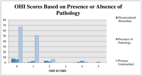
Twenty-two individuals within the study sample were recorded with distinct indications of secondary postmortem manipulation (disarticulation/dismemberment). Sixteen individuals within the sample have had their heads moved, removed or were headless. Eight were disarticulated or had limbs removed. There were two individuals in the sample who had their head and limbs removed (Sk. 15640 and Sk. 17412). Of the sample whose body parts were removed, nine were female, six were male and seven were indeterminate. Postmortem manipulation of the limbs and skull occurred in all ages of the sample, with preference in the adult category (subadult: n = 8; adult: n = 14).
Fifteen individuals of the sample were documented as having pathological conditions, such as degenerative joint disease and cribra orbitalia. Four of these were assigned an OHI score of four or five (%3E85% of bone intact), all were male. The subsample of individuals recorded with indicators of pathological conditions included eight males, two females and five individuals who were indeterminate. Pathologies were primarily present in the adult sample (adult: n = 11 and subadult: n = 4). No individuals with documented pathological indications showed evidence of dismemberment with the exception of Sk. 2056, a mature adult male with degenerative changes to the spine and hand. He was recorded with possible disarticulation of the left leg.
Bacterial microbioerosion in the study sample was common and consistent with bones of intact bodies that had been interred peri-mortem (Booth, 2015; Hedges, 2002; Jans et al., 2004; Nielsen-Marsh et al., 2007; White & Booth, 2014). Immediate burial protects the body from invertebrates that expedite the decomposition process and ensures maximum levels of putrefaction (Breitmeier et al., 2005; Campobasso, Di Vella, & Introna, 2001; Mann, Bass, & Meadows, 1990; Rodriguez & Bass, 1983, 1985; Simmons, Cross, Adlam, & Moffatt, 2010; White & Booth, 2014; Zhou & Bayard, 2011). Generalized bacterial destructive change was observed throughout the sample in the form of blackened remineralized osteons with the destructive change appearing to originate from the Haversian canals into a hypermineralized rim. The majority of the sample demonstrated a generalized loss of recognizable microstructural features including lamellar structure, osteocyte lacunae and canaliculi, and the disintegration, disaggregation and dissociation of osteons. Within the disaggregated osteons, small blackened foci were occasionally present and are interpreted as microfocal destructive tunnels, although differentiation of tunnel type was not assessed. In addition, the presence of externally derived inclusions was observed lying within Haversian canals, osteocyte lacunae and canaliculi. There was a noticeable reduction in birefringence. The decrease in birefringence is assumed to be linked to the deterioration of collagen and/or the changes in the orientation of hydroxyapatite crystals from diagenesis (Schoeninger, Moore, Murray, & Kingston, 1989). Cracked osteons were few, and there was no visible external cracking observed on the periosteal surface.
5 DISCUSSION
There are two primary hypotheses on the causality of bacterial microbioerosion to the bone tissue microstructure. First, that the tissue alteration is commenced by soil microorganisms in the burial environment, postskeletonization (Grupe & Dreses-Werringloer, 1993; Hackett, 1981; Hanson & Buikstra, 1987; Marchiafava et al., 1974; Piepenbrink, 1986; Piepenbrink, 1989; Yoshino, Kimijima, Miyasaka, Sato, & Seta, 1991). Self-destructive enzymes are released following autolysis, opening the soft tissues and dependent on the chemistry and biochemistry of the surrounding soil macroflora and microflora, microorganisms gain access to penetrate the bone (Child, 1995).
The second hypothesis suggests bacterial microbioerosion in the bone is produced by endogenous transmigration of gut bacteria into the postmortem vasculature to the internal cortical structures of the bone (Bell et al., 1996; Guarino, Angelini, Vollono, & Orefice, 2006; Hollund et al., 2012; Jans et al., 2004; Nielsen-Marsh et al., 2007; White & Booth, 2014). Within the first few days after death, the decay of an organism's immune system and mucosal membranes facilitate putrefaction and the transmigration of endogenous bacteria (Gill-King, 1997; Janaway, 1987; Polson & Goe, 1985). Differing postmortem treatments, such as rapid extraneous soft tissue loss, can expose the skeleton to diverse levels of putrefaction, reducing the autolytic decomposition experienced by the bone and amount of bacterial microbioerosion (Bell et al., 1996; Hollund et al., 2012; Jans et al., 2004; Nielsen-Marsh et al., 2007; Parker Pearson et al., 2005; White & Booth, 2014). Findings from an experimental study of buried neonate and juvenile pig carcasses reveal the bone microstructure in neonatal carcasses to be absent from microbioerosional change, whereas the older carcasses showed intensive microbioerosional diagenesis (White & Booth, 2014). This is likely related to the development of the gut microbiome developed at birth (Mackie, Sghir, & Gaskins, 1999; White & Booth, 2014).
Significant differences in microstructural preservation were observed between the adult and subadult samples, and there are a number of possible reasons these differences are being observed. First, it is probable differential interment treatments of juvenile remains influenced the progression of microbioerosion. According to Boz and Hager (2013, 2014), both infants and neonates had the most variability of interment location compared with adults. Neonates were found interred in less common areas, where few adults have been found, such as side-rooms, foundational layers of buildings and near ovens. No adults have been found in side-rooms or building foundation layers. Their analysis revealed that the majority of the side room burials were neonates (83%) and a high proportion of juveniles in foundation layers (73%) (Boz & Hager, 2014). External areas such as middens have yielded several neonates and few adults. Neonates were frequently found interred in baskets or containers and not disturbed postinterment evidenced by the lack of commingled remains. Analyses of headless skeletons excavated at Çatalhöyük show no evidence for taking the skulls of neonatal or preterm skeletons (Boz & Hager, 2014).
Differences in microstructural preservation between subadult and adult age groups could also be attributed to variations in mineral densities of the bone between juvenile and adult remains. The bone mineral content in children tends to regress at the beginning of the postnatal period and develop with age (Guy, Masset, & Baud, 1997; Saunders & Hoppa, 1993). In a comparative study of human and animal bones by Robinson, Nicholson, Pollard, and O'Connor (2003), human rib tissues were observed to have greater porosity measurements than the animal tissues, with the exception of immature domestic animal bone which exhibited higher porosity than their adult counterparts. It is possible that the increase of diagenesis in the juvenile remains is related to the differences in pore network microarchitecture (Bala et al., 2016). In a study by Lefèvre et al. (2019), significant differences between bone mineralization, crystallinity and carbonation were observed between fibulas of adult and juvenile cortical bone samples with these properties inferior in juvenile tissues. Demineralization during diagenesis disrupts the protein mineral, rendering the collagen susceptible to hydrolysis, leading to a general increase in porosity and average pore size. Due to the delicate fragility and small size of subadult tissues, it is likely that their microstructure may be more susceptible to diagenesis and taphonomic weathering (Hanson & Buikstra, 1987; Von Endt & Ortner, 1984; Zapata, Pèrez-Sirvent, Martínez-Sánchez, & Tovar, 2006). Additionally, the difference in preservation between old age adults and the younger adult categories can be associate to the findings of age-related bone loss in ribs as endosteal resorption exceeds periosteal formation (Pavón, Cucina, & Tiesler, 2010; Pirok, Ramser, Takahashi, Villanueva, & Frost, 1966; Sedlin, Frost, & Villanueva, 1963) and decrease in intracortical porosity (Agnew & Stout, 2012).
Our results indicate a lack of statistically significant differences in the preservation of the bone tissue between males and females. This may be supported by previous findings that postmortem manipulation in both sexes is an interment feature of the site (Boz & Hager, 2014; Hodder, 2005). The small sample size of old age adults, comprised of five males and five females, is a limitation to the study. It is possible that no statistically significant differences were observed between these groups because the sample size was too small and lacked statistical power. There is the potential that differences in preservation between males and females of the old age category may become more distinct with a larger sample. All old age males and females were assigned scores that ranged between zero to two. The young adult, and mature adult categories tended to be better preserved than the old adult group. All individuals assigned a score of 4 or above (%3E85% bone preserved) were within the younger adult age categories.
The majority (n = 156) of the sample was poorly preserved and assigned an OHI score of two or less (%3C33% preservation). Only six individuals in the sample received an OHI score four or five (%3E85% preservation). Four of these individuals had the presence of pathological conditions, and one had their head removed. A sixth individual (Sk. 1424) who was assigned an OHI score of four had the presence of black staining and fibres on the skeleton. It is possible that the poor preservation of the sample can be attributed to the fragile nature of rib elements and is a limitation to the study. The rib may be more sensitive to diagenetic processes due to lower levels of calcium and phosphorus than other, more robust, long bones like the femur (Lambert, Vlasak, Thometz, & Buikstra, 1982). Archaeological microbioerosional studies may favour the utilization of robust long bones such as the femur, due to their thick cortex, high cortical content, survival rate in the archaeological record and ability for DNA and isotope sampling. However, in the interest of conservation and the preservation of materials, destructive histological analysis of long bones is not ideal. The histological study of ribs is useful, as it requires minimal tissue sampling and specimen preparation. Furthermore, the examination of ribs facilitates age assessment using histological protocols (Stout, 1986; Stout, Dietze, Işcan, & Loth, 1994; Stout & Lueck, 1995; Stout & Paine, 1992; Stout & Teitelbaum, 1976), as well as more detailed microstructural analyses of metabolic health (Agarwal & Stout, 2003; Agnew & Stout, 2012).
With the knowledge that individuals in this study were subject to variable early postmortem treatments (Boz & Hager, 2013, 2014; Haddow & Knüsel, 2017; Mellaart, 1964; Pilloud et al., 2016), the skeletal microstructure would be expected to have diverse levels of bacterial attack if an individual was not interred immediately after death. The six individuals with the best preservation, relative to the rest of the sample, were documented with indications of pathological conditions or had body parts moved with the exception of Sk. 1424.
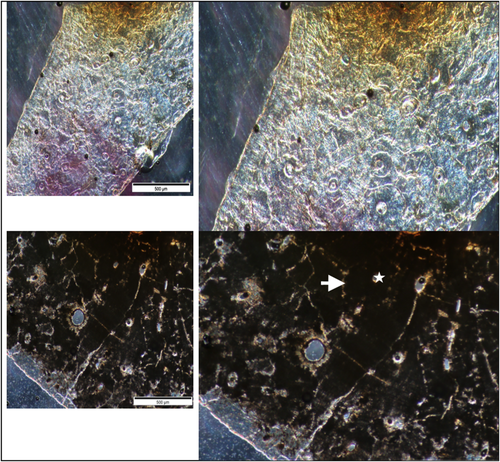
There were no significant preservational differences observed in the bone microstructure of individuals displaying by pathology or dismemberment. Only five individuals of the sample had intact bone microstructure. Four of the six individuals assigned a score of four or greater were documented with the presence of pathological conditions including congenital or neuromechanical abnormalities of the skeleton, inflammatory bone deposits and degenerative diseases (Sk. 10813, Sk. 8410, Sk. 2056 and Sk. 2886). Sk. 10813 was a large male interred with burial goods and had the presence of a bony growth at bregma, received an OHI score of five (Figure 6). The three remaining individuals were documented as having a pathological condition and received a score of four. Sk. 8410, a mature adult male with degenerative joint disease of the spine, pelvis and hands, was interred with four others. Sk. 2056, a mature adult male, was recorded with the left leg possibly disarticulated, osteophyte growth, eburnation and degeneration on cervical vertebrae (C3-C4), and degenerative changes on the hamate and metacarpals indicating hand injury. He was interred above a female (Sk. 2058). Sk. 2886, a young adult male, had the presence of fractures to multiple bones of the upper thorax and lateral incisor and Scheuermann's asymmetry of dorsal vertebrae. It is possible that the presence of pathological conditions acted as a safeguard against bacterial microdiagenesis in these individuals. Archaeological research of pagetic bone tissue microstructure showed that extensive regions were unaffected by bacterial microbioerosion (Bell & Jones, 1991), and chronic inflammatory diseases and bone forming pathologies, such as treponemal disease, tend to stimulate osteoblastic action increasing preservation in archaeological bone. Additionally, it has been observed that when mineralization is impaired by pathological attack, the density of the bone microstructure is impacted (Boyde, Hendel, Hendel, Maconnachie, & Jones, 1990; Boyde & Jones, 1983; Boyde, Maconnachie, Reid, Delling, & Mundy, 1986). As there was no distinct signature in dismembered, pathological or primary undisturbed burials of the sample, additional research is necessary to define the causative agents of bacterial microbioerosion.
6 CONCLUSION
The objective of this study was to compare preservational differences between groups demarcated by biological sex and age and between primary undisturbed burials and individuals with indications of dismemberment, decapitation or pathological condition. Our findings reveal differential preservation rates between adult and juvenile age groups with less bone microstructure intact in the juvenile remains. Investigation between individuals with indication of body parts removed or pathological condition did not find differential preservation when compared with the primary undisturbed interment. These findings may assist our interpretation of the lifeways at Çatalhöyük and of the postmortem burial history of an individual in a terrestrial environment. Further research into the mechanisms driving bacterial microbioerosion is needed to confirm the source of bacterial microbioerosion.
In histological studies such as this one, it is important to develop an understanding of whether observed differences in the bone microstructure can be attributed to differences in mortuary treatments and if the integrity of the bone microstructure is influenced by age or pathology. A limitation and future research objective is the further analysis of the whole sample, through use of a more robust specimen, such as the femur. It is possible that the poor preservation observed in the rib is due to the fragility of the bones low mineral content and high porosity; therefore, ribs may not fully represent the degree of postmortem change of an individual due to their delicate cortical structure. Further research of the rib sections in this sample has the potential for additional analysis such as the identification of specific inclusive materials that have penetrated into the bone microstructure using scanning electron microscopy, application of X-ray computed microtomography to estimate percentage of remineralization, mercury intrusion porosimetry to quantify the amount of microporosity and application of the Cracking Index to quantify cracked osteons to provide a more robust review of diagenesis at Çatalhöyük.
ACKNOWLEDGEMENTS
We thank the Çatalhöyük Research Project, UNESCO and the University of California, Berkeley, for providing the material to study. We also thank Scott Haddow for providing additional contextual burial data of individuals excavated at the site.


