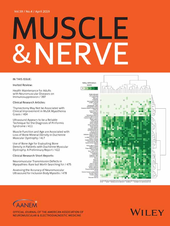
Disease duration and disability in dysfeRlinopathy can be described by muscle imaging using heatmaps and random forests
Corresponding Author
David Gómez-Andrés MD, PhD
Paediatric Neurology, Vall d'Hebron University Hospital and VHIR (Euro-NMD, ERN-RND), Barcelona, Spain
Correspondence to: D.Gómez-Andrés; e-mail: [email protected] and Jorge A. Bevilacqua e-mail: [email protected]Search for more papers by this authorJorge Díaz MD
Medical Imaging Center, University of Chile Clinical Hospital, Santiago, Chile
Search for more papers by this authorFrancina Munell MD, PhD
Paediatric Neurology, Vall d'Hebron University Hospital and VHIR (Euro-NMD, ERN-RND), Barcelona, Spain
Search for more papers by this authorÁngel Sánchez-Montáñez MD
Paediatric Neuroradiology, Vall d'Hebron University Hospital (Euro-NMD, ERN-RND), Barcelona, Spain
Search for more papers by this authorIrene Pulido-Valdeolivas MD, PhD
Visual Pathway Laboratory, Neuroimmunology Center and Neurology Department, Biomedical Research Center August Pi i Sunyer (IDIBAPS), Hospital Clínic Barcelona, Spain
Search for more papers by this authorLionel Suazo MD
Medical Imaging Center, University of Chile Clinical Hospital, Santiago, Chile
Search for more papers by this authorCristián Garrido MT
Medical Imaging Center, University of Chile Clinical Hospital, Santiago, Chile
Search for more papers by this authorSusana Quijano-Roy MD, PhD
APHP–Neurology and Intensive Care Department. University Hospital Raymond Poincaré, Garches, U1179 Versailles University, Neuromuscular Disorders Reference Center of Nord-Est-Île de France, ERN Neuro-NMD, France
Search for more papers by this authorCorresponding Author
Jorge A. Bevilacqua MD, PhD
Neuromuscular Unit, Department of Neurology and Neurosurgery, University of Chile Clinical Hospital
Department of Anatomy and Legal Medicine, Faculty of Medicine, University of Chile, Santiago, Chile
Correspondence to: D.Gómez-Andrés; e-mail: [email protected] and Jorge A. Bevilacqua e-mail: [email protected]Search for more papers by this authorCorresponding Author
David Gómez-Andrés MD, PhD
Paediatric Neurology, Vall d'Hebron University Hospital and VHIR (Euro-NMD, ERN-RND), Barcelona, Spain
Correspondence to: D.Gómez-Andrés; e-mail: [email protected] and Jorge A. Bevilacqua e-mail: [email protected]Search for more papers by this authorJorge Díaz MD
Medical Imaging Center, University of Chile Clinical Hospital, Santiago, Chile
Search for more papers by this authorFrancina Munell MD, PhD
Paediatric Neurology, Vall d'Hebron University Hospital and VHIR (Euro-NMD, ERN-RND), Barcelona, Spain
Search for more papers by this authorÁngel Sánchez-Montáñez MD
Paediatric Neuroradiology, Vall d'Hebron University Hospital (Euro-NMD, ERN-RND), Barcelona, Spain
Search for more papers by this authorIrene Pulido-Valdeolivas MD, PhD
Visual Pathway Laboratory, Neuroimmunology Center and Neurology Department, Biomedical Research Center August Pi i Sunyer (IDIBAPS), Hospital Clínic Barcelona, Spain
Search for more papers by this authorLionel Suazo MD
Medical Imaging Center, University of Chile Clinical Hospital, Santiago, Chile
Search for more papers by this authorCristián Garrido MT
Medical Imaging Center, University of Chile Clinical Hospital, Santiago, Chile
Search for more papers by this authorSusana Quijano-Roy MD, PhD
APHP–Neurology and Intensive Care Department. University Hospital Raymond Poincaré, Garches, U1179 Versailles University, Neuromuscular Disorders Reference Center of Nord-Est-Île de France, ERN Neuro-NMD, France
Search for more papers by this authorCorresponding Author
Jorge A. Bevilacqua MD, PhD
Neuromuscular Unit, Department of Neurology and Neurosurgery, University of Chile Clinical Hospital
Department of Anatomy and Legal Medicine, Faculty of Medicine, University of Chile, Santiago, Chile
Correspondence to: D.Gómez-Andrés; e-mail: [email protected] and Jorge A. Bevilacqua e-mail: [email protected]Search for more papers by this authorABSTRACT
Introduction: The manner in which imaging patterns change over the disease course and with increasing disability in dysferlinopathy is not fully understood.
Methods: Fibroadipose infiltration of 61 muscles was scored based on whole-body MRI of 33 patients with dysferlinopathy and represented in a heatmap. We trained random forests to predict disease duration, Motor Function Measure dimension 1 (MFM-D1), and modified Rankin scale (MRS) score based on muscle scoring and selected the most important muscle for predictions.
Results: The heatmap delineated positive and negative fingerprints in dysferlinopathy. Disease duration was related to infiltration of infraspinatus, teres major–minor, and supraspinatus muscles. MFM-D1 decreased with higher infiltration of teres major–minor, triceps, and sartorius. MRS related to infiltration of vastus medialis, gracilis, infraspinatus, and sartorius.
Discussion: Dysferlinopathy shows a recognizable muscle MRI pattern. Fibroadipose infiltration in specific muscles of the thigh and the upper limb appears to be an important marker for disease progression. Muscle Nerve 59:436–444, 2019
Supporting Information
| Filename | Description |
|---|---|
| mus26403_sup-0001-FigureS1.pdfPDF document, 28.6 KB | Figure S1 Heatmaps that show the relationship between disease duration (S1a), MFM-D1 (S1b) and modified Rankin scale (S1c) and every muscle selected in the corresponding random forest. |
| mus26403_sup-0002-FigureS2.pdfPDF document, 21.5 KB | Figure S2 Adjusted marginal prediction of disease duration according to the 6 most important muscles in the random forest. Infiltration score are translated into ordinals (0 is equal to 0 score, 1 is equal to 1, 2 is equal to 2a, 3 is equal to 2b score and 4 is equal to 3 score) |
| mus26403_sup-0003-FigureS3.pdfPDF document, 24.8 KB | Figure S3 Adjusted marginal prediction of MFM-D1 according to the 6 most important muscles in the random forest |
| mus26403_sup-0004-FigureS4.pdfPDF document, 24.1 KB | Figure S4 Adjusted marginal prediction of modified Rankin scale according to the 6 most important muscles in the random forest |
Please note: The publisher is not responsible for the content or functionality of any supporting information supplied by the authors. Any queries (other than missing content) should be directed to the corresponding author for the article.
REFERENCES
- 1Bashir R, Britton S, Strachan T, Keers S, Vafiadaki E, Lako M, et al. A gene related to Caenorhabditis elegans spermatogenesis factor fer-1 is mutated in limb-girdle muscular dystrophy type 2B. Nat Genet 1998; 20: 37–42.
- 2Liu J, Aoki M, Illa I, Wu C, Fardeau M, Angelini C, et al. Dysferlin, a novel skeletal muscle gene, is mutated in Miyoshi myopathy and limb girdle muscular dystrophy. Nat Genet 1998; 20: 31–36.
- 3Aoki M, Liu J, Richard I, Bashir R, Britton S, Keers SM, et al. Genomic organization of the dysferlin gene and novel mutations in Miyoshi myopathy. Neurology 2001; 57: 271–278.
- 4Illa I, Serrano-Munuera C, Gallardo E, Lasa A, Rojas-Garcia R, Palmer J, et al. Distal anterior compartment myopathy: a dysferlin mutation causing a new muscular dystrophy phenotype. Ann Neurol 2001; 49: 130–134.
- 5Barthelemy F, Wein N, Krahn M, Levy N, Bartoli M. Translational research and therapeutic perspectives in dysferlinopathies. Mol Med 2011; 17: 875–882.
- 6Park HJ, Hong JM, Suh GI, Shin HY, Kim SM, Sunwoo IN, et al. Heterogeneous characteristics of Korean patients with dysferlinopathy. J Korean Med Sci 2012; 27: 423–429.
- 7Cupler EJ, Bohlega S, Hessler R, McLean D, Stigsby B, Ahmad J. Miyoshi myopathy in Saudi Arabia: clinical, electrophysiological, histopathological and radiological features. Neuromuscul Disord 1998; 8: 321–326.
- 8Katz JS, Rando TA, Barohn RJ, Saperstein DS, Jackson CE, Wicklund M, et al. Late-onset distal muscular dystrophy affecting the posterior calves. Muscle Nerve 2003; 28: 443–448.
- 9Ro LS, Lee-Chen GJ, Lin TC, Wu YR, Chen CM, Lin CY, et al. Phenotypic features and genetic findings in 2 Chinese families with Miyoshi distal myopathy. Arch Neurol 2004; 61: 1594–1599.
- 10Brummer D, Walter MC, Palmbach M, Knirsch U, Karitzky J, Tomczak R, et al. Long-term MRI and clinical follow-up of symptomatic and presymptomatic carriers of dysferlin gene mutations. Acta Myol 2005; 24: 6–16.
- 11Illa I, De Luna N, Dominguez-Perles R, Rojas-Garcia R, Paradas C, Palmer J, et al. Symptomatic dysferlin gene mutation carriers: characterization of two cases. Neurology 2007; 68: 1284–1289.
- 12Paradas C, Llauger J, Diaz-Manera J, Rojas-Garcia R, De Luna N, Iturriaga C, et al. Redefining dysferlinopathy phenotypes based on clinical findings and muscle imaging studies. Neurology 2010; 75: 316–323.
- 13Diaz J, Woudt L, Suazo L, Garrido C, Caviedes P, Cardenas AM, et al. Broadening the imaging phenotype of dysferlinopathy at different disease stages. Muscle Nerve 2016; 54: 203–210.
- 14Jin S, Du J, Wang Z, Zhang W, Lv H, Meng L, et al. Heterogeneous characteristics of MRI changes of thigh muscles in patients with dysferlinopathy. Muscle Nerve 2016; 54: 1072–1079.
- 15Woudt L, Di Capua GA, Krahn M, Castiglioni C, Hughes R, Campero M, et al. Toward an objective measure of functional disability in dysferlinopathy. Muscle Nerve 2016; 53: 49–57.
- 16Diaz-Manera J, Fernandez-Torron R, Lauger J, James MK, Mayhew A, Smith FE, et al. Muscle MRI in patients with dysferlinopathy: pattern recognition and implications for clinical trials. J Neurol Neurosurg Psychiatry 2018; 89: 1071–1081.
- 17Chen X, Ishwaran H. Random forests for genomic data analysis. Genomics 2012; 99(6): 323-329.
- 18Berard C, Payan C, Hodgkinson I, Fermanian J, Group MFMCS. A motor function measure for neuromuscular diseases. Construction and validation study. Neuromusc disord 2005; 15(7): 463-470.
- 19van Swieten JC, Koudstaal PJ, Visser MC, Schouten HJ, van Gijn J. Interobserver agreement for the assessment of handicap in stroke patients. Stroke 1988; 19(5): 604-607.
- 20 Aids to the examination of the peripheral nervous system, Memorandum No. 45. London: Medical Research Cuoncil; 1976.
- 21Kornblum C, Lutterbey G, Bogdanow M, Kesper K, Schild H, Schroder R, Wattjes MP. Distinct neuromuscular phenotypes in myotonic dystrophy types 1 and 2 : a whole body highfield MRI study. Journal of neurology 2006; 253(6): 753-761.
- 22Gower JC. A general coefficient of similarity and some of its properties. Biometrics 1971; 27: 857–871.
- 23Ishwaran H, Kogalur UB, Gorodeski EZ, Minn AJ, Lauer MS. High dimensional variable selection for survival data. J Am Stat Assoc 2010; 105: 205–217.


