Volume 72, Issue 2 p. 602
Erratum
Free Access

Erratum to Dynamic contrast-enhanced MRI in mice at high field: Estimation of the arterial input function can be achieved by phase imaging (Magn Reson Med 2014;71:544–550)

Anne-Catherine Fruytier

Anne-Catherine Fruytier

Université catholique de Louvain, Biomedical Magnetic Resonance Unit, Brussels, Belgium

Search for more papers by this author
Julie Magat

Julie Magat

Université catholique de Louvain, Biomedical Magnetic Resonance Unit, Brussels, Belgium

Search for more papers by this author
Benedicte Jordan

Benedicte Jordan

Université catholique de Louvain, Biomedical Magnetic Resonance Unit, Brussels, Belgium

Search for more papers by this author
Greg Cron

Greg Cron

Ottawa Hospital Research Institute, Diagnostic Imaging, 501 Smyth Road, Ottawa, K1H 8L6 Ontario, Canada

Search for more papers by this author
Florence Colliez

Florence Colliez

Université catholique de Louvain, Biomedical Magnetic Resonance Unit, Brussels, Belgium

Search for more papers by this author
Bernard Gallez

Corresponding Author

Bernard Gallez

Université catholique de Louvain, Biomedical Magnetic Resonance Unit, Brussels, Belgium

Correspondence to: Bernard Gallez, Université catholique de Louvain, Biomedical Magnetic Resonance Unit, Av. Mounier 73.40, B-1200 Brussels, Belgium. [email protected]Search for more papers by this author
First published: 20 May 2014
Citations: 1

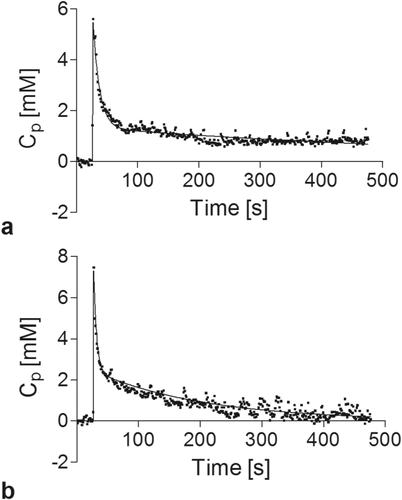
In the article “Dynamic contrast-enhanced MRI in mice at high field: estimation of the arterial input function can be achieved by phase imaging,” by Fruytier et al. (Magn Reson Med. 2014;71:544–550), Figure 1 presents a scaling error. The time course of concentration in plasma (Cp) is not expressed in mM as mentioned on the axis, but in M.

Here you will find the corrected Figure 1, expressed in mM.
Details are in the caption following the image
Typical time course of concentration in plasma (CP) extracted from phase imaging over 8 minutes (mM) : (a) CP obtained from aorta/vena cava of FBV/N mice and (b) from iliac artery/vein of NMRI mice with phase imaging (b). Experimental data represented by (•) were modeled with a biexponential fitting (–). Note that for the two mice models, we obtain positive CP with AIF extracted from phase imaging.

    The full text of this article hosted at iucr.org is unavailable due to technical difficulties.