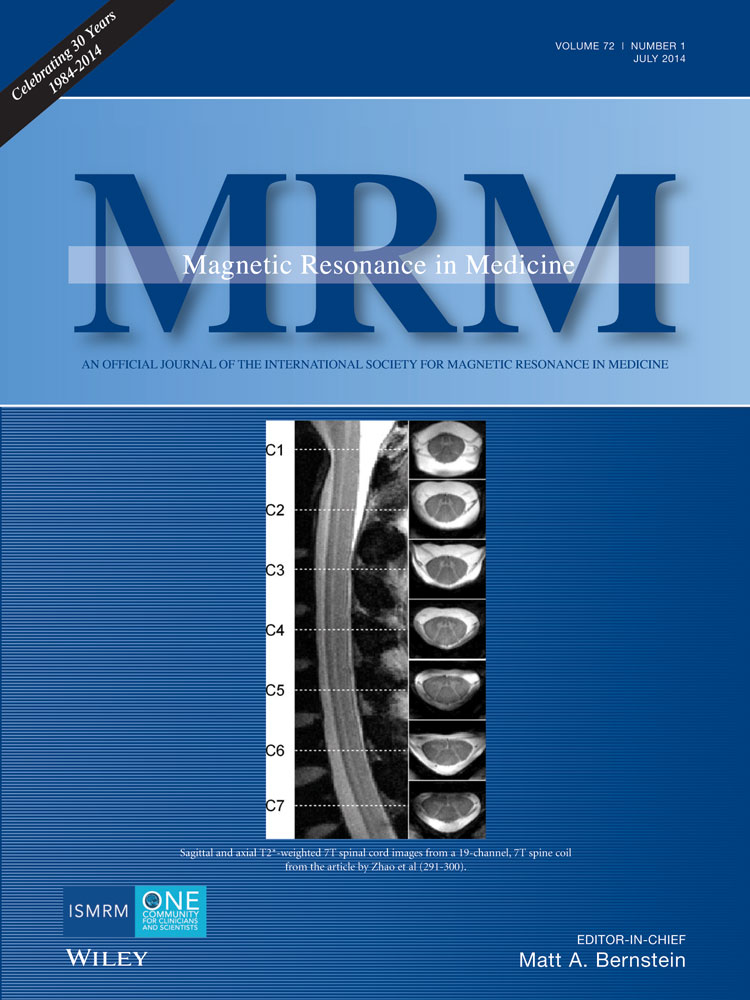
Nineteen-channel receive array and four-channel transmit array coil for cervical spinal cord imaging at 7T
Wei Zhao
A. A. Martinos Center for Biomedical Imaging, Department of Radiology, Massachusetts General Hospital, Charlestown, Massachusetts, USA
Harvard Medical School, Boston, Massachusetts, USA
Search for more papers by this authorJulien Cohen-Adad
Department of Electrical Engineering, Ecole Polytechnique de Montreal, Montreal, Quebec, Canada
Search for more papers by this authorJonathan R. Polimeni
A. A. Martinos Center for Biomedical Imaging, Department of Radiology, Massachusetts General Hospital, Charlestown, Massachusetts, USA
Harvard Medical School, Boston, Massachusetts, USA
Search for more papers by this authorBoris Keil
A. A. Martinos Center for Biomedical Imaging, Department of Radiology, Massachusetts General Hospital, Charlestown, Massachusetts, USA
Harvard Medical School, Boston, Massachusetts, USA
Search for more papers by this authorBastien Guerin
A. A. Martinos Center for Biomedical Imaging, Department of Radiology, Massachusetts General Hospital, Charlestown, Massachusetts, USA
Harvard Medical School, Boston, Massachusetts, USA
Search for more papers by this authorKawin Setsompop
A. A. Martinos Center for Biomedical Imaging, Department of Radiology, Massachusetts General Hospital, Charlestown, Massachusetts, USA
Harvard Medical School, Boston, Massachusetts, USA
Search for more papers by this authorPeter Serano
A. A. Martinos Center for Biomedical Imaging, Department of Radiology, Massachusetts General Hospital, Charlestown, Massachusetts, USA
Harvard Medical School, Boston, Massachusetts, USA
Search for more papers by this authorAzma Mareyam
A. A. Martinos Center for Biomedical Imaging, Department of Radiology, Massachusetts General Hospital, Charlestown, Massachusetts, USA
Harvard Medical School, Boston, Massachusetts, USA
Search for more papers by this authorPhilipp Hoecht
Siemens AG, Healthcare Sector, Magnetic Resonance, Erlangen, Germany
Search for more papers by this authorCorresponding Author
Lawrence L. Wald
A. A. Martinos Center for Biomedical Imaging, Department of Radiology, Massachusetts General Hospital, Charlestown, Massachusetts, USA
Harvard Medical School, Boston, Massachusetts, USA
Harvard-MIT Division of Health Sciences and Technology, Massachusetts Institute of Technology, Cambridge, Massachusetts, USA
Correspondence to: Wei Zhao, Ph.D., A. A. Martinos Center for Biomedical Imaging, Department of Radiology, Massachusetts General Hospital, Charlestown, MA. E-mail: [email protected]Search for more papers by this authorWei Zhao
A. A. Martinos Center for Biomedical Imaging, Department of Radiology, Massachusetts General Hospital, Charlestown, Massachusetts, USA
Harvard Medical School, Boston, Massachusetts, USA
Search for more papers by this authorJulien Cohen-Adad
Department of Electrical Engineering, Ecole Polytechnique de Montreal, Montreal, Quebec, Canada
Search for more papers by this authorJonathan R. Polimeni
A. A. Martinos Center for Biomedical Imaging, Department of Radiology, Massachusetts General Hospital, Charlestown, Massachusetts, USA
Harvard Medical School, Boston, Massachusetts, USA
Search for more papers by this authorBoris Keil
A. A. Martinos Center for Biomedical Imaging, Department of Radiology, Massachusetts General Hospital, Charlestown, Massachusetts, USA
Harvard Medical School, Boston, Massachusetts, USA
Search for more papers by this authorBastien Guerin
A. A. Martinos Center for Biomedical Imaging, Department of Radiology, Massachusetts General Hospital, Charlestown, Massachusetts, USA
Harvard Medical School, Boston, Massachusetts, USA
Search for more papers by this authorKawin Setsompop
A. A. Martinos Center for Biomedical Imaging, Department of Radiology, Massachusetts General Hospital, Charlestown, Massachusetts, USA
Harvard Medical School, Boston, Massachusetts, USA
Search for more papers by this authorPeter Serano
A. A. Martinos Center for Biomedical Imaging, Department of Radiology, Massachusetts General Hospital, Charlestown, Massachusetts, USA
Harvard Medical School, Boston, Massachusetts, USA
Search for more papers by this authorAzma Mareyam
A. A. Martinos Center for Biomedical Imaging, Department of Radiology, Massachusetts General Hospital, Charlestown, Massachusetts, USA
Harvard Medical School, Boston, Massachusetts, USA
Search for more papers by this authorPhilipp Hoecht
Siemens AG, Healthcare Sector, Magnetic Resonance, Erlangen, Germany
Search for more papers by this authorCorresponding Author
Lawrence L. Wald
A. A. Martinos Center for Biomedical Imaging, Department of Radiology, Massachusetts General Hospital, Charlestown, Massachusetts, USA
Harvard Medical School, Boston, Massachusetts, USA
Harvard-MIT Division of Health Sciences and Technology, Massachusetts Institute of Technology, Cambridge, Massachusetts, USA
Correspondence to: Wei Zhao, Ph.D., A. A. Martinos Center for Biomedical Imaging, Department of Radiology, Massachusetts General Hospital, Charlestown, MA. E-mail: [email protected]Search for more papers by this authorAbstract
Purpose
To design and validate a radiofrequency (RF) array coil for cervical spinal cord imaging at 7T.
Methods
A 19-channel receive array with a four-channel transmit array was developed on a close-fitting coil former at 7T. Transmit efficiency and specific absorption rate were evaluated in a B1+ mapping study and an electromagnetic model. Receive signal-to-noise ratio (SNR) and noise amplification for parallel imaging were evaluated and compared with a commercial 3T 19-channel head–neck array and a 7T four-channel spine array. The performance of the array was qualitatively demonstrated in human volunteers using high-resolution imaging (down to 300 μm in-plane).
Results
The transmit and receive arrays showed good bench performance. The SNR was approximately 4.2-fold higher in the 7T receive array at the location of the cord with respect to the 3T coil. The g-factor results showed an additional acceleration was possible with the 7T array. In vivo imaging was feasible and showed high SNR and tissue contrast.
Conclusion
The highly parallel transmit and receive arrays were demonstrated to be fit for spinal cord imaging at 7T. The high sensitivity of the receive coil combined with ultra-high field will likely improve investigations of microstructure and tissue segmentation in the healthy and pathological spinal cord. Magn Reson Med 72:291–300, 2014. © 2013 Wiley Periodicals, Inc.
REFERENCES
- 1Gilmore CP, Geurts JJG, Evangelou N, Bot JCJ, van Schijndel RA, Pouwels PJW, Barkhof F, Bü L. Spinal cord grey matter lesions in multiple sclerosis detected by post-mortem high field MR imaging. Mult Scler 2009; 15: 180–188.
- 2Bü L and Geurts J. Magnetic resonance imaging as a tool to examine the neuropathology of multiple sclerosis. Neuropathol Appl Neurobiol 2004; 106–117.
- 3Bachmann R, Reilmann R, Schwindt W, Kugel H, Heindel W, Krämer S. FLAIR imaging for multiple sclerosis: a comparative MR study at 1.5 and 3.0 Tesla. Eur Radiol 2006; 16: 915–921.
- 4Sicotte NL, Voskuhl RR, Bouvier S, Klutch R, Cohen MS, Mazziotta JC. Comparison of multiple sclerosis lesions at 1.5 and 3.0 Tesla. Invest Radiol 2003; 38: 423–427.
- 5Wu B, Wang C, Krug R, Kelley DA, Xu D, Pang Y, Banerjee S, Vigneron DB, Nelson SJ, Majumdar S, et al. 7 T human spine imaging arrays with adjustable inductive decoupling. IEEE Trans Biomed Eng 2010; 57: 397–403.
- 6Zhang X, Ugurbil K, Chen W. Microstrip RF surface coil design for extremely high-field MRI and spectroscopy. Magn Reson Med 2001; 46: 443–450.
- 7Kraff O, Bitz AK, Kruszona S, Orzada S, Schaefer LC, Theysohn JM, Maderwald S, Ladd ME, Quick HH. An eight-channel phased array RF coil for spine MR imaging at 7 T. Invest Radiol 2009; 44: 734–740.
- 8Sigmund EE, Suero GA, Hu C, McGorty K, Sodickson DK, Wiggins GC, Helpern JA. High-resolution human cervical spinal cord imaging at 7 T. NMR Biomed 2012; 25: 891–899.
- 9Keltner J, Carlson J. Electromagnetic fields of surface coil in vivo NMR at high frequencies. Magn Reson Med 1991; 22: 467–480.
- 10Tropp J. Reciprocity and gyrotropism in magnetic resonance transduction. Phys Rev A 2006; 74: 1–13.
- 11Wiggins GC, Triantafyllou C, Potthast A, Reykowski A, Nittka M, Wald LL. 32-channel 3 Tesla receive-only phased-array head coil with soccer-ball element geometry. Magn Reson Med 2006; 56: 216–223.
- 12Cohen-Adad J, Mareyam A, Keil B, Polimeni JR, Wald LL. 32-channel RF coil optimized for brain and cervical spinal cord at 3 T. Magn Reson Med 2011; 66: 1198–208.
- 13Wiggins GC, Potthast A, Triantafyllou C, Wiggins CJ, Wald LL. Eight-channel phased array coil and detunable TEM volume coil for 7 T brain imaging. Magn Reson Med 2005; 54: 235–240.
- 14Adriany G, Auerbach EJ, Snyder CJ, Gözübüyük A, Moeller S, Ritter J, Van de Moortele P-F, Vaughan T, Uğurbil K. A 32-channel lattice transmission line array for parallel transmit and receive MRI at 7 tesla. Magn Reson Med 2010; 63: 1478–1485.
- 15Duan Q, Sodickson DK, Lattanzi R, Zhang B, Wiggins GC. Optimizing 7 T Spine Array Design through Offsetting of Transmit and Receive Elements and Quadrature Excitation. In Proceedings of the Joint Annual Meeting ISMRM-ESMRMB 2010, Stockholm, Sweden, 2010. p. 51.
- 16Vossen M, Teeuwisse W, Reijnierse M, Collins CM, Smith NB, Webb AG. A radiofrequency coil configuration for imaging the human vertebral column at 7 T. J Magn Reson 2011; 208: 291–297.
- 17Roemer PB, Edelstein WA, Hayes CE, Souza SP, Mueller OM. The NMR phased array. Magn Reson Med 1990; 16: 192–225.
- 18Possanzini C, Boutelje M. Influence of magnetic field on preamplifiers using GaAs FET technology. In Proceedings of the 16th Annual Meeting of ISMRM, Toronto, Canada, 2008. p. 1123.
- 19Jevtic J. Ladder Networks for Capacitive Decoupling in Phased-Array Coils. In Proceedings of the 9th Annual Meeting of ISMRM, Glasgow, Scotland, UK, 2001. p. 17.
- 20Lee RF, Giaquinto RO, and Hardy CJ. Coupling and decoupling theory and its application to the MRI phased array. Magn Reson Med 2002; 48: 203–213.
- 21Peterson DM, Wolverton BL. Simulation and Analysis of Balanced Matching Circuits at 3 Tesla. In Proceedings of the 10th Annual Meeting of ISMRM, Honolulu, Hawaii, USA, 2002. p. 885.
- 22Reykowski A, Wright SM, Porter JR. Design of matching networks for low noise preamplifiers. Magn Reson Med 1995; 33: 848–852.
- 23Kozlov M, Turner R. Fast MRI coil analysis based on 3-D electromagnetic and RF circuit co-simulation. J Magn Reson 2009; 200: 147–152.
- 24Setsompop K, Alagappan V, Gagoski B, Witzel T, Polimeni J, Potthast A, Hebrank F, Fontius U, Schmitt F, Wald LL, et al. Slice-selective RF pulses for in vivo B1+ inhomogeneity mitigation at 7 tesla using parallel RF excitation with a 16-element coil. Magn Reson Med 2008; 60: 1422–1432.
- 25Nishimura DG. Principles of Magnetic Resonance Imaging, Stanford University: Self-Published; 1996. 155 p.
- 26Adriany G, Van de Moortele P-F, Wiesinger F, Moeller S, Strupp JP, Andersen P, Snyder C, Zhang X, Chen W, Pruessmann KP, et al. Transmit and receive transmission line arrays for 7 Tesla parallel imaging. Magn Reson Med 2005; 53: 434–445.
- 27Pruessmann KP, Weiger M, Scheidegger MB, Boesiger P. SENSE: sensitivity encoding for fast MRI. Magn Reson Med 1999; 42: 952–962.
10.1002/(SICI)1522-2594(199911)42:5<952::AID-MRM16>3.0.CO;2-S CAS PubMed Web of Science® Google Scholar
- 28Wiesinger F, Van de Moortele P-F, Adriany G, De Zanche N, Ugurbil K, Pruessmann KP. Parallel imaging performance as a function of field strength–an experimental investigation using electrodynamic scaling. Magn Reson Med 2004; 52: 953–964.
- 29Kraff O, Wrede KH, Schoemberg T, Dammann P, Noureddine Y, Orzada S, Ladd ME, Bitz AK. MR safety assessment of potential RF heating from cranial fixation plates at 7 T. Med Phys 2013; 40: 042302.
- 30Cohen-Adad J, Zhao W, Keil B, Ratai E-M, Lawson R, Dheel C, Wald LL, Rosen BR, Cudkowicz M, Atassi N. 7 T MRI of the spinal cord can detect corticospinal tract abnormality in ALS. Muscle Nerve 2013; 47: 760–762.
- 31Cohen-Adad J, Zhao W, Wald LL, Oaklander AL. 7 T MRI of spinal cord injury. Neurology 2012; 79: 2217.


