Morphological and Antimicrobial Study of Silica-Based Hybrids Synthesized via Sol–Gel Chemistry
Abstract
The aim of this study was to describe two series of organic–inorganic silica-based hybrid materials synthesized via the sol–gel method, with 0, 5, 10, and 15 wt% of quercetin, respectively, and 0, 6, 12, 24, and 50 wt% of polyethylene glycol for each quercetin wt% composition. The hybrid materials proprieties were characterized using Fourier-transform infrared (FT-IR) spectroscopy and scanning electron microscopy (SEM). Antibacterial tests were performed on the hybrid materials to evaluate their ability to inhibit the bacterial growths. The used strains were Escherichia coli and Pseudomonas aeruginosa (as Gram-negative), and Enterococcus faecalis and Staphylococcus aureus (as Gram-positive). FT-IR analysis demonstrated the formation of the hybrid materials by H-bonds and the SEM images proved an excellent homogenization of the materials enclosed in the prepared composites. Finally, antibacterial tests revealed a decrease in antibacterial properties with an increment of PEG percentage.
1 Introduction
Quercetin is a natural compound that belongs to the flavonoids class. It is the most fully studied due to its antiallergic, antibacterial, antioxidant, antiviral, anti-inflammatory, and anticarcinogenic properties.[1, 2] It has been demonstrated that through the sol–gel chemistry, several organic–inorganic hybrids can be synthesized.[3-5] This is because of the possibility to synthesize pure glass-based material at room temperature.[6] The study aims to synthesize, via the sol–gel methods, biomaterials usable in the medical field observing how different concentrations ratios of PEG and quercetin can affect the material as well as its antibacterial activity.
Here, the silica/PEG/quercetin systems were synthesized as a function of different concentrations of PEG (0, 6, 12, 24, and 50 wt%) and quercetin (0, 5, 10, and 15 wt%), respectively. Fourier transform-infrared spectroscopy (FT-IR), scanning electron microscopy (SEM), and Kirby–Bauer analyses were used to assess the properties of the synthesized hybrid materials. The hybrid formation and the influence of both PEG and quercetin in the hybrid materials were evaluated by FT-IR measurements, while the SEM was used to evaluate the morphological properties. Finally, the Kirby–Bauer test was used to understand the ability of the materials to inhibit bacteria (Escherichia coli, Pseudomonas aeruginosa, Enterococcus faecalis, and Staphylococcus aureus), that were chosen because of their ability to cause nosocomial infections.[7, 8]
2 Results and Discussion
2.1 FT-IR Analysis of Silica/PEG/Quercetin Systems
FT-IR analysis was used to investigate the chemical composition of the materials and identify the interactions among their components. The FT-IR spectra (peaks summarized in Table 1) of the hybrid materials show the Si─O─Si asymmetric stretching at 1090 cm−1 with a shoulder at 1200 cm−1 and the Si─O bending at 470 cm−1, suggesting the formation of the SiO2 matrix.[9, 10] Moreover, the co-presence of quercetin, PEG, and silica signals indicate the formation of the hybrids, in which both the organic and inorganic components could interact with hydrogen bonds. Indeed, the bands at 3600–3200 cm−1 and 2880 cm−1, assigned to ─OH and ─CH2 stretching and the ─OH bending vibration at 1650 cm−1, possessed intense absorption with the increase in PEG amount in the system.[11] Furthermore, an increase in quercetin amount led to an increase in the absorption band at 1740 cm−1 due to the C═O vibration.[12]
| Samples | Peak Position: Wavenumber (cm−1) | |||||
|---|---|---|---|---|---|---|
| O─H Stretching | O─H Bending | CH2, CH3 Stretching |
Si─O─Si Asym. Stretching |
C ═ O Stretching |
Si─OH Bending |
|
| Silica | 3600–3200 | 1650 | – | 1200–1090 | – | 470 |
| Silica/PEG (6, 12, 24, and 50 wt%) | 3600–3200 | 1650 | 2950–2880 | 1200–1090 | – | 472–460 |
| Silica/Quercetin (5, 10, and 15 wt%) | 3600–3200 | 1650 | 2950–2880 | 1200–1090 | 1740 | 472–460 |
| Silica/PEG (6, 12, 24 and 50 wt%)/ Quercetin (5, 10, and 15 wt%) | 3600–3200 | 1650 | 2950–2880 | 1200–1090 | 1740 | 472–460 |
2.2 SEM Analysis of Silica/PEG/Quercetin Systems
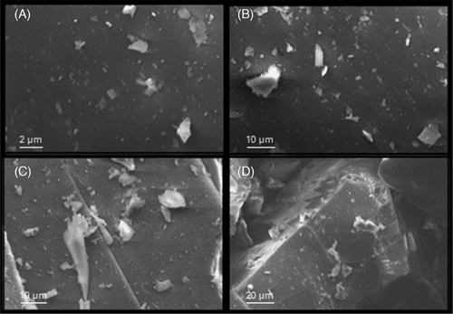
Scanning electron microscopy was carried out to obtain information on the samples' morphologies. SEM examination (Figure 1) indicates that both PEG and quercetin included in the silica matrix do not alter the morphology of the obtained hybrid composites. Indeed, both organic and inorganic phases seem to be well-homogenized, thus confirming the hybrid feature of the synthesized sol–gel materials.

2.3 Antimicrobial Properties of Silica/PEG/Quercetin Systems
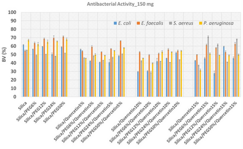
Figure 2 reports the antimicrobial activity of all the synthesized materials. The strong reduction in the bacterial viability for both Gram-positive and -negative bacteria assayed suggests the high antimicrobial activity of the hybrid materials. This is due to the combination of PEG and quercetin properties as they both possess antimicrobial activity.[13, 14]

3 Conclusions
The antimicrobial susceptibility test demonstrated the antibacterial propriety of the samples. Distinctly from each component concentration, both Gram-positive (S. aureus and E. faecalis) and Gram-negative (E. coli and P. aeruginosa) bacteria are inhibited. These results remarked that the existence of both quercetin and PEG in the silica matrix, strongly influences the antibacterial properties of the materials. Indeed, there is no linear decrease of bacterial viability by the increment of the quercetin in presence of PEG. This might be associated to the entrapment of the quercetin into the porous of the systems that could slow down the release. Finally, the FT-IR spectra detected the formation of the hybrid materials by H-bonds, while the SEM analysis established a good homogenization of the materials enclosed in the prepared composites. Despite the microbial activity, further investigations need to be conducted to assess the kinetic of quercetin release, the cytotoxicity, the biocompatibility of the synthesized hybrids, and their suitability to be used as biomaterials for tissue engineering.
4 Experimental Section
Sol–gel synthesis: Silica hybrid materials were synthesized with different percentages of PEG (0, 6, 12, 24, and 50 wt%) and quercetin (0, 5, 10, and 15 wt%) by the sol–gel technique. First, an inorganic silicate solution was obtained by adding tetraethyl orthosilicate (TEOS, Sigma–Aldrich, Darmstadt, Germany) to Ethanol (EtOH, Sigma–Aldrich) and distillate water under continuous magnetic stirring. Consequently, a solution of quercetin followed by a solution of polyethylene glycol (PEG, MW = 400, Sigma–Aldrich), both dissolved in ethanol, was added to the system. Lastly, nitric acid (<65%, Sigma–Aldrich) was added, and a clear and homogeneous solution was obtained after about 10 min of stirring. The solutions prepared were put at room temperature until the gelation process had been reached. The gelled samples were dried at 50 °C in the oven for 24 h, allowing the removal of the solvent residue.[13]
FT-IR analysis: Transmittance spectra were recorded in the range of 400–4000 cm−1 with a resolution of 2 cm−1 (60 scans), by using the Prestige21 Shimadzu system, equipped with a DTGS KBr sensor (deuterated triglycine sulfate with potassium bromide windows). The analysis procedure utilized KBr disks in which 2 mg of sample were combined with 200 mg of KBr.[15] The elaboration of the FT-IR spectra was made by IR Solution and Origin 8 software.
SEM analysis: A morphology study of the prepared samples was carried out by SEM (ZEISS EVO MA 15, EVO-ZEISS, Cambridge, UK). The samples were gold-sputtered up to a thickness of 20 nm by means of an Emitech K-550 sputter coater (Ashford Kent, UK). In order to perform the analysis, an accelerating voltage of 20.00 kV and a working distance of 15 mm was used. Scans were carried out at different magnifications ranging from 500× to 25.000×.
Antimicrobial properties: The antibacterial analysis of silica/PEG/quercetin systems was performed by Kirby–Bauer method. Both Gram-negative and Gram-positive bacteria were grown in the absence and in the presence of each sample. Specifically, Escherichia coli (ATCC 25 922) and Pseudomonas aeruginosa (ATCC 27 853) as Gram-negative bacteria and Staphylococcus aureus (ATCC 25 923) and Enterococcus faecalis (ATTC 29 212) as Gram-positive bacteria were used. For each strain, a culture medium was prepared. The sample disks (150 mg) were obtained by grounding and pressing the synthesized hybrid materials and placing them in the middle of the petri plate. Before the analysis, these disks were sterilized under UV light for 1 h.[16]
Acknowledgements
Open access publishing facilitated by Universita degli Studi della Campania Luigi Vanvitelli, as part of the Wiley - CRUI-CARE agreement.
Conflict of Interest
The authors declare no conflict of interest.
Author Contributions
The authors have contributed equally to the work.
Open Research
Data Availability Statement
The data presented in this study are available on request from corresponding author.


