Preparation of Polymeric Nanoparticles by Photo-Crosslinking of an Acryloylated Polyaspartamide in w/o Microemulsion
Abstract
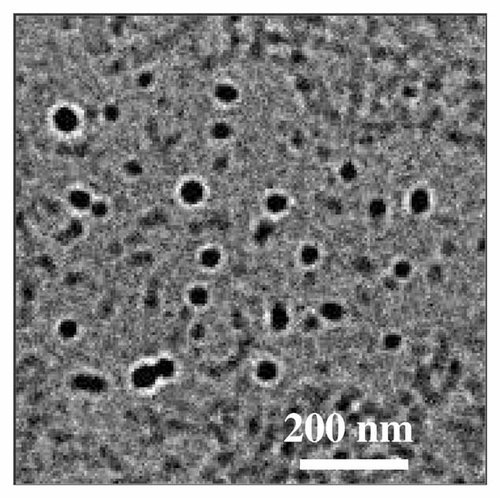
Summary: Biodegradable polymeric nanoparticles have been prepared by UV irradiation of an acryloylated water soluble polymer by an inverse microemulsion. The starting polymer was a α,β-poly(N-2-hydroxyethyl)-D,L-aspartamide (PHEA) partially functionalized with glycidyl methacrylate (GMA) in order to introduce reactive vinyl groups in the side chain. The PHEA-GMA copolymer obtained (PHG) was crosslinked by UV irradiation of the inverse microemulsion prepared by mixing an aqueous solution of PHG with propylene carbonate (PC)/ethyl acetate (EtOAc) in the presence of sorbitan trioleate (SPAN 85) as surfactant. Nanoparticles obtained were characterized by FTIR spectrophotometry, transmission electron microscopy, size distribution analysis and zeta potential measurements. Nanoparticles investigated revealed spherical and homogeneous shading, the particle size having a mean diameter of 88 ± 13 nm (PDI = 0.21) and a negative surface charge in several aqueous media. Moreover, in vitro chemical and enzymatic hydrolysis studies evidenced the partial biodegradability of PHG nanoparticles, which is more evident after incubation with enzymes such as esterases. PHG nanoparticles were loaded during UV irradiation process with Cytarabine, chosen as a model drug, and Cyt-loaded PHG nanoparticles were able to release it in a simulated physiological fluid (phosphate buffer at pH 7.4) and in blood plasma.


