Prevention of hepatitis C virus infection using a broad cross-neutralizing monoclonal antibody (AR4A) and epigallocatechin gallate
Michael Houghton was funded by the Canada Excellence in Research Chair program. Adrian Egli is supported by a Swiss National Fund (PBBSP3-130963) and a Lichtenstein Foundation grant. Mansun Law and Dennis R. Burton were supported in part by grants R01AI079031 and R01AI071084, respectively, from the National Institute for Allergy and Infectious Disease. D. L. J. Tyrrell is supported by a Canadian Institutes of Health Research grant.
Potential conflict of interest: Nothing to report.
Abstract
The anti–hepatitis C virus (HCV) activity of a novel monoclonal antibody (mAb; AR4A) and epigallocatechin gallate (EGCG) were studied in vitro using a HCV cell culture system and in vivo using a humanized liver mouse model capable of supporting HCV replication. Alone, both exhibit reliable cross-genotype HCV inhibition in vitro, and combination therapy completely prevented HCV infection. In vitro AR4A mAb (alone and combined with EGCG) robustly protects against the establishment of HCV genotype 1a infection. EGCG alone fails to reliably protect against an HCV challenge. In conclusion, AR4A mAb represents a safe and efficacious broadly neutralizing antibody against HCV applicable to strategies to safely prevent HCV reinfection following liver transplantation, and it lends further support to the concept of HCV vaccine development. The poor bioavailability of EGCG limits HCV antiviral activity in vitro. Liver Transpl 22:324–332, 2016. © 2015 AASLD.
Abbreviations
-
- DAA
-
- direct-acting antiviral
-
- Di-MeEGCG
-
- dimethylated epigallocatechin gallate
-
- EGCG
-
- epigallocatechin gallate
-
- GT
-
- genotype
-
- hAAT
-
- human alpha-1-antitrypsin
-
- HCV
-
- hepatitis C virus
-
- HCVcc
-
- cell culture–produced hepatitis C virus
-
- HIV
-
- human immunodeficiency virus
-
- IC50
-
- 50% inhibitory concentration
-
- IFN
-
- interferon
-
- IG
-
- intragastric
-
- IgG
-
- immunoglobulin G
-
- IP
-
- intraperitoneal
-
- JFH
-
- Japanese fulminant hepatitis
-
- mAb
-
- monoclonal antibody
-
- MeEGCG
-
- methylated epigallocatechin gallate
-
- MOI
-
- multiplicity of infection
-
- NS5A
-
- nonstructural protein 5A
-
- SCID/uPA
-
- severe combined immunodeficiency/albumin/urokinase plasminogen activator
-
- SEM
-
- standard error of the mean
The management of chronic hepatitis C virus (HCV) infection continues to rapidly evolve. Novel direct-acting antivirals (DAAs) capable of achieving universally high cure rates carry much promise.1 Equitable access to these agents represents a global challenge with a large proportion of those chronically infected worldwide likely to have very limited access for the considerable future.2, 3
DAAs will also feature prominently in the global resolve to eradicate HCV infection. HCV prevention, however, will remain fundamental to realizing this aspiration.3, 4 Primary prevention via vaccination has been undermined by the lack of a convenient animal model and the challenge presented by the diversity among HCV genotypes (GTs) and quasispecies.4-6 Likewise, capitalizing on the unique opportunity afforded by liver transplant to prevent HCV reinfection has been unsuccessful to date.7-9
HCV-associated liver disease (cirrhosis or hepatocellular carcinoma) is long established as the leading indication for liver transplantation worldwide.10 In those undergoing this lifesaving intervention, HCV reinfection is presently universal and drives inferior posttransplant outcomes.10-12 A window exists peritransplantation to optimize posttransplant outcomes by preventing allograft HCV infection. A precedent exists within liver transplantation whereby hepatitis B virus reinfection can be prevented after transplant.13, 14 To date, replicating this in relation to HCV has proven elusive.15
Strategies to employ existing therapies (pegylated interferon/ribavirin with or without first generation protease inhibitors) in a preemptive role before transplant or soon after transplant have been unsuccessful. The intolerability and contraindications of these agents in highly complex and medically unstable patients severely limits the applicability of such approaches.7, 8 A randomized trial using hepatitis C immune globulin concluded that this was a safe and tolerable agent in liver transplant recipients, but no beneficial effect on the rate of HCV recurrence was observed.9
Very recently, limited data in select populations employing sofosbuvir and ribavirin pretransplantation have proven the feasibility of this approach.16 Generalizing this approach to complex and often compromised patients before and after liver transplant is, however, the subject of ongoing studies.
Many monoclonal antibodies (mAbs) and polyclonal antibodies targeting linear or conformational epitopes within the HCV surface glycoprotein (E1-E2) have been described that affect HCV neutralization in vitro.17-19 E1/E2 interacts with a number of cell-surface molecules to mediate HCV entry, eg, C-type lectins, CD81, Scavenger receptor-B1.20 The in vitro performance of HCV-neutralizing antibody has been more variable.21 Law et al.22 characterized a number of antigenic regions on E2 and identified numerous human mAbs with cross-neutralizing activity. This group has identified further antigenic regions of E1/E2 and generated a human mAb (AR4A) targeting a discontinuous epitope outside the CD81 binding site on HCV E1/E2. This epitope is highly conserved across GTs, and AR4A demonstrates potent, cross-neutralizing activity in vitro.23, 24
Epigallocatechin gallate (EGCG) is the most abundant catechin present in green tea extract. Green tea extracts have a long history of safe human consumption and have many purported benefits.25, 26 Green tea extracts are safe, widely available, and inexpensive.27 Two groups independently published data reporting an in vitro antiviral effect of EGCG against HCV.28, 29 This was primarily attributed to prevention of initial attachment of the viral particle to hepatocytes. EGCG, however, also inhibited the direct cell-cell mode of HCV transmission.28 In vitro evidence of the anti-HCV effect of EGCG is lacking.
Using the HCV cell culture system and the severe combined immunodeficiency/albumin/urokinase plasminogen activator (SCID/uPA) humanized liver mouse model, we examined the anti-HCV effect of AR4A and EGCG alone and in combination. We hypothesized that combining safe, tolerable, and effective HCV therapies acting at different sites of HCV cell entry could reliably protect against HCV challenge in vivo.
MATERIALS AND METHODS
Cells and Viruses
Huh7.5 cells were maintained in complete Dulbecco's modified Eagle's medium supplemented with 10% (vol/vol) fetal bovine serum, 0.1 mM of nonessential amino acids, 100 U/mL of penicillin, and 100 mg/mL of streptomycin. Cells were incubated at 37°C in conditions of 5% CO2.30
Plasmids encoding chimeric HCV genomes representing GTs 1-6 were used to generate cell culture–derived hepatitis C virus (HCVcc) as previously described.31
Compounds
EGCG (95% purity, Sigma-Aldrich, St Louis, MO) was dissolved in double distilled H2O and stored at –20°C. Recombinant human interferon (IFN) alfa-2a (positive control) was obtained from PBL (Piscataway, NJ) and stored as instructed. Human anti-E1/E2 mAb AR4A has previously been characterized in detail.23 Murine immunoglobulin G (IgG) obtained from BD Biosciences (Franklin Lakes, NJ) was used as an isotype control antibody for in vitro assays. Mouse monoclonal anti-human CD81 antibody from BD Biosciences represented a positive antibody control. An isotype human anti–human immunodeficiency virus (HIV)–1 IgG was administered to the control group of mice during in vivo studies. All antibodies were stored at 4°C.
HCV Inhibition Assays
Ninety-six well plates were coated with polylysine (Trevigen, Gaithersburg, MD) before plating with 104 Huh7.5 cells in 100 μL of growth media and incubated overnight. Serially diluted concentrations of EGCG were added as appropriate before, simultaneous with, or after inoculation with HCVcc at a multiplicity of infection (MOI) of 0.01. For antibody-neutralization assays, the relevant antibody was serially diluted to the required concentrations and preincubated with HCVcc for 1 hour before addition to Huh7.5 cells. IFN-alfa-2a 100 IU/mL and anti-human CD81 antibody (2.5 μg/mL) served as positive controls. Murine IgG (10 μg/mL) served as an isotype antibody control. After 10 hours of incubation with HCVcc, the cells were washed to remove unbound virus and fresh media or the relevant concentration of the investigational agents was added. HCV infectivity was determined at 48 hours using nonstructural protein 5A (NS5A) immune-histostaining (mouse monoclonal NS5A antibody [9E10], Charles Rice).
Cell Viability Assay
Cell Proliferation Kit I assay (Roche, Basel, Switzerland) was performed as recommended by the manufacturer.
Animal Care
Homozygous SCID/uPA mice were kept virus and antigen free and housed in the provincial laboratory vivarium at the University of Alberta.32, 33 The University of Alberta Health Sciences Animal Welfare Committee approved experimental protocols, and animals were cared for in accordance with the 1993 guidelines of the Canadian Council on Animal Care. Mice were anesthetized for transfer of human hepatocytes via intrasplenic injection. Full ethical approval for the use of human tissue was obtained from the human research ethics board of the Faculty of Medicine, University of Alberta. Informed consent was obtained from all donors.
Human Alpha-1-Antitrypsin (hAAT) Levels
The extent and stability of human liver chimerism can be assessed by serial measurements of hAAT. Serum hAAT levels were analyzed as previously described.34 Animals with low serum hAAT levels were used in tolerability/toxicology studies; high hAAT mice were used in the HCV challenge studies.
Dosing and Administration of Agents
Anti E1/E2 mAb (AR4A)
An initial dose of 200 mg/kg was administered via intraperitoneal (IP) injection 24 hours before HCV inoculation. Prior studies have shown that this dose yields mAb serum values in excess of in vitro 90% neutralization titers.22 A further four mAb doses of 50 mg/kg were administered via IP injections at intervals of 5 days throughout the experiment. Mice in the control group received equivalent doses of an isotype antibody: human anti-HIV-1 IgG.
Epigallocatechin Gallate (EGCG)
Considering the available information on in vivo EGCG pharmacokinetics, toxicity, and efficacy, a dosing schedule of 100 mg/kg twice daily by gavage (intragastric [IG]) was employed.35 This dose was higher than that which had demonstrated efficacy in previous mouse studies. Importantly, it was also lower than doses where toxicity in mice had been observed with repeated dosing.36 Furthermore, this dosing schedule was within the range tolerated by SCID/uPA mice with reference to volume and frequency of gavage. EGCG dosing began 48 hours before HCV inoculation and continued for 14 days.
EXPERIMENTAL PROTOCOL
- Group 1: Control mice receiving isotype antibody IP injection and water IG.
- Group 2: Mice receiving EGCG IG only.
- Group 3: Mice receiving AR4A mAb IP injection only.
- Group 4: Mice receiving both AR4A IP injection and EGCG IG.
The study outline and protocol employed is illustrated in Fig. 1.
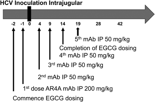
HCV inoculation was administered by intrajugular injection. The inoculum used (50 μL, 1.5 × 10⁷ IU/mL) was patient-derived HCV GT 1a serum. Blood sampling was conducted weekly by drawing 100 μL via tail bleeds for measurement of HCV titers and serum hAAT levels.
EGCG Tolerability/Toxicology
EGCG 200 mg/kg/day was administered to uninfected low hAAT level animals. Control animals received an equivalent volume of water. The health status of the animals was monitored by their general condition and weight change. At the end of the 14-day dosing period, the animals were euthanized. Cardiac puncture was performed to obtain serum for measurement of EGCG levels, and liver tissue was snap-frozen and stored at −80°C for later measurement of EGCG.
HCV RNA Quantification
Viral RNA was extracted from aliquots of mouse serum using a guanidine extraction method (Buffer AVL, Qiagen, Valencia, CA) as per the manufacturer's instructions and quantitated as described previously.34 The lower limit of quantification of this assay is 300 IU/mL.
EGCG Quantification
Plasma and liver tissue levels of EGCG were analyzed using the high-performance liquid chromatography methodology as previously published.35
Statistical Analysis


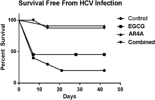
In the HCV challenge experiments, animals with HCV RNA detectable above the threshold (1000 IU/mL) by polymerase chain reaction at day 7 or thereafter were considered “infected.” Animals not reaching this threshold were “censored” and considered free from HCV infection. A Kaplan-Meier survival curve (“survival free from HCV infection”) was thus generated. Statistical significance between the groups was calculated using a 2-tailed log-rank test.
RESULTS
AR4A Demonstrates Effective HCV Neutralizing Activity In Vitro
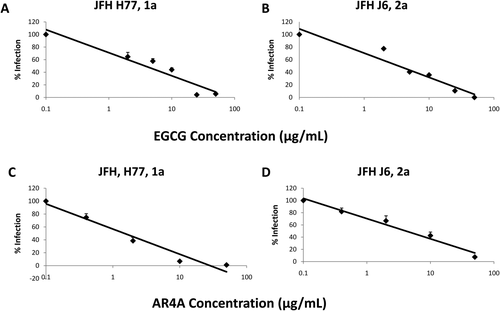
The in vitro neutralizing capability of AR4A against HCVcc expressing surface glycoproteins of GTs 1-6 was examined. Focusing on GT 1a (GT used for mouse challenge experiments) and 2a (parent Japanese fulminant hepatitis [JFH] GT), AR4A exhibited superior neutralizing activity against HCV GT 1a when compared with GT 2a, which is consistent with prior reports.23, 37 IC50 estimates of 1.28 μg/mL and 4.37 μg/mL were obtained for GTs 1a and 2a, respectively (Fig. 2C,D).
EGCG Inhibits HCV Infection In Vitro
A dose-dependent decline in HCV infection was observed when Huh7.5 cells had been simultaneously treated with various concentrations of EGCG (0-100 μg/mL; Fig. 2A,B). The EGCG IC50 was similar for HCVcc GT 1a and 2a infections (6.6 versus 5.6 μg/mL, respectively). At concentrations greater than 25 μg/mL, EGCG consistently achieved almost complete inhibition of HCVcc infection. This inhibitory effect of EGCG was independently confirmed using a firefly luciferase reporter virus, and EGCG did not negatively impact cellular viability (Fig. S1). Time of addition assays confirmed that optimal HCV inhibition was critically dependent on the presence of EGCG at the time of infection (not shown).
Additive Reduction in HCV Infection Combining AR4A and EGCG
AR4A and EGCG primarily act early in the HCV life cycle to inhibit HCV cellular attachment and entry. Using the HCVcc system, we studied the anti-HCV activity of both agents in combination.
Using GT 1a HCVcc, AR4A (2 μg/mL) combined with EGCG (10 μg/mL) demonstrated significantly increased inhibition compared to either agent alone; 88.7% inhibition compared to 61% for AR4A alone (P = 0.01), 56% for EGCG alone (Fig. 3A). Additive efficacy was again demonstrated with HCV GT 2a (Fig. 3B).

High-Dose AR4A With Low-Dose EGCG Completely Inhibits HCV Infection In Vitro
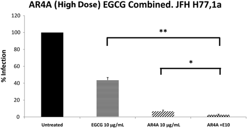
To replicate the clinical scenario whereby therapeutic antibody products are administered at high concentrations and knowing that the in vivo bioavailability of EGCG may prove to be a limiting factor, we next titrated AR4a and EGCG to identify conditions capable of completely inhibiting HCV infection. The mAb AR4A, at a concentration of 10 μg/mL, consistently achieved approximately 95% neutralization of GT 1a HCV. Even at the highest dose of AR4A used (50 μg/mL), residual infection could be detected. Despite 95% neutralization with AR4A (10 μg/mL) alone, the addition of EGCG at 10 μg/mL resulted in a significant further inhibition of HCV (P < 0.001 for AR4A alone versus combination), with complete inhibition of HCV infection attained in a number of experimental repeats (Fig. 4). The activity of AR4A (10 μg/mL) and EGCG (10 μg/mL) in combination was further assessed against JFH-1 chimeric constructs expressing the structural proteins of GTs 2–6. High titer AR4A strongly neutralized GTs 4a, 5a, and 6a, which is a finding that is consistent with prior reports.23
Animal HCV Challenge Experiments
All therapeutic regimens were well tolerated by the animals. Serum hAAT levels in mice were evenly distributed through the 4 groups and remained broadly stable throughout the experiment (Fig. S2). Two mice did not recover following the intrajugular HCV inoculation procedure: 1 animal in each of the control and combination groups. Additionally, 2 mice from the EGCG alone and the combination groups became morbid following the day 7 blood draw, and 1 animal in the AR4A alone arm was lost after day 14.
AR4A Containing Treatment Arms Robustly Protected Against HCV Infection in SCID/uPA Mice; EGCG Alone Has No Apparent Protective Effect
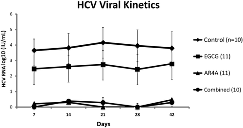
HCV infection was established in 8 of 10 control animals, with 5 progressing to high-level replication over 42 days of follow-up (Fig. S3).
AR4A demonstrated clear efficacy. Only 2/11 mice receiving AR4A alone and 1/10 mice in the combination arm had HCV RNA detected above the threshold of 1000 IU/mL throughout study follow-up. “Survival free from HCV infection” was significantly increased in AR4A-treated groups. Given the small number of events in these study groups, no difference was demonstrable between the groups receiving AR4A alone or both AR4A and EGCG.
EGCG monotherapy failed to reliably protect from HCV infection, and observed outcomes were no different from the control group. The viral kinetics and a Kaplan-Meier curve of “survival free from HCV infection” are illustrated in Figs. 5 and 6, respectively.
EGCG Levels in Plasma and Liver Tissue of Mice Following 14 Consecutive Days of Administration
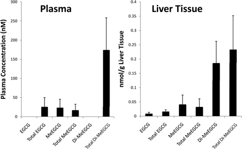
In order to assess if repeated high doses of EGCG 200 mg/kg/day were capable of achieving sufficient levels of EGCG (and its metabolites) in plasma and liver, we collected samples after 14 consecutive days of dosing. For this analysis, samples were obtained 4 hours following the final dose. Figure 7 illustrates the levels of detection of EGCG and its metabolites. The parent compound itself or its metabolites were detectable in all treated animals; however, importantly, the levels in both plasma and liver tissue were low (in the nanomolar range) when measured 4 hours after administration.
DISCUSSION
HCV-related liver disease is the leading indication for liver transplantation. Reinfection continues to drive inferior outcomes.12 Numerous unsuccessful attempts to address this discrepancy have been made. Recently, novel approaches provide some cause for optimism,16, 38 potentially shifting the paradigm whereby the treatment of HCV after liver transplantation is restricted to a delayed phase after transplantation when reinfection and histological disease have already been established.15, 39, 40
There is a need for safe, effective, and tolerable therapies that can use the window of opportunity provided by liver transplantation to prevent HCV reinfection. Complex drug-drug interactions require due consideration as does treatment durability in the face of a dynamic virus with high replicative capacity in the setting of impaired host humoral and cell-mediated immunity. Effective inhibition of HCV entry into “naïve” allograft hepatocytes represents a primary objective of preventative strategies.
In this work, we investigated agents acting primarily to inhibit HCV cell entry. Consistent with prior reports, AR4A and EGCG both reliably demonstrate cross-GT inhibition of HCV in vitro. Using these agents in combination was additive and the value of EGCG maintained significance even at high AR4A titers.
SCID/uPA mice receiving AR4A mAb by IP injection were significantly less likely to develop established infection following HCV challenge with a GT 1a inoculum. This is the first data reporting the anti-HCV efficacy of AR4A monotherapy in an animal model capable of sustaining HCV replication. AR4A clearly provides robust protection against the initial establishment of infection, demonstrating durable activity, which compares favorably with prior studies employing immunotherapy.21, 22
Despite clear in vitro inhibition, administration of EGCG alone, although well tolerated by SCID/uPA mice, did not offer protection against HCV challenge in vivo. This is the first report concerning the in vivo anti-HCV activity of EGCG. This lack of efficacy was likely driven by the low bioavailability of orally administered EGCG.41, 42 Previous studies have shown that peak plasma and liver concentrations of unconjugated EGCG in mice were 40 nmol/L and 3.5 nmol/g, respectively, following treatment with IG EGCG (75 mg/kg). EGCG undergoes extensive phase 2 metabolism, resulting in the formation of glucuronide conjugates and methylated metabolites. The levels of these metabolites have been shown in animal models to exceed the levels of the parent compound.41, 43 Studies have shown that EGCG is rapidly metabolized by catechol-O-methyltransferase resulting in the formation of methylated epigallocatechin gallate and dimethylated epigallocatechin gallate.44 These methylated metabolites have been shown to have reduced biological activity in a number of systems compared to the unmetabolized parent compound.45, 46
To date, no studies have examined the effects of methylation on the antiviral activity of EGCG. A recent study of influenza virus reported that methylated (-)-epigallocatechin has reduced inhibitory potency (IC50 = 33.4 μg/mL) compared to unmetabolized (-)-epigallocatechin (IC50 = 13.5 μg/mL) in vitro.47 Given the extensive phase 2 biotransformation of EGCG in vivo, further studies on the antiviral activity of the major metabolites are warranted.
Calland et al.48 have recently proposed a novel mechanism whereby EGCG and related natural compounds disrupt initial HCV attachment in vitro. Through an interaction with surface glycoproteins, such compounds alter the shape of HCV viral particles inhibiting efficient cellular attachment. With a proposed mechanism of action reliant on direct disruption of the initial stages of HCV cellular attachment and direct cell-cell transmission, failing to maintain adequate local concentrations will clearly limit the efficacy of EGCG in vivo.
Prior clinical studies administering passive immunotherapy to HCV patients undergoing liver transplantation have been disappointing. Polyclonal immunoglobulin failed to prevent HCV reinfection.9 Similarly, an anti-E2 mAb (HCV-AbxTL68), although effecting some decline in titers, did not prevent HCV recurrence in patients undergoing liver transplantation.49 Another group administered an anti-E2 mAb (MBL-HCV1) to 6 HCV transplant recipients. All eventually experienced reinfection with viral species harboring mutations in the target epitope.50
AR4A consistently demonstrated a robust ability to protect against HCV challenge with a patient-derived GT 1a inoculum, and its in vitro characteristics compare very favorably with those of other anti-HCV mAbs.23, 37, 51 The increased efficacy of AR4A mAb can be proposed by it uniquely targeting an epitope region abridging both E1 and E2, and containing a residue which is highly conserved across HCV species.
The SCID/uPA humanized liver mouse model is an extremely useful tool for conducting in vivo HCV studies.32, 33, 37 These mice lack an adaptive immune response, thus caution is required when translating findings to the clinical situation, considering that the preexisting host antibody bound to HCV viral particles can competitively inhibit effective neutralization by anti-HCV mAbs.52, 53 Likewise, it is possible that the native viral particle in the experimental systems used is physically different to that which circulates in association with human lipoproteins. An overestimation of efficacy could contribute to the comparatively poor activity observed in patient studies to date.9, 49, 50
Another limitation is the use of a patient-derived GT 1a inoculum only. GT 1 is the predominant HCV GT in Europe and North America; however, the immediate generalizability to individuals with HCV of diverse GTs awaiting liver transplant is somewhat limited. However, clear in vitro cross-GT activity of AR4A was demonstrated. Furthermore, the patient inoculum provided a heterologous HCV species challenge against which AR4A demonstrated impressive preventative capacity.
Despite these limitations, this study has yielded a number of important findings. We have demonstrated the proficiency of the anti-E1/E2 mAb AR4A to prevent the establishment of replicating HCV infection in vivo. On the other hand, EGCG, though an effective in vitro inhibitor, demonstrated a lack of definitive efficacy to protect SCID/uPA mice against HCV infection.
The results identify next generation anti-E1/E2 monoclonal antibodies as a potential therapeutic advance, which can constitute a primary component of future preventative approaches. The cross neutralizing ability of AR4A targeting a highly conserved epitope also provides further proof of principle that effective preformed antibody can contribute to protection against HCV challenge. A candidate vaccine has been shown to elicit such antibodies in vaccinated human volunteers, providing encouragement for demonstration of future preventive efficacy.54
To optimize the efficacy of this mAb in liver transplantation, additional agents to protect against virologic breakthrough and resistant mutants will be a prerequisite. Future clinical studies of anti-HCV mAbs incorporating novel pan-genotypic DAAs in complex transplant candidates with advanced HCV-related liver disease are required.
The application of EGCG, though eminently more available worldwide than DAAs, is limited by poor bioavailability. Further characterization of the interaction between the HCV viral particle and EGCG carries the potential to identify new agents targeting HCV cell entry.48 Moreover, capitalizing upon the unique circumstances of liver transplantation, novel applications of nontoxic compounds such as EGCG or derivatives could provide an opportunity to prime liver allografts ex vivo against HCV reinfection following implantation.
Employing therapies before or immediately at transplantation stands to deliver timely, effective, and tolerable HCV prophylactic combinations. This provides considerable optimism that in the future routine prevention of HCV reinfection after liver transplantation is in fact attainable. In achieving this goal, outcomes for patients with HCV undergoing liver transplant will be brought back in line with that of their non-HCV counterparts while ensuring optimal use of a highly valuable resource—human livers.
ACKNOWLEDGEMENTS
We thank Chelcey Dibben and Brandie Greaves for provision of expert animal care. Additional thanks to Erick Giang of The Scripps Research Institute for antibody production.


