Volume 129, Issue 5 pp. 1041-1045
Allergy/Rhinology

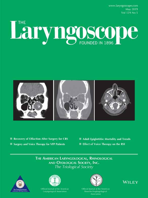
Diagnosis of a maxillary sinus fungus ball without intralesional hyperdensity on computed tomography

Che-Fang Ho MD

Che-Fang Ho MD

Division of Rhinology, Department of Otolaryngology, Chang Gung Memorial Hospital and Chang Gung University, Taoyuan, Taiwan

Department of Otolaryngology–Head and Neck Surgery, Chang Gung Memorial Hospital and Chang Gung University, Keelung, Taiwan

Search for more papers by this author
Ta-Jen Lee MD

Ta-Jen Lee MD

Division of Rhinology, Department of Otolaryngology, Chang Gung Memorial Hospital and Chang Gung University, Taoyuan, Taiwan

Search for more papers by this author
Pei-Wen Wu MD

Pei-Wen Wu MD

Division of Rhinology, Department of Otolaryngology, Chang Gung Memorial Hospital and Chang Gung University, Taoyuan, Taiwan

Department of Otolaryngology–Head and Neck Surgery, Chang Gung Memorial Hospital and Chang Gung University, Keelung, Taiwan

Search for more papers by this author
Chi-Che Huang MD

Chi-Che Huang MD

Division of Rhinology, Department of Otolaryngology, Chang Gung Memorial Hospital and Chang Gung University, Taoyuan, Taiwan

the Graduate Institute of Clinical Medical Sciences, College of Medicine, Chang Gung University, Taoyuan, Taiwan

Search for more papers by this author
Po-Hung Chang MD

Po-Hung Chang MD

Division of Rhinology, Department of Otolaryngology, Chang Gung Memorial Hospital and Chang Gung University, Taoyuan, Taiwan

the Graduate Institute of Clinical Medical Sciences, College of Medicine, Chang Gung University, Taoyuan, Taiwan

Search for more papers by this author
Yen-Lin Huang MD, PhD

Yen-Lin Huang MD, PhD

Department of Anatomic Pathology, Chang Gung Memorial Hospital and Chang Gung University, Taoyuan, Taiwan

Search for more papers by this author
Yueh-Lin Lee MD

Yueh-Lin Lee MD

Department of Radiology, Chang Gung Memorial Hospital and Chang Gung University, Keelung, Taiwan

Search for more papers by this author
Chien-Chia Huang MD

Corresponding Author

Chien-Chia Huang MD

Division of Rhinology, Department of Otolaryngology, Chang Gung Memorial Hospital and Chang Gung University, Taoyuan, Taiwan

the Graduate Institute of Clinical Medical Sciences, College of Medicine, Chang Gung University, Taoyuan, Taiwan

Send correspondence to Chien-Chia Huang, MD, Division of Rhinology, Department of Otolaryngology, Chang Gung Memorial Hospital and Chang Gung University, No. 5, Fu-Shin Street, Kweishan, Taoyuan 333, Taiwan. E-mail: [email protected]Search for more papers by this author
First published: 24 December 2018
Citations: 20
chien-chia.h. designed the study. c.-f.h., p.-w.w., y.-l.h., and y.-l.l. contributed to collection of data. chien-chia.h., t.-j.l., chi-che.h., and p.-h.c. analyzed the data. c.-f.h., and chien-chia.h. wrote the article with input from all authors.
This work was funded by a research grant from the Chang Gung Memorial Hospital (CMRPG3G0061 and CMRPG3G0671). The funder had no role in the study design, data collection, or analysis; decision to publish; or preparation of the manuscript.
The authors have no other funding, financial relationships, or conflicts of interest to disclose.

Abstract

Objectives/Hypothesis

Maxillary sinus fungus ball (MSFB) is the most common type of noninvasive fungal rhinosinusitis. Surgical removal of the ball achieves good outcomes. Making a rapid and accurate diagnosis is important to avoid unnecessary medical therapy. Intralesional hyperdensity (IH) on computed tomography (CT) is reportedly a good indicator. The aim of this study was to evaluate the diagnostic features of MSFB without IH on preoperative CT images.

Study Design

Retrospective database review.

Methods

Two hundred fifty-eight patients with histopathological evidence of a sinus fungal ball were retrospectively investigated. Forty-seven of 222 patients with MSFB did not show IH on preoperative CT images and were enrolled in the MSFB group. Forty-one patients with unilateral nonfungal chronic rhinosinusitis were enrolled in a control group. CT features previously reported to have diagnostic significance were evaluated.

Results

Sclerosis of the lateral sinus wall, erosion of the inner sinus wall, and irregular surface of the material were significantly more common in the MSFB group than in the control group. In the subgroup of patients with total opacification in the maxillary sinus, the sensitivity, specificity, and positive and negative predictive values for erosion of the inner sinus wall were more than 90%. In the subgroup with partial opacification, the sensitivity, specificity, and positive predictive value of an irregular surface of the material were more than 80%.

Conclusions

We have devised an algorithm to help diagnose MSFB without IH on preoperative CT images. Use of this algorithm would improve the diagnostic accuracy and ensure appropriate treatment.

Level of Evidence

4 Laryngoscope, 129:1041–1045, 2019

The full text of this article hosted at iucr.org is unavailable due to technical difficulties.