Isolated lung involvement in IgG4-related disease
Immunoglobulin G4 (IgG4)-related disease (IgG4-RD) is a chronic autoimmune disorder characterized by mass-forming sclerosing lesions, elevated IgG4 levels, and tissue infiltration by IgG4-positive plasma cells. While commonly affecting multiple organs, sole lung involvement in IgG4-RD, is relatively rare or underreported. We present an uncommon case of IgG4-RD with isolated lung involvement, manifesting as a pseudotumor in the patient's lung.
We report a 76-year-old Asian male with a history of spontaneous secondary pneumothorax, related to bilateral bullous emphysema, and chronic obstructive pulmonary disease (COPD). He was seen in our emergency department with acute dyspnea, general weakness, and right chest pain. Chest x-ray confirmed recurrent right-side pneumothorax, requiring emergent surgical decompression.
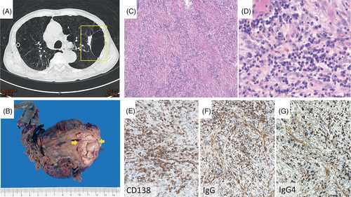
Preoperative images of contrast-enhanced chest computed tomography (CT) revealed a nodule with calcified flecks, adjacent to a cyst, raising the suspicion of lung cancer (Figure 1A). Bilateral pulmonary wedge resection was performed in order to treat recurrent pneumothorax and acquire a pathologic diagnosis of the nodule abutting the cyst (Figure 1B). Histopathological analysis revealed lung parenchyma with cystic change and a dense infiltration of lymphoplasmacytic cells, stromal fibrosis, and obliterative phlebitis (Figure 1C, D). Immunohistochemistry demonstrated 155 IgG4-positive cells per high power field (HPF) and an IgG4/IgG ratio of 55% (Figure 1E–G). Laboratory findings showed increased serum levels of IgG (1897 mg/dL; normal: 540–1822 mg/dL), IgG4 (616 mg/dL; normal: 3–201 mg/dL), and immunoglobulin E (IgE) (281 IU/mL; normal: <87 IU/mL). Complement components C3 and C4 were within normal range. The systematic survey of other organs has not shown other manifestations of IgG4-RD, solely lungs were involved. The patient was then initiated on first-line therapy with oral dexamethasone, and anti-rheumatic with oral hydroxychloroquine. Over a 3-month follow-up at rheumatology outpatient department, his IgG and IgG4 serum levels have declined, showing no signs of disease relapse.

Isolated lung IgG4-RD is rarely reported, with only less than 20 published cases thus far. Similar to previously reported cases, correctly diagnosing IgG4-related lung disease (RLD) can be challenging especially when the disease manifests as a pseudotumor. It is crucial for physicians to correctly recognize this underestimated diagnosis, to minimize excessive resection and to avoid unnecessary surgeries. The 2019 ACS/EULAR classification provides a comprehensive scoring system based on clinical, biological, imaging, and histological findings, giving us characterization of IgG4-RLD.1 Umehera et al.'s diagnostic criteria established three definitive features of IgG4-RLD: (1) CT-objectified thoracic organ involvement, (2) IgG4 levels above 135 mg/dL, and (3) IgG4+ cell infiltration >10/HPF with an IgG4+/IgG+ ratio >40%.2 Our case fulfills all three Umehera's criteria, making the diagnosis of IgG4-RLD certain.3 First-line therapy with corticosteroids is usually effective for disease control and to prevent disease relapse, while early administration of anti-rheumatic drugs remains a subject of divided consensus.4, 5
IgG4-RLD, especially sole involvement of the lungs, is a relatively rare manifestation, and is effectively treated by steroid therapy. The IgG4-RLD should be taken into a possible entity in differential diagnosis of mass-forming lung lesion, even when no other organ manifestation is clinically apparent at the time of diagnosis.
CONFLICT OF INTEREST STATEMENT
The authors declare no conflict of interest.


