MiR-451a ameliorates alcoholic hepatitis via repressing HDAC8-mediated proinflammatory response
Abstract
Alcoholic hepatitis (AH) is identified as an inflammatory syndrome with high morbidity and mortality as a result of severe hepatocellular dysfunction and liver injury. Accumulated studies indicated that miRNAs are involved in AH. The potential effect of miR-451a in AH mice was examined in the current study. A mice AH model was established and the miR-451a expression in AH mice compared with the sham group was tested by real-time polymerase chain reaction (qRT-PCR). AH mice were injected with pre-miR-451a lentivirus for miR-451a overexpression and histone deacetylase (HDAC8) lentivirus for HDAC8 overexpression in AH mice. The underlying mechanisms were explored by searching the potential target genes of miR-451a in miRanda database and then we confirmed this. We found that miR-451a expression was significantly decreased in AH mice compared with the sham group. Moreover, miR-451a overexpression alleviated alcohol-induced liver inflammation and injuries of AH mice. Additionally, further mechanism exploration disclosed that HDAC8 was a target of miR-451a. The protective effect of miR-451a on AH in AH mice was abolished by HDAC8 overexpression. In summary, miR-451a ameliorates AH via repressing HDAC8-mediated proinflammatory response.
1 INTRODUCTION
Alcoholic hepatitis (AH) is identified as an inflammatory syndrome owing to alcohol abuse that can be life-threatening.1-3 Most patients diagnosed with AH are found to be addicted to long-standing alcohol consumption.4 AH is characterized by liver inflammation resulting from hepatic neutrophil infiltration and macrophage recruitment and activation, which leads to hepatocellular dysfunction and severe liver injury.5, 6 AH can still develop even after abstaining from alcohol and it is essential to give prompt medical interventions. However, clinically available drugs like corticosteroids and pentoxifylline only have a survival rate of less than 50%.7, 8 Liver transplantation seems to be promising but this may be just available to a small minority of AH patients.9 New treatments are urgent to be developed and largely depend on clearly recognizing the disease's pathogenesis.
MicroRNAs (miRNAs) are identified as a class of 18 to 22 nucleotides noncoding short RNAs which could specifically complement and bind to the 3′-UTR of the target gene and likely to be involved in the pathogenesis of various diseases.10-12 Recent studies have shown that miRNAs are implicated in liver disease.13, 14 Ambade et al reported that AH accelerated the occurrence of early tumors by upregulating miR-122.15 Additionally, the overexpression of miR-155 aggravated liver inflammation and promoted liver fibrosis and alcohol-induced steatohepatitis in mice.16 MiR-34a could partly alleviate the liver injury caused by alcohol17 and overexpression of miR-21 was reported to be a protective factor of AH by reducing the ethanol-induced apoptosis.18 Moreover, the expression of miR-182 was increased in AH mice and inhibition of miR-182 could alleviate inflammatory response and have a protective effect on liver injury.19 Previous studies have demonstrated that miR-451a was decreased in mice with nonalcoholic steatohepatitis and overexpression of miR-451a significantly suppressed inflammation caused by fatty acids via the AMPK/AKT pathway.20 However, no studies have disclosed the potential role of miR-451a in AH. Thus, a mice AH model was established in the present study and the effect of miR-451a on AH was examined. Then the underlying mechanisms were explored by searching the miRanda database to predict the targets of miR-451a.
2 MATERIALS AND METHODS
2.1 Animals and establishment of mice AH model
C57BL/6J mice (6 weeks old) were purchased from Beijing Vital River Laboratory Animal Technology Company (Beijing, China) and fed in a standard laboratory animal center. To establish an AH model, mice were fed with 0.1% 3,5-diethoxycarbonyl-1,4-dihydrocollidine (DDC) diet for consecutive 4 weeks to induce chronic liver damage. Then the mice were sacrificed and liver tissues and blood samples were collected for the further analysis. For miR-451a overexpression or histone deacetylase (HDAC8) overexpression, AH mice were injected with pre-miR-451a lentivirus or HDAC8 lentivirus purchased from Genechem (Shanghai, China) through tail vein, respectively. There are six mice per group. All the experimental procedures and animal care were approved by the Ethics Committee of Inner Mongolia Medical University and performed according to the Guide for the Care and Use of Laboratory Animals.21
2.2 Cell culture
AML-12 cell line was used in the current study. For in-vitro miR-451a overexpression, cells were transfected with miR-451a mimics or the corresponding negative control by FuGENE (Roche Molecular Biochemicals) in AML-12 cells. Cell samples were then harvested for the further detection of mRNA and protein levels.
2.3 Immunohistochemistry and H&E staining
Immunostaining of F4/80 and myeloperoxidase (MPO) and H&E staining were performed as described before.22, 23 F4/80 antibody (1:200, #70076) and MPO antibody (1:500, #14569) used in this study were purchased from Cell Signaling Technology (Beverly, MA).
2.4 Real-time polymerase chain reaction
The miR-451a-specific forward primer sequence was designed based on the miRNA sequence according to the method previously described and qRT-PCR was performed.20, 24 U6 snRNA was used as internal control for miRNA. For HDAC8 quantification, the primer sequences used in this study are as follows: mouse HDAC8, 5′-CTGATGTTGGCCTGGGGAAA-3′ (forward) and 5′-AGGGAGTTCTGGTGAAACAGG-3′ (reverse); and GAPDH 5′-ACCCTTAAGAGGGATGCTGC-3′ (forward) and 5′-CGGGACGAGGAAACACTCTC-3′ (reverse).
2.5 Western blotting
Western blotting was performed as the protocol previously described.25 Primary antibodies used in this study include HDAC8 (SAB4502369, 1:1000, Sigma-Aldrich, St. Louis, MO) and GAPDH (#5174, 1:1000, Cell Signaling Technology).
2.6 Dual-luciferase reporter assay
To confirm HDAC8 was a target of miR-451a, dual-luciferase reporter assay was performed as the protocol similarly previously described.26
2.7 ELISA and measurement of aspartate aminotransferase, alanine aminotransferase, and lactate dehydrogenase levels
The levels of TNF-α (# KRC3011, Thermo Fisher scientific, Waltham, MA) and IL-1β (# BMS6002, Thermo Fisher scientific) in the liver tissues were detected by using an ELISA kit according to the manufacturer's instruction. Serum aspartate aminotransferase (AST), alanine aminotransferase (ALT), and lactate dehydrogenase (LDH) were detected by the kits from Nanjing Jiancheng (C010-1-1, C009-3-1, A020-1-2, Nanjing, China) according to the corresponding kit instructions.
2.8 Statistical analysis
Results (mean ± SD) were analyzed by an independent Student's t test to compare two groups, and one-way ANOVA with Bonferroni's correction was used to compare multiple groups by the GraphPad Prism 6.0 software. P < .05 was considered to be statistically significant.
3 RESULTS
3.1 MiR-451a was downregulated in AH mice
To examine the role of miR-451a in AH, an AH model was established by feeding mice with 0.1% DDC diet for consecutive 4 weeks to induce chronic liver damage. Then mice were sacrificed and liver tissues and blood samples were collected for the further analysis. Results of immunohistochemistry demonstrated that hepatic F4/80+ macrophages and the number of myelopeoxidase (MPO)+ neutrophils in the liver were significantly increased in AH mice compared with the mice in sham group and the H&E staining results demonstrated the chronic alcoholic liver injury in the AH group compared with the Sham group (Figure 1A). No significant liver steatosis was observed in the liver tissues in AH group by detection of triglyceride (TG) and total cholesterol (TC) (Figure S1A). Hepatic expression of the proinflammatory mediators including tumor necrotic factor-α (TNF-α), interleukin 1 beta (IL-1β) were also examined and data suggested that TNF-α and IL-1β were significantly higher in AH mice than the mice in sham group (TNF-α, 60.67 ± 15.91 versus 297.33 ± 39.27 pg/mL; IL-1β, 52 ± 12.41 versus 275.5 ± 49.88 pg/mL) (Figure 1B). Moreover, levels of AST (50.33 ± 11.20 versus 523.17 ± 78.75 U/L), ALT (61.83 ± 12.01 versus 212 ± 35.49 U/L), and LDH (1279.83 ± 264.04 versus 6592.83 ± 803.17 U/L) were significantly increased in AH mice than the mice in sham group (Figure 1C). Additionally, miR-451a level was significantly decreased in AH mice compared with the mice in sham group (Figure 1D). These results demonstrated that the AH model was successfully established and miR-451a was downregulated in AH mice.

3.2 MiR-451a overexpression alleviates alcohol-induced liver inflammation and injury of AH mice
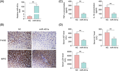
To further clarify the effect of miR-451a overexpression on AH, AH mice were injected with pre-miR-451a lentivirus through tail vein. As shown in Figure 2A, the miR-451a expression level was significantly increased in the DDC-fed mice with pre-miR-451a lentivirus injection. Hepatic F4/80+ macrophages and myelopeoxidase (MPO)+ neutrophils in the liver were significantly decreased as a result of miR-451a overexpression in AH mice (Figure 2B). MiR-451a overexpression also significantly alleviated alcohol-induced liver inflammation with decreased inflammatory cytokine such as TNF-α and IL-1β in AH mice (TNF-α, 295.00 ± 54.55 versus 100.17 ± 18.82 pg/mL; IL-1β, 258.33 ± 35.02 versus 101.33 ± 23.96 pg/mL) (Figure 2C). In addition, the AH mice with overexpressed miR-451a had lower serum AST (587.00 ± 37.98 versus 268.33 ± 36.97 U/L), ALT (239.83 ± 24.55 versus 129.00 ± 26.83 U/L), and LDH (6341.50 ± 695.35 versus 2599.67 ± 353.15 U/L) levels than those AH mice injected with negative control (NC) lentivirus (Figure 2D). These results suggested that miR-451a overexpression alleviates alcohol-induced liver inflammation and injury of AH mice.

3.3 HDAC8 is a target of miR-451a
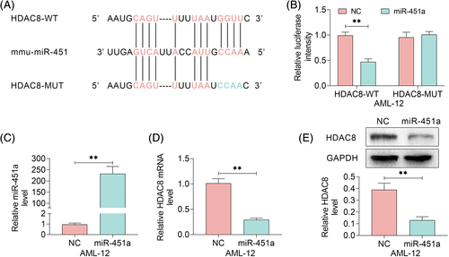
The target genes of miR-451a were predicted by searching in miRanda database. The complementary sequence of miR-451a was observed in 3′-UTR of HDAC8 mRNA (Figure 3A). Further results disclosed that luciferase activity of HDAC8 with wild-type (WT) 3′-UTR was significantly decreased owing to miR-451a overexpression by treatment of miR-451a mimics whereas HDAC8 with mutant 3′-UTR was not influenced (Figure 3B,C). Moreover, the HDAC8 mRNA and protein expression levels were detected in AML-12 cells treated with NC mimic, or miR-451a mimics. MiR-451a overexpression significantly decreased the HDAC8 expression level (Figure 3D,E and Figure S1B). These data indicated that HDAC8 is a target of miR-451a.

3.4 MiR-451a alleviates AH mice alcohol-induced liver inflammation and injury by targeting HDAC8
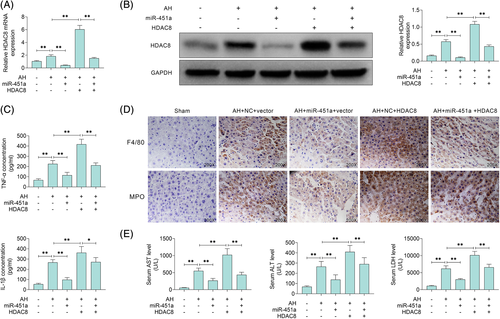
To further examine whether HDAC8 mediated the protective effect of miR-451a in AH mice, AH mice were administrated with pre-miR-451a lentivirus or lentivirus of HDAC8 plasmids through tail vein for the overexpression of miR-451a or HDAC8, respectively (Figure 4A,B). We found that HDAC8 overexpression in AH mice significantly exacerbated the liver inflammation with increased TNF-α (67.5 ± 12.82, 225.50 ± 32.65, 114.67 ± 28.40, 417.67 ± 49.01, 211.33 ± 24.34 pg/mL in the indicated groups) and IL-1β (54.167 ± 9.28, 266.67 ± 27.12, 97.17 ± 22.06, 362.00 ± 60.67, 271.17 ± 43.13 pg/mL in the indicated groups) whereas this effect was reversed by miR-451a overexpression (Figure 4C). Moreover, consistent changes of hepatic F4/80+ macrophages and myelopeoxidase (MPO)+ neutrophils in the liver, in addition to serum AST (56.33 ± 6.68, 554.00 ± 80.04, 268.33 ± 63.24, 1022.67 ± 183.09, 438.67 ± 74.39 U/L in the indicated groups), ALT (69.00 ± 9.61, 265.50 ± 51.04, 138.50 ± 46.89, 410.00 ± 62.50, 291.00 ± 62.29 U/L in the indicated groups), and LDH (1128.67 ± 127.15, 6187.67 ± 830.72, 2989.00 ± 283.82, 10 126.83 ± 1129.78, 6588.83 ± 887.42 U/L in the indicated groups) levels, were observed when AH mice were injected with pre-miR-451a lentivirus or lentivirus of HDAC8 plasmids (Figure 4D,E). Taken together, miR-451a alleviates AH mice alcohol-induced liver inflammation and injury by targeting HDAC8.

4 DISCUSSION
Alcoholic hepatitis is an inflammatory syndrome characterized by high morbidity and mortality as a result of severe hepatocellular dysfunction and liver injury.3, 5 Liver inflammation owing to hepatic neutrophil infiltration and macrophage recruitment and activation leads to severe liver damage.27 Emerging studies have reported that the pathophysiology of AH is related to varieties of miRNAs including miR-122, miR-155, miR-34a, miR-182, and so on,15-17, 19 and targeting miRNAs was confirmed to be a potential therapeutic strategy. In the present study, miR-451a was proved to be involved in AH with decreased expression in AH mice. MiR-451a overexpression alleviates alcohol-induced liver inflammation and injury of AH mice. Additionally, HDAC8 was found to be a potential target of miR-451a by searching in the miRanda database and further studies clarified that HDAC8 overexpression abolished the protective effect of miR-451a on alcohol-induced liver inflammation and injury in AH mice.
Accumulating evidence have reported that miR-451a has a significant suppressive effect on the inflammation. Previous studies suggested that miR-451 acted as an anti-inflammatory factor by inhibiting the NF-κB signaling pathway in diabetic nephropathy.28 MiR-451 inhibited p38 MAPK signaling pathway and suppressed proinflammatory cytokines production in rheumatoid arthritis.29 Macrophage migration inhibitory factor has been identified as a proinflammatory cytokine and miR-451a could directly downregulate its expression in psychiatric symptoms.30 Additionally, miR-451a, as an immune suppressive microRNA, inhibited proinflammatory cytokine secretion by targeting 14-3-3.31 Moreover, miR-451a negatively regulate inflammatory cytokine secretion including IL-8 and TNF-α in nonalcoholic steatohepatitis through the AMPK/AKT pathway and miR-451 has potential therapeutic applications for preventing the progression of nonalcoholic steatohepatitis.20 In our study, miR-451a overexpression suppressed the alcohol-induced liver inflammation and alleviates liver injury of AH mice.
Histone deacetylases (HDACs) are involved in various cellular processes by removal the acetyl group from lysine residues of target proteins.32-34 Small-molecule inhibitors of HDACs are utilized to treat cancer in clinic by inhibiting cancer cell proliferation and inducing apoptosis.35 In addition, HDAC inhibitors have been proved to be a potential therapeutic via inhibiting proinflammatory cytokines production in vivo and in vitro.36 In the present study, the expression of HDAC8 was increased in AH mice and HDAC8 overexpression exacerbated the inflammatory response and liver injury. MiR-451a overexpression downregulated HDAC8 and exerted a protective effect on the alcohol-induced hepatic dysfunction and liver damage through suppressing the secretion of TNF-α and IL-β. Epigenetic abnormalities were involved in cancer initiation and progression, posttranslational modifications of histones regulate chromatin remodeling, and downstream gene expression.37 In this study, HDAC8 was identified as a downward target and overexpressed HDAC8 aggravated AH in mice. However, it is not clearly clarified how does HDAC8 work in AH and the target genes of HDAC8 has not been elucidated in this article. Taken together, the anti-inflammatory effect of miR-451a in AH mice was verified in our study. Moreover, miR-451a could be a promising target to alleviate the AH by inhibiting inflammatory response induced by alcohol.
CONFLICT OF INTEREST
The authors declare no potential conflict of interests.


