Sonography of Dermatologic Emergencies
Abstract
Dermatologic conditions may be the subjects of potential emergency consultations, and the knowledge of their sonographic appearance can facilitate an early diagnosis and management. In this pictorial essay, the sonographic dermatologic anatomy, technique, and conditions that can be supported by a prompt sonographic diagnosis are reviewed. The sonographic signs that may help diagnose these entities are discussed with a practical approach.
Nowadays, sonography is a technique available worldwide that has found applications in almost all medical specialties. Furthermore, sonography is currently used at all levels, from medical students to subspecialties.1 It can support treatment of a wide variety of conditions ranging from the detection of appendicitis2 to the resection of a brain tumor.3 Moreover, emergency departments in several countries have fast access to sonography, a field of ultrasound imaging that has been called point-of-care ultrasound.4
There are also dermatologic conditions that may generate emergency consultations, some of them due to the sudden appearance or abrupt change in the clinical appearance or size of a dermatologic condition or the addition or worsening of symptoms such as cutaneous or ungual pain, drainage, ulceration, and edema. The aim of this pictorial is to provide an idea of the dermatologic entities that may be the subject of emergency consultations and can be supported by the prompt use of sonography. The requirements and the sonographic technique as well as the normal anatomy of the skin, nail, and hair are also briefly reviewed.
Requirements and Examination Technique
Multichannel color Doppler ultrasound equipment with high- and variable-frequency transducers working with upper frequencies of 15 MHz or higher are recommended. A copious amount of gel is applied to the surface of the lesion, and a sonographic sweep is performed. Side-to-side or lesional-perilesional comparisons may support the examination. The acquisition sequence starts with a grayscale evaluation, then color Doppler imaging, and, last, a spectral curve analysis of the regional vessels. All lesions are examined in at least two perpendicular axes, and extended fields of view may also help show the extent. Three-dimensional reconstructions are considered optional; however, they may provide a more understandable view of the conditions to physicians who are not familiar with this technique.5
The cases that are shown here were extracted from the database of the Institute of Diagnostic Imaging and Research of the Skin and Soft Tissues. All were examined by the same radiologist using the same equipment (LOGIQ E9, XD Clear, GE Healthcare, Milwaukee, WI) with variable linear and compact linear high-frequency transducers with upper frequency ranges of 16 and 18 MHz. All examinations followed the Helsinki principles of medical ethics.
Normal Sonographic Anatomy of the Skin, Nail, and Hair
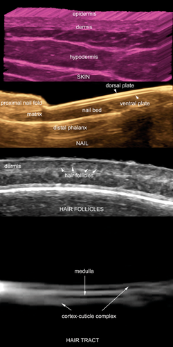
The upper layer of the skin is called the epidermis and shows in nonglabrous skin (ie, all but the palms and soles), as a hyperechoic monolaminar layer. In glabrous skin (ie, palms and soles), the epidermis presents a bilaminar appearance. The morphology of the epidermis is mainly due to its keratin content. The next lower layer is the dermis, which appears as a hyperechoic band that is less bright than the epidermis. The echogenicity of the dermis is due to its collagen content. The next layer, the hypodermis, appears as a hypoechoic layer due to the presence of fatty tissue with hyperechoic linear fibrous septa in between the fat.
The upper dermis in adults may show lower echogenicity due to deposits of glycosaminoglycans produced by photoaging, something that has been called the subepidermal low-echogenicity band.
The nails show a hypoechoic ungual bed that may vary to slightly hyperechoic at the proximal region. The ungual plate appears as a bilaminar hyperechoic structure with two parallel lines called the dorsal (external) and ventral (internal) plates.
Underneath the nail bed, the hyperechoic line of the cortex of the distal phalanx is seen. The periungual tissue, referred to as the proximal and lateral nail folds, is seen as a nonglabrous type of skin, albeit lacking subcutaneous fat. However, the pulp of the finger, which is close to the lateral nail folds, contains prominent fatty tissue.
The hair has two parts: the follicle that is located in the dermis and the tracts or shafts that correspond to the visible part. The hair follicle appears as a hypoechoic oblique band in the dermis, which may reach down to the upper hypodermis. The hair tract can appear as a trilaminar or bilaminar hyperechoic structure. Eighty percent of the hair tracts of the scalp have a trilaminar appearance with two layers of a cortex-cuticle complex and a central layer of medulla; the rest shows up as bilaminar. This bilaminar hair corresponds to a villous type without the medullar part and is also seen in the rest of the body. At all sites, hypodermal or subungual low-velocity arterial or venous blood flow is usually detected (Figure 1).5, 6
Dermatologic Emergencies on Sonography
For academic purposes, the dermatologic conditions are separated into skin, nail, and scalp entities.
Skin
Ruptured Epidermal Cysts
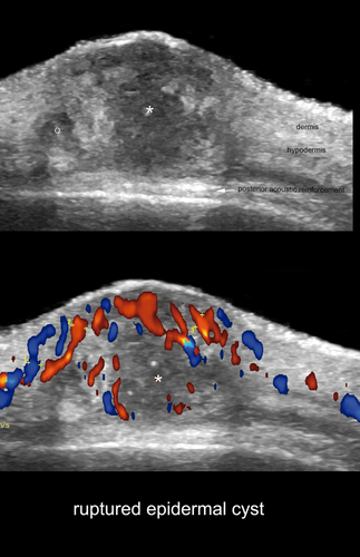
Epidermal cysts are common dermatologic lesions and have a keratin content. Their echogenicity varies according to the phase of the cyst (ie, intact, inflamed, partially ruptured, or totally ruptured). In the presence of rupture, the usually well-defined anechoic or hypoechoic round or oval structure located in the dermis or hypodermis turns into a partially or totally poorly defined or lobulated hypoechoic or heterogeneous structure. Whatever the stage and appearance of the cyst, a posterior acoustic reinforcement artifact is usually present and may support the diagnosis. In some cases, it is possible to detect hypoechoic sites of leakage of keratin at the periphery of the cyst, which usually cause a foreign body–like type of reaction with increased echogenicity of the hypodermis. Increased vascularity at the periphery of the cyst is also a frequent finding due to the inflammation (Figure 2).5-7
Plantar Warts
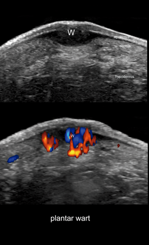
These are caused by the human papilloma virus and usually cause pain in the plantar region. The differential diagnoses are callouses (keratomas), foreign bodies, and Morton neuromas. The sonographic appearance of plantar warts is characterized by a hypoechoic fusiform structure involving the epidermis and dermis. Color Doppler imaging may support the differential diagnosis, and commonly, there is increased blood flow in the dermal part of the wart, particularly in patients with painful warts.8 In 54% of cases, plantar bursitis can be detected underneath the wart (Figure 3).9
Panniculitis
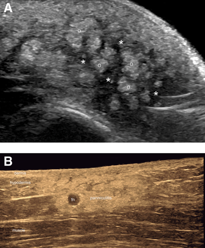
This condition is an inflammatory process of the fatty tissue of the hypodermis. It can be separated into lobular, septal, and mixed types according to the main location of the inflammatory components. Rarely, panniculitis is pure in its involvement, and it usually affects both the fatty lobules and septa; however, the purpose of sonography is to identify the predominant location of the involvement. Examples of mainly lobular panniculitis are found in lupus and neonatal fat necrosis. The most common form of mainly septal panniculitis is erythema nodosum, which mainly affects the anterior part of the legs. On sonography, lobular panniculitis appears as a diffuse increase in echogenicity of the fatty lobules. In septal panniculitis, there is hypoechoic thickening of the septa between the hyperechoic fatty lobules. Increased hypodermal vascularity can be also detected in all forms (Figure 4).10, 11
Hidradenitis Suppurativa
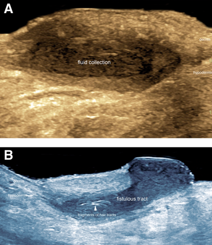
This inflammatory disease seems to originate in the hair follicle and commonly affects intertriginous regions such as the axillary and groin areas. Clinically, it shows draining fluid collections and fistulous tracts. Moreover, the fistula may be multiple and communicating in the same region. On sonography, the fluid collections appear as anechoic or hypoechoic saclike structures connected to the widened base of the hair follicles. Fistulous tracts appear as anechoic or hypoechoic bandlike structures communicating with the dilated hair follicles. Commonly, retained hyperechoic bilaminar or monolaminar fragments of hair tracts are detected within the fluid collections and fistulous tracts. Increased vascularity in the periphery of or within these structures can also be found (Figure 5).12-14
Ulcerated Hemangiomas
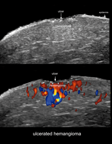
Hemangiomas are the most common benign tumors in infancy. These have a classic fast postnatal growth period called the proliferative phase, and then they start a period of slow regression. During the proliferative phase, the growth of the neovessels in the dermis can be so prominent that they may erode the epidermis and cause ulceration. Among the complications of infantile hemangiomas, ulceration is the most common; it occurs in approximately 15% to 25% of cases and can produce bleeding in 40% with a subsequent scar.15, 16 Preterm neonates have been reported to have a higher risk of ulceration in hemangiomas.17 On sonography, there is an abrupt loss of the hyperechoic line of the epidermis and poorly defined decreased echogenicity of the dermis, increased echogenicity of the hypodermis, or both. On color Doppler imaging, prominent vascularity with arterial and venous vessels is usually detected (Figure 6 and Video 1).
Thrombosed Vascular Malformations
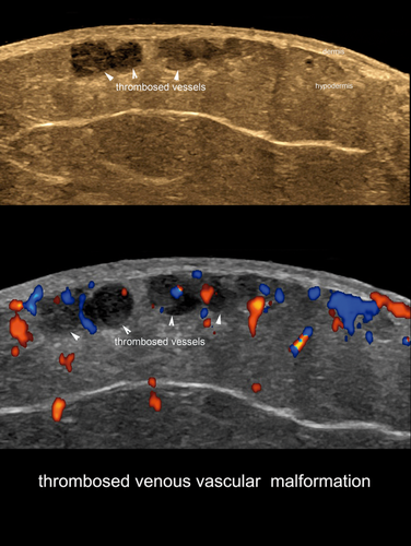
Vascular malformations are errors of morphogenesis that have abnormal proportions of vessels with normal epithelium. According to the type of vessel present in the malformation, they can be classified as arterial, venous, capillary, lymphatic, or mixed. Also, according to the velocity of the flow, they can be named high flow (arterial or arteriovenous) or low flow (venous, lymphatic, capillary, or a mixture of the latter subtypes). According to our experience, a low-flow venous malformation is the most common type that shows thrombosis. On sonography, the anechoic tubules or lacunar areas become filled with hypoechoic material and lose their compressibility and flow. Deep venous thrombosis as a complication of percutaneous sclerotherapy of vascular malformations has been reported to occur in 4% of cases (Figure 7).18
Myiasis
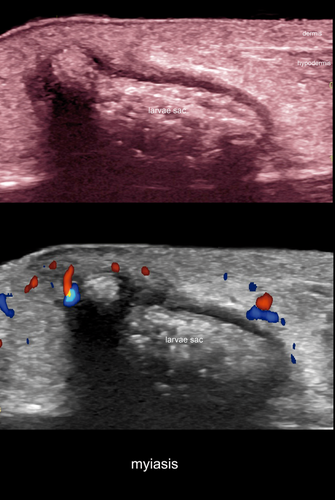
This parasitic condition is caused by infestation of skin by a fly larva. The most common type in Central and South America is Dermatobia hominis; however, other types have been reported, such as Cordylobia anthropophaga from Africa. On sonography, it frequently appears as an oval structure with a hypoechoic rim and hyperechoic center with spontaneous movement and peripheral blood flow (Figure 8 and Video 2).19-21
Mycetomas
These are chronic granulomatous infections of the dermis, hypodermis, or both. Depending on their cause, they can be separated into actinomycetomas (filamentous bacteria) or eumycetomas (fungus). These infections are more common in tropical countries and rural regions and commonly affect the lower limbs, particularly the feet.22 On sonography, they appear as dermal or hypodermal hypoechoic focal areas or tracts with or without communication between each other and sometimes with increased vascularity in the periphery. Dispersed and single hyperechoic dots within round hypoechoic structures, also called “dot-in-circle” signs, have also been reported on sonography and magnetic resonance imaging (Figure 9).23
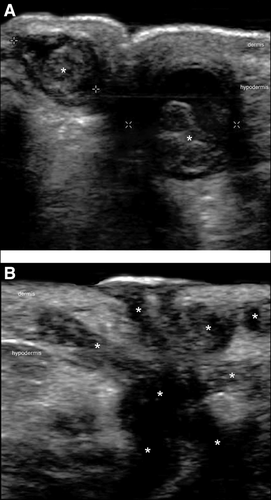
Filler Complications
Filler agents are used for treating wrinkles or sagging skin. The sudden appearance of facial erythema, edema, lumps, or bumps may cause an emergency consultation. Also, entrance of these exogenous materials into the bloodstream during the injection procedure may cause acute thrombosis, which could be critical in the face. On sonography, the most common forms of cosmetic fillers can be identified. For example, hyaluronic acid shows up as anechoic or hypoechoic round or oval pseudocystic structures, and polymethilmetacrilate is hyperechoic and shows a posterior mini–comet tail artifact. Calcium hydroxyapatite is hyperechoic and produces a posterior acoustic shadowing artifact. Although silicone oil is not approved by the US Food and Drug Administration as a cosmetic filler, it may be detected in several countries. It appears as hyperechoic deposits with a posterior reverberance that produces a “snowstorm” artifact.24, 25 When there are suspicions of thrombosis, a color Doppler examination of the vessels that feed the face should be performed, particularly the facial, angular, alar nasal and labial arteries. In our experience, most of the thrombotic episodes affect small dermal vessels and cause hypovascular regions in the skin. Sonography may also support the percutaneous injection of hyaluronidase for dissolving hyaluronic acid deposits (Figure 10).26
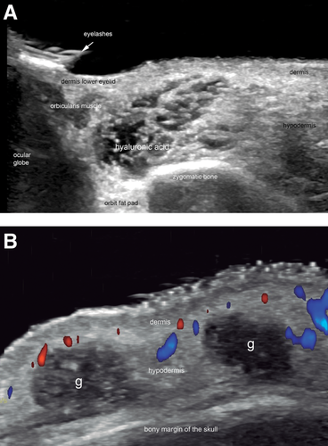
Nail
Subungual Abscesses
These appear as an anechoic fluid collection in the nail bed that may contain hyperechoic spots due to air bubbles. Increased vascularity may be observed in the periphery of the fluid collection (Figure 11).27
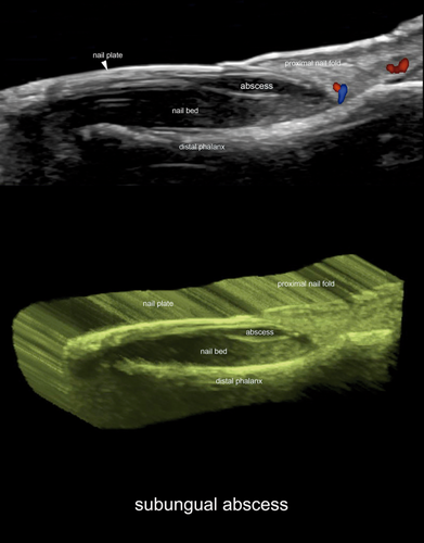
Glomus Tumors
The nail bed is the most common location for these benign tumors derived from the neuromyoarterial plexus. Clinically, they produce exquisite pain, commonly aggravated by cold. On sonography, the most common form of presentation of glomus tumors is a single well-defined hypervascular hypoechoic nodule that produces scalloping of the bony margin of the distal phalanx (Figure 12).27-29
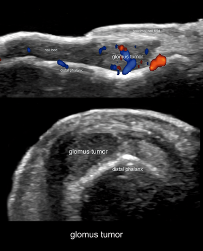
Onychocryptosis
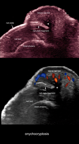
Also called ingrowing nail, this condition is the embedding of the nail plate into the periungual tissues. It commonly affects the big toes and may be related to congenital or acquired malalignment of the nail. On sonography, its appears as a hyperechoic bilaminar fragment in the periungual nail fold. Hypoechoic inflamed granulomatous dermal tissue as well as hypervascularity with low-flow vessels usually surround the nail plate fragment (Figure 13).27
Scalp and Hair
Dissecting Cellulitis of the Scalp
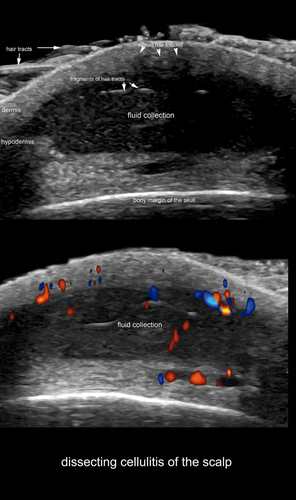
Also called “perifolliculitis capitis abscedens et suffodiens,” this condition clinically produces focal or patchy alopecia, palpable and draining lumps, as well as scarring in the scalp. Draining of aseptic or purulent material may be observed and may cause an emergency consultation. On sonography, it appears as single or multiple dermal or hypodermal anechoic or hypoechoic fluid collections, fistulous tracts in the scalp region, or both, some of them communicating between each other and connected to the base of widened hair follicles. Hyperechoic fragments of hair tracts may be detected within the structures.30 These lesions resemble the alterations described for hidradenitis suppurativa and perhaps correspond to a presentation variant of hidradenitis (Figure 14).13, 14
Conclusions
There are dermatologic lesions that may generate an emergency consultation and can be studied with sonography. Awareness of the sonographic appearance of these conditions may support early diagnosis and management in these entities.


