Volume 56, Issue 1 pp. 121-133
Research Article

In Vivo Absolute Metabolite Quantification Using a Multiplexed ERETIC-RX Array Coil for Whole-Brain MR Spectroscopic Imaging

Bijaya Thapa PhD

Bijaya Thapa PhD

A. A. Martinos Center for Biomedical Imaging, Department of Radiology, Massachusetts General Hospital, Charlestown, Massachusetts, USA

Harvard Medical School, Boston, Massachusetts, USA

Search for more papers by this author
Azma Mareyam MS

Azma Mareyam MS

A. A. Martinos Center for Biomedical Imaging, Department of Radiology, Massachusetts General Hospital, Charlestown, Massachusetts, USA

Search for more papers by this author
Jason Stockmann PhD

Jason Stockmann PhD

A. A. Martinos Center for Biomedical Imaging, Department of Radiology, Massachusetts General Hospital, Charlestown, Massachusetts, USA

Harvard Medical School, Boston, Massachusetts, USA

Search for more papers by this author
Bernhard Strasser PhD

Bernhard Strasser PhD

A. A. Martinos Center for Biomedical Imaging, Department of Radiology, Massachusetts General Hospital, Charlestown, Massachusetts, USA

Harvard Medical School, Boston, Massachusetts, USA

Search for more papers by this author
Boris Keil PhD

Boris Keil PhD

Institute of Medical Physics and Radiation Protection, TH-Mittelhessen University of Applied Sciences (THM), Giessen, Germany

Search for more papers by this author
Philipp Hoecht PhD

Philipp Hoecht PhD

Siemens Healthcare GmbH, Erlangen, Germany

Search for more papers by this author
Stefan A. Carp PhD

Stefan A. Carp PhD

A. A. Martinos Center for Biomedical Imaging, Department of Radiology, Massachusetts General Hospital, Charlestown, Massachusetts, USA

Harvard Medical School, Boston, Massachusetts, USA

Search for more papers by this author
Xianqi Li PhD

Xianqi Li PhD

A. A. Martinos Center for Biomedical Imaging, Department of Radiology, Massachusetts General Hospital, Charlestown, Massachusetts, USA

Harvard Medical School, Boston, Massachusetts, USA

Search for more papers by this author
Zhe Wang PhD

Zhe Wang PhD

Siemens Medical Solutions USA, Boston, Massachusetts, USA

Search for more papers by this author
Yulin V. Chang PhD

Yulin V. Chang PhD

Siemens Medical Solutions USA, Boston, Massachusetts, USA

Search for more papers by this author
Jorg Dietrich MD, PhD

Jorg Dietrich MD, PhD

Harvard Medical School, Boston, Massachusetts, USA

Division of Neuro-Oncology, Department of Neurology, Massachusetts General Hospital, Boston, Massachusetts, USA

Search for more papers by this author
Erik Uhlmann MD

Erik Uhlmann MD

Harvard Medical School, Boston, Massachusetts, USA

Department of Neurology, Beth Israel Deaconess Medical Center, Boston, Massachusetts, USA

Search for more papers by this author
Daniel P. Cahill MD, PhD

Daniel P. Cahill MD, PhD

Harvard Medical School, Boston, Massachusetts, USA

Department of Neurosurgery, Massachusetts General Hospital, Boston, Massachusetts, USA

Search for more papers by this author
Tracy Batchelor MD

Tracy Batchelor MD

Harvard Medical School, Boston, Massachusetts, USA

Department of Neurology, Brigham's and Women Hospital, Boston, Massachusetts, USA

Dana Farber Cancer Institute, Boston, Massachusetts, USA

Search for more papers by this author
Lawrence Wald PhD

Lawrence Wald PhD

A. A. Martinos Center for Biomedical Imaging, Department of Radiology, Massachusetts General Hospital, Charlestown, Massachusetts, USA

Harvard Medical School, Boston, Massachusetts, USA

Search for more papers by this author
Ovidiu C. Andronesi MD, PhD

Corresponding Author

Ovidiu C. Andronesi MD, PhD

A. A. Martinos Center for Biomedical Imaging, Department of Radiology, Massachusetts General Hospital, Charlestown, Massachusetts, USA

Harvard Medical School, Boston, Massachusetts, USA

Address reprint requests to: O.C.A., Suite 2301, Building 149, 13th Street, Charlestown, MA, USA. E-mail [email protected]

Search for more papers by this author
First published: 27 December 2021
[Correction added on 07 January 2022, after first online publication: Middle initial of author Stefan A. Carp has been added in this version.]

Abstract

Background

Absolute quantification of metabolites in MR spectroscopic imaging (MRSI) requires a stable reference signal of known concentration. The Electronic REference To access In vivo Concentrations (ERETIC) has shown great promise but has not been applied in patients and 3D MRSI. ERETIC hardware has not been integrated with receive arrays due to technical challenges, such as coil combination and unwanted coupling between multiple ERETIC and receive channels, for which we developed mitigation strategies.

Purpose

To develop absolute quantification for whole-brain MRSI in glioma patients.

Study Type

Prospective.

Population

Five healthy volunteers and three patients with isocitrate dehydrogenase mutant glioma (27% female). Calibration and coil loading phantoms.

Field Strength/Sequence

A 3 T; Adiabatic spin-echo spiral 3D MRSI with real-time motion correction, Fluid Attenuated Inversion Recovery (FLAIR), Gradient Recalled Echo (GRE), Multi-echo Magnetization Prepared Rapid Acquisition of Gradient Echo (MEMPRAGE).

Assessment

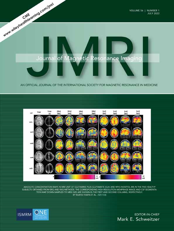
Absolute quantification was performed for five brain metabolites (total N-acetyl-aspartate [NAA]/creatine/choline, glutamine + glutamate, myo-inositol) and the oncometabolite 2-hydroxyglutarate using a custom-built 4x-ERETIC/8x-receive array coil. Metabolite quantification was performed with both EREIC and internal water reference methods. ERETIC signal was transmitted via optical link and used to correct coil loading. Inductive and radiative coupling between ERETIC and receive channels were measured.

Statistical Tests

ERETIC and internal water methods for metabolite quantification were compared using Bland–Altman (BA) analysis and the nonparametric Mann–Whitney test. P < 0.05 was considered statistically significant.

Results

ERETIC could be integrated in receive arrays and inductive coupling dominated (5–886 times) radiative coupling. Phantoms show proportional scaling of the ERETIC signal with coil loading. The BA analysis demonstrated very good agreement (3.3% ± 1.6%) in healthy volunteers, while there was a large difference (36.1% ± 3.8%) in glioma tumors between metabolite concentrations by ERETIC and internal water quantification.

Conclusion

Our results indicate that ERETIC integrated with receive arrays and whole-brain MRSI is feasible for brain metabolites quantification. Further validation is required to probe that ERETIC provides more accurate metabolite concentration in glioma patients.

Evidence Level

2

Technical Efficacy

Stage 1

The full text of this article hosted at iucr.org is unavailable due to technical difficulties.