TGFβ Inducible Early Gene-1 Plays an Important Role in Mediating Estrogen Signaling in the Skeleton
ABSTRACT
TGFβ Inducible Early Gene-1 (TIEG1) knockout (KO) mice display a sex-specific osteopenic phenotype characterized by low bone mineral density, bone mineral content, and overall loss of bone strength in female mice. We, therefore, speculated that loss of TIEG1 expression would impair the actions of estrogen on bone in female mice. To test this hypothesis, we employed an ovariectomy (OVX) and estrogen replacement model system to comprehensively analyze the role of TIEG1 in mediating estrogen signaling in bone at the tissue, cell, and biochemical level. Dual-energy X-ray absorptiometry (DXA), peripheral quantitative computed tomography (pQCT), and micro-CT analyses revealed that loss of TIEG1 expression diminished the effects of estrogen throughout the skeleton and within multiple bone compartments. Estrogen exposure also led to reductions in bone formation rates and mineralizing perimeter in wild-type mice with little to no effects on these parameters in TIEG1 KO mice. Osteoclast perimeter per bone perimeter and resorptive activity as determined by serum levels of CTX-1 were differentially regulated after estrogen treatment in TIEG1 KO mice compared with wild-type littermates. No significant differences were detected in serum levels of P1NP between wild-type and TIEG1 KO mice. Taken together, these data implicate an important role for TIEG1 in mediating estrogen signaling throughout the mouse skeleton and suggest that defects in this pathway are likely to contribute to the sex-specific osteopenic phenotype observed in female TIEG1 KO mice. © 2014 American Society for Bone and Mineral Research.
Introduction
TGFβ Inducible Early Gene-1 (TIEG1) is a Krüppel-like transcription factor (KLF10), which was originally identified by our laboratory in human osteoblasts.1 Since its discovery, we and others have demonstrated that TIEG1 is expressed in a variety of cell and tissue types,2, 3 directly regulates the TGFβ signaling pathway,4, 5 functions as a tumor suppressor in cancer cells by inhibiting proliferation and inducing apoptosis,6-9 and regulates a multitude of genes and biological processes.3, 10 Importantly, allelic variations in the TIEG1 gene,11 and altered TIEG1 expression levels,12 have been identified in patients with osteoporosis.
Through the development of knockout (KO) mice, we have elucidated essential roles for TIEG1 in regulating bone homeostasis and maintenance.13 Specifically, calvarial osteoblasts derived from TIEG1 KO mice display decreased expression of important osteoblast marker genes including Runx2 and Osterix and exhibit reduced and delayed mineralization relative to wild-type controls.13, 14 TIEG1 KO osteoblasts are also unable to fully support osteoclast differentiation in vitro,13 likely because of the decreased ratio of receptor activator of NF-κB ligand (RANKL) to osteoprotegerin (OPG) produced by these cells.13, 15 Osteoclasts isolated from TIEG1 KO mice similarly exhibit delayed differentiation, which appears to be explained in part by defects in RANKL and NFATc1 signaling.16
Detailed examination of the TIEG1 KO mouse skeleton has revealed a sex-specific osteopenic phenotype characterized by decreased bone mineral density, bone mineral content, and bone mineral area only in female animals compared with wild-type littermates.17, 18 Three-point bending tests demonstrated that the femurs of female KO mice have reduced strength and increased flexibility compared with wild-type controls,18 whereas histomorphometric analyses indicated decreased cancellous bone area and trabecular number.18 At the cellular level, TIEG1 KO mice also exhibit reduced bone formation rates compared with wild-type littermates,18 as well as decreased numbers of osteocytes embedded within the bone matrix.17, 19 Based on the fact that these defects are only observed in female animals, a potential role for estrogen in mediating this phenotype was suspected.
Estrogen is known to play an essential role in regulating bone turnover and skeletal homeostasis,20 and it has long been understood that loss of estrogen action leads to bone loss and the development of osteoporosis in women.21 Estrogen replacement therapies have been shown to increase bone mineral density, decrease bone turnover,22, 23 and reduce the risk of fractures24, 25 in postmenopausal women and are therefore indicated for the prevention of osteoporosis. Although estrogen has been established as a predominant factor in maintaining skeletal health, the exact mechanisms by which it functions have yet to be fully understood. Elucidation of the specific biological pathways, molecular mechanisms, and individual genes that are necessary for mediating the estrogenic effects on the skeleton will undoubtedly improve our ability to therapeutically modulate these processes and treat bone diseases.
The aim of the present study was to examine the role of TIEG1 in mediating the effects of estrogen on the mouse skeleton and determine if deficient estrogen signaling contributes to the female-specific osteopenic phenotype observed in TIEG1 KO mice. Here, we detail the effects of ovariectomy (OVX) and estrogen replacement on the skeletons of wild-type and TIEG1 KO mice through the use of dual-energy X-ray absorptiometry (DXA), peripheral quantitative computed tomography (pQCT), micro-CT, and histomorphometric analyses. Additionally, we examine alterations in biochemical markers of bone formation and turnover in response to OVX and estrogen replacement. The outcomes of these studies have revealed that loss of TIEG1 expression significantly diminishes the effects of estrogen on bone and suggests that defects in the estrogen signaling pathway contribute to the low bone mass phenotype of female TIEG1 KO mice.
Materials and Methods
Animals and study design
The generation and characterization of TIEG1 KO mice has been described previously.13 For this study, wild-type and TIEG1 KO littermates on a congenic C57BL/6 background were utilized.18 At approximately 5 weeks of age, female wild-type and TIEG1 KO mice underwent either sham or OVX surgeries. Surgeries were conducted at 5 weeks of age to reduce the effects of endogenous estrogen production on the skeleton. This time point was chosen based on a previous publication that demonstrated that estradiol levels do not begin to significantly rise in mice until after 5 weeks of age.26 Immediately after surgeries, either 30-day slow-releasing placebo or 17β-estradiol (1.88 µg) pellets (Innovative Research of America, Sarasota, FL, USA) were implanted subcutaneously between the shoulder blades of mice. This study design resulted in the generation of 6 groups of mice with each group containing 10 to 12 animals: group 1: wild-type/sham/placebo; group 2: wild-type/OVX/placebo; group 3: wild-type/OVX/estrogen; group 4: TIEG1 KO/sham/placebo; group 5: TIEG1 KO/OVX/placebo; and group 6: TIEG1 KO/OVX/estrogen. All mice were housed in a temperature-controlled room (22 ± 2°C) with light/dark cycle of 12 hours. Animals had free access to water and were fed standard laboratory chow ad libitum. The Institutional Animal Care and Use Committee (IACUC) approved all animal care and experimental procedures.
DXA and pQCT analyses
Thirty days after pellet implantation, all mice underwent DXA and pQCT scans as follows. For DXA analysis, a Lunar PIXImus densitometer (software version 2.10) was calibrated using a hydroxyapatite phantom provided by the manufacturer (Lunar Corp., Madison, WI, USA). After calibration, mice were anesthetized and whole body scans were conducted to obtain whole body lean mass (g), fat mass (g), bone mineral density (BMD, g/cm2), and bone mineral content (BMC, g). Bone mineral density and content was also determined for the lumbar portion of the vertebral column. For pQCT, mice were placed in supine position on a gantry using the Stratec XCT Research SA + , software version 6.20C (Stratec Medizintechnik Gmbh, Pforzheim, Germany) immediately after DXA scans. Slice images were measured at 1.9 mm (corresponding to the proximal tibial metaphysis) and 9 mm (corresponding to the tibial diaphysis) from the proximal end of the tibia to obtain trabecular and cortical parameters, respectively, as described previously.17
Micro-CT analyses
After DXA and pQCT scans, mice were euthanized using CO2 and the left femora and 5th lumbar vertebrae (LV5) were removed and fixed in 10% neutral-buffered formalin overnight. Formalin-fixed bones were transferred to 70% ethanol and stored at 4°C until time of analysis. Micro-CT was used for nondestructive three-dimensional evaluation of bone volume and cortical and cancellous bone architecture. Total femora and total LV5 were scanned at a voxel size of 12 × 12 × 12 µm using a Scanco µCT40 scanner (Scanco Medical AG, Brüttisellen, Switzerland). The threshold for analysis was determined empirically and set at 245 (0 to 1000 range). Total femur bone volume was determined followed by evaluation of 20 slices (0.32 mm) of cortical bone in the femoral midshaft. Direct midshaft cortical measurements included cross-sectional volume (volume of cortical bone and bone marrow, mm3), cortical volume (mm3), marrow volume (mm3), cortical thickness (µm), and polar moment of inertia (IPolar). Total bone volume was also determined for LV5. This was followed by evaluation of cancellous bone (entire region of secondary spongiosa between the cranial and caudal growth plates) in the vertebral body. Cancellous bone measurements included bone volume/tissue volume (%), trabecular number (mm−1), trabecular thickness (µm), and trabecular spacing (µm).
Bone histomorphometry
At 4 days and 1 day before euthanization, mice were injected subcutaneously with calcein (10 mg/kg) to label mineralizing bone. Distal femora were processed for static and dynamic histomorphometry as previously described.18 Sections were stained according to the Von Kossa method with a tetrachrome counter stain (Polysciences, Warrington, PA, USA) for assessment of cell-based measurements. Unstained sections were used for assessment of bone area and dynamic histomorphometry. Bone area/tissue area (%), trabecular number (mm−1), trabecular thickness (µm), trabecular spacing (µm), mineralizing perimeter/bone perimeter (double-labeled perimeter + ½ single-labeled perimeter, %), mineral apposition rate (µm/day), bone formation rate/bone perimeter (µm2/µm/year), bone formation rate/bone area (%/year), and osteoclast perimeter/bone perimeter (%) were determined using the Osteomeasure image analysis system (OsteoMetrics Inc., Atlanta, GA, USA) as described.18 Additionally, mineralizing perimeter/bone perimeter (%), mineral apposition rate (µm/d), and bone formation rate/bone perimeter (µm2/µm/year) were determined on the endocortex of the femoral diaphysis. All data are reported using standardized units and nomenclature for bone histomorphometry.27
Biochemical markers of bone turnover
At the time of death, serum was isolated from terminal blood draws of all mice. The serum levels of a bone formation marker, procollagen type 1 amino-terminal propeptide (P1NP), and a bone resorption marker, C-Telopeptide of Type I Collagen (CTX-1), were quantitated using ELISA kits from ImmunoDiagnostic Systems (Fountain Hills, AZ, USA) as described by the manufacturer. All assays were performed in duplicate and group averages were calculated.
Statistical analyses
All measurements were determined 30 days after surgeries and pellet implantation. The goal of these studies was to determine if loss of TIEG1 expression affected the actions of estrogen on the murine skeleton. Therefore, our primary statistical endpoint was to determine if there was an interaction between estrogen treatment and mouse genotype. Because a number of parameters analyzed in this study were not normally distributed, we utilized a nonparametric test of interaction, namely, the adjusted rank transform test, as previously described28 and reviewed in Sawilowsky.29 In this analysis, four groups of animals were compared and included: 1) ovariectomized placebo-treated wild-type mice; 2) ovariectomized estrogen-treated wild-type mice; 3) ovariectomized placebo-treated TIEG1 KO mice; and 4) ovariectomized estrogen-treated TIEG1 KO mice. Where an interaction between genotype and treatment was identified, no further tests were conducted. If an interaction was not present, a secondary set of analyses was conducted to determine if differences between mouse genotypes, or differences between treatments, existed for a given parameter.28 After a correction for multiple comparisons, p values ≤ 0.01 were considered to be significant. All analyses were conducted using the SAS 9.3 (SAS Institute, Inc., Cary, NC, USA). Percent changes between ovariectomized placebo-treated mice and ovariectomized estrogen-treated mice were calculated for both genotypes and indicated on each of the graphs depicted throughout this manuscript. Median values for each of these four groups of mice, as well as their corresponding interquartile range, are presented in Supplemental Table S1 for all parameters. Data for sham-operated wild-type and TIEG1 KO mice are depicted on each graph solely for comparison purposes and were not included in the statistical analysis.
Results
DXA analysis of wild-type and TIEG1 KO mice after OVX and estrogen replacement
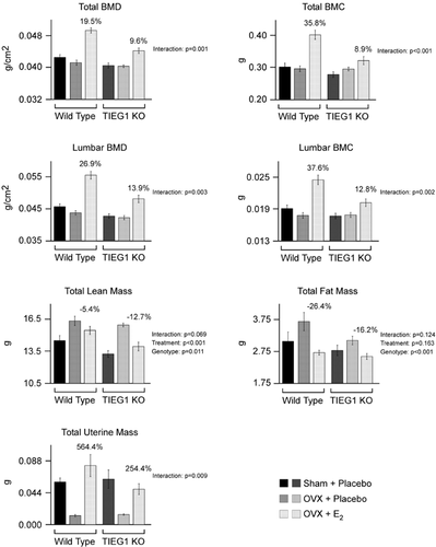
As a first step toward analyzing the role of TIEG1 in mediating estrogen action in the skeleton, DXA analyses were used to determine differences in the total body BMD and BMC resulting from estrogen replacement in ovariectomized wild-type and TIEG1 KO mice. For both of these parameters, a significant difference in the response to estrogen replacement was identified between the two genotypes with wild-type mice exhibiting a three- to fourfold greater response to estrogen compared with that of TIEG1 KO animals (Fig. 1). A nearly identical pattern of response to estrogen was observed for BMD and BMC in the lumbar vertebrae with wild-type mice exhibiting a two- to threefold greater response than TIEG1 KO littermates (Fig. 1). An interaction between mouse genotype and treatment was not detected for total body lean or fat mass as both wild-type and TIEG1 KO mice displayed a decrease in both of these parameters after estrogen exposure (Fig. 1). Total body lean mass was decreased twice as much in TIEG1 KO mice compared with wild-type controls and was determined to be significantly repressed in both genotypes (Fig. 1). Total body fat mass was shown to be affected by estrogen similarly in both genotypes of mice but was significantly decreased in TIEG1 KO animals regardless of treatment, implicating an important biological role for TIEG1 in fat tissue (Fig. 1). Uterine weights were determined in all groups of mice to ensure successful estrogen depletion and replacement within our model system. As expected, uterine weights were elevated in estrogen-treated wild-type and KO mice compared with OVX placebo-treated animals (Fig. 1). Interestingly, the magnitude of response was more than doubled in wild-type animals and an interaction between genotype and treatment was also detected for this parameter, suggesting that TIEG1 may mediate estrogen signaling in other target tissues (Fig. 1).
pQCT analysis of the proximal tibial metaphysis and diaphysis of wild-type and TIEG1 KO mice after OVX and estrogen replacement
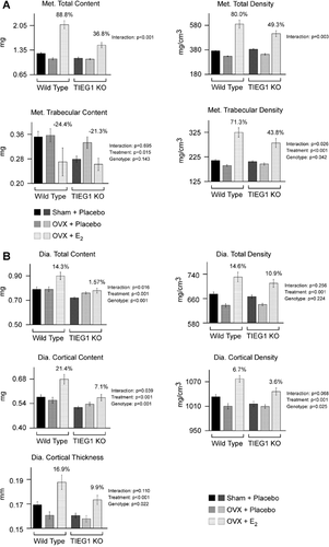
To identify differences in the response of a weight-bearing long bone to OVX and estrogen replacement between wild-type and TIEG1 KO mice, the left tibias were analyzed via pQCT. In the proximal tibial metaphysis, an interaction between genotype and treatment was identified for total bone content and total bone density, indicating a role for TIEG1 in mediating estrogenic responses at this bone site (Fig. 2A). As with much of the data presented in Fig. 1, wild-type mice exhibited a two- to threefold greater estrogenic response with regard to these two parameters compared with TIEG1 KO littermates (Fig. 2A). Similar responses to estrogen treatment were observed for trabecular content and density in wild-type and TIEG1 KO animals and estrogen treatment was shown to result in significant increases in trabecular density regardless of TIEG1 expression (Fig. 2A). A nearly significant decrease in trabecular content was also observed in estrogen-treated animals (Fig. 2A). At the tibial diaphysis, the magnitude of response to estrogen was appreciably different for total mineral content, cortical mineral content, and cortical mineral density between wild-type and TIEG1 KO mice, although no significant interactions were found for any of these parameters (Fig. 2B). Estrogen treatment was shown to lead to significant increases in total mineral content, total mineral density, cortical content, cortical density, and cortical thickness at this bone site regardless of mouse genotype (Fig. 2B). Total content and cortical content were also shown to be significantly decreased in TIEG1 KO mice regardless of treatment (Fig. 2B).
Micro-CT analysis of the femur and LV5 vertebrae
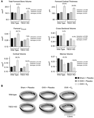
In light of the DXA and pQCT results, we next sought to identify differences in the microarchitecture of bones isolated from TIEG1 KO mice after OVX and estrogen replacement relative to wild-type controls. With regard to the femur midshaft, the response to estrogen treatment was at least twofold greater in wild-type mice compared with TIEG1 KO animals with regard to total femoral bone volume and femoral IPOLAR (Fig. 3A). However, no significant differences in the response to estrogen were detected between wild-type and TIEG1 KO mice for any of the parameters analyzed at this bone site (Fig. 3A). Estrogen treatment resulted in significant increases in total femoral bone volume, cortical thickness, femoral IPOLAR, cortical volume, and marrow volume regardless of genotype when compared with placebo-treated control animals (Fig. 3A). TIEG1 KO mice exhibited significant decreases in total femoral bone volume, femoral IPOLAR, cross-sectional volume, cortical volume, and marrow volume regardless of treatment when compared with wild-type animals (Fig. 3A). A representative micro-CT image of the femur diaphysis is shown for each genotype and treatment group in Fig. 3B.
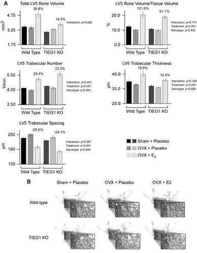
We next analyzed differences in trabecular bone using the LV5 vertebrae. At this bone site, total bone volume was found to significantly differ between wild-type and TIEG1 KO mice after estrogen treatment with an approximately 2.5-fold greater response observed in wild-type animals (Fig. 4A). Estrogen treatment also resulted in significant increases in bone volume/tissue volume, trabecular number, and trabecular thickness, with a concomitant decrease in trabecular spacing, in both wild-type and TIEG1 KO mice (Fig. 4A). The magnitude of change for these four parameters was nearly identical in both genotypes of animals. Additionally, trabecular thickness was significantly decreased in TIEG1 KO mice compared with wild-type controls regardless of treatment (Fig. 4A). A representative micro-CT image of the LV5 vertebrae is shown for each genotype and treatment group in Fig. 4B.
Histomorphometric analysis of the distal femur metaphysis and endocortical diaphysis
Static histomorphometric analyses were next performed on the distal femur metaphysis of wild-type and TIEG1 KO mice after OVX and estrogen replacement. In agreement with the micro-CT data, estrogen treatment resulted in significant increases in bone area/tissue area and trabecular number, as well as decreased trabecular spacing, in both wild-type and TIEG1 KO mice (Fig. 5A). Although a statistically significant difference was not detected between the two genotypes of mice for any of the four parameters examined at this bone site, the magnitude of response to estrogen was approximately two- to threefold greater in wild-type mice for each of these measurements with the exception of trabecular spacing (Fig. 5A). With regard to dynamic histomorphometric parameters at this bone site, no significant differences were detected with regard to estrogen treatment or mouse genotype (Fig. 5B). Interestingly, mineralizing perimeter/bone perimeter, bone formation rate/bone perimeter, bone formation rate/bone area, and osteoclast perimeter/bone perimeter were differentially affected by estrogen treatment as each of these parameters were decreased in wild-type mice but increased in TIEG1 KO animals (Fig. 5B).
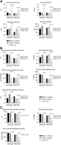
In light of the increased cortical thickness and concomitant decreased marrow volume observed at the femur mid-diaphysis in both wild-type and TIEG1 KO mice after estrogen replacement (Fig. 3A), dynamic histomorphometry was also performed on the endocortical surface of the femur diaphysis. Because of the extensive rates of mineralization occurring at this site, it was impossible to obtain accurate measurements of osteoclast perimeter/bone perimeter. Unlike that of the femur metaphysis, mineralizing perimeter/bone perimeter and bone formation rate/bone perimeter exhibited significant differences in the response to estrogen treatment between wild-type and TIEG1 KO mice at this bone site with wild-type animals exhibiting a more robust decrease in these two parameters relative to placebo-treated controls (Fig. 5C). Although no interaction was detected between treatment and mouse genotype for mineral apposition rate, estrogen treatment was shown to significantly decrease this parameter in both wild-type and TIEG1 KO animals (Fig. 5C).
Effects of estrogen replacement on biochemical markers of bone turnover
Based on the differential effects of estrogen replacement in wild-type and TIEG1 KO mice on many bone parameters as determined by DXA, pQCT, micro-CT, and histomorphometry, we next examined alterations in the serum levels of biochemical markers of bone turnover. Specifically, P1NP, an indicator of bone formation, and CTX-1, a marker of bone resorption, were determined by ELISA. A trend toward a significant difference in the response to estrogen between wild-type and TIEG1 KO mice was observed for CTX-1 but not P1NP (Fig. 6). In agreement with the osteoclast perimeter/bone perimeter shown in Fig. 5B, estrogen treatment resulted in decreased levels of CTX-1 in wild-type mice but a slight increase in this biochemical marker in TIEG1 KO animals (Fig. 6). A treatment effect was not detected for either of these markers; however, CTX-1 levels were determined to be significantly elevated in KO mice relative to wild-type controls regardless of treatment (Fig. 6).

Discussion
In this study, we performed a detailed comparison of the skeletal responses of wild-type and TIEG1 KO mice after OVX and estrogen replacement. The results of this study have revealed that TIEG1 plays an important role in mediating the effects of estrogen throughout the skeleton. Specifically, DXA, pQCT, and micro-CT analyses demonstrate that TIEG1 KO mice exhibit significantly diminished responses to estrogen replacement with regard to multiple skeletal parameters and bone sites when compared with wild-type littermates. Histomorphometric analyses also revealed either significant increases, or a trend toward greater estrogenic responsiveness, for trabecular thickness, bone formation rates, and mineralizing perimeter per bone perimeter in wild-type mice compared with TIEG1 KO animals. Analysis of biochemical markers of bone turnover revealed that loss of TIEG1 expression increases bone resorption in vivo as KO mice exhibit higher serum CTX-1 levels than wild-type littermates. Furthermore, a trend toward a differential response of CTX-1 levels after estrogen replacement was detected between the two genotypes, suggesting an important role for TIEG1 in regulating this process. Taken together, these data identify TIEG1 as an important mediator of estrogenic signaling in the mouse skeleton.
We have previously demonstrated that loss of TIEG1 expression in mice results in a sex-specific osteopenic phenotype characterized by low bone mineral density, bone mineral content, and overall loss of bone strength only in female animals.18 These data suggested that defects in estrogen signaling in the bones of TIEG1 KO mice may play an important role in mediating this phenotype. It is well accepted that the skeleton is a direct estrogen target organ and estrogen deficiency is one of the primary causes of osteoporosis in women21 because it is essential for regulating bone homeostasis30-32 and maintaining bone health.20 Estrogen classically signals through two primary receptors, estrogen receptor-alpha (ERα) and estrogen receptor-beta (ERβ). The presence of estrogen receptors in bone was first described in 1988,33, 34 and since then numerous studies have sought to elucidate their roles in mediating the effects of estrogen on the skeleton. The development of ERα and ERβ KO mice has confirmed that both of these receptors play important roles in bone.35-37 A more recent study has suggested that ERα is the only receptor responsible for mediating the protective effects of estrogen on the skeleton in male mice, whereas in female mice, ERβ also appears to play a role.36
We have previously reported that estrogen increases TIEG1 expression in both human38 and mouse osteoblasts.39 Interestingly, this estrogenic regulation appears to be primarily mediated by ERβ through the recruitment of steroid receptor co-activator-1 (SRC-1) to estrogen response elements located in the first intron of the TIEG1 gene.39 SRC-1 was one of the first receptor co-activators to be cloned and is known to play important roles in enhancing estrogen action in many tissues.40 Similar to the results presented here, deletion of SRC-1 in mice also diminishes the effects of estrogen on the skeleton.41 Taken together, these studies suggest that regulation of TIEG1 expression by estrogen, at least in part through the actions of ERβ and SRC-1, is necessary for optimal estrogen signaling in bone.
At the cellular level, estrogen exhibits differential effects on osteoblasts and osteoclasts. It has been shown both in vitro and in vivo that estrogen suppresses apoptosis and increases the lifespan of osteoblasts.42 Estrogen supplementation in OVX rats has previously been shown to decrease mineral apposition rate, double-labeled perimeter, and osteoblast number in cancellous bone.43 Treatment of postmenopausal women with estrogen also reduces osteoblast numbers, bone formation rates, and serum markers of bone formation.44, 45 Similar observations were made in the present study in OVX wild-type mice as 30 days of estrogen supplementation led to a decrease in mineralizing perimeter/bone perimeter and bone formation rates in the femur. Interestingly, these estrogen-induced changes were generally not observed in TIEG1 KO mice, further suggesting that TIEG1 plays a role in mediating the effects of estrogen in osteoblast cells.
With regard to osteoclasts, many studies have demonstrated that estrogen decreases osteoclast numbers and induces osteoclast apoptosis,46, 47 both of which lead to suppression of bone resorption.47-50 A recent study has shown that estrogen treatment of rats leads to increased expression of ERβ in osteoclasts that are undergoing apoptosis,51 suggesting that ERβ, at least in part, mediates this process. This is of particular interest because we have shown that estrogen induces the expression of TIEG1 through the actions of ERβ.39 Furthermore, we have demonstrated that loss of TIEG1 expression delays osteoclast differentiation and supresses their rates of apoptosis.16 Estrogen treatment of OVX wild-type mice in the present study led to a decrease in osteoclast perimeter per bone perimeter with a numerical, but nonsignificant, reduction in serum levels of CTX-1 compared with placebo-treated OVX animals. In contrast, estrogen treatment of TIEG1 KO mice did not result in reduced osteoclast perimeter per bone perimeter or circulating levels of CTX-1, suggesting a role for TIEG1 in mediating estrogenic effects on osteoclast cells.
In summary, we have implicated a role for TIEG1 in mediating estrogen signaling in the mouse skeleton. As demonstrated throughout our studies, TIEG1 KO mice exhibit an approximately two- to threefold attenuated estrogenic response throughout the skeleton at the tissue, cellular, and biochemical level compared with that of wild-type littermates. These data suggest that much of the sex-specific osteopenic phenotype observed in female TIEG1 KO mice may be explained by defects in the estrogen signaling pathway. Although we have not yet determined the precise mechanisms by which TIEG1 mediates estrogen signaling in bone or determined the exact contribution of osteoblasts, osteoclasts, and osteocytes to the observed defects in TIEG1 KO mice, our present and past studies suggest that all of these cell types are likely involved. Future studies are necessary to address the exact roles of TIEG1 and its involvement in mediating estrogen signaling in osteoblasts, osteoclasts, and osteocytes.
Disclosures
All authors state that they have no conflicts of interest.
Acknowledgments
The authors thank Sherry Linander for her excellent clerical assistance. The work presented here was supported by NIH grants DE14036 (TCS and JRH), T32AR056950 (AG), and F32AR063596 (AG), as well as a Paul Calabresi K12 award NCI CA90628 (JRH) and the Mayo Foundation (TCS).
Authors' roles: Study design: JRH, FAS, UTI, RTT, TCS, and MS. Study conduct: JRH, KSP, MC, KAP, KDP, and MS. Data collection: JRH, KSP, MC, KAP, KDP, UTI, RTT, and MS. Data analysis: JRH, KSP, MC, KAP, KDP, VJS, UTI, RTT, TCS, and MS. Drafting manuscript: JRH, TCS, and MS. Revising manuscript: JRH, KSP, AG, FAS, JNI, VJS, UTI, RTT, TCS, and MS. Approving final version of manuscript: all authors. JRH and MS take responsibility for the integrity of the data analysis.


