CT imaging character of different brain regions in different ages of Diannan small-ear pigs
Abstract
Objective
To collect and establish normal data of the brain regions of Diannan small-ear (DSE pigs, basically throughout measuring and comparing computed tomography values of the barin.
Methods
12 ordinary DSE pigs were divided into juvenile, adult, old groups based on the physiological ages (n=4) A SOMATOM Definition AS 64-row 128-slice 4D spiral CT scanner (SOMATOM Definition AS, Germany) was used to collect CT images of DSE pigs, record and analyze the scanning data.
Results
Compared with the juvenile group, the CT values of the right frontal lobe, right parietal lobe, right temporal lobe, left temporal lobe, and right occipital lobe were significantly higher in the elderly group. Compared with the adult group, the CT values of the right frontal lobe, left frontal lobe, right parietal lobe, right temporal lobe, left temporal lobe, and right occipital lobe of the elderly group were significantly higher.
Conclusion
The results of the study can be used to evaluate the changes in CT values of various brain regions in piglets of different ages and future pig head injury models or other studies that require CT-based analysis.
Introduction
Animal in vivo experiment is the most important link from designing to clinical application. Currently, Diannan small-ear (DSE) pigs possess the characteristics of easy breeding, genetic stability (Liu C, et al., 2020; Wu F, et al., 2020), short body, thin hair, etc., at the same time they have many similarities with humans in terms of tissue anatomy, physiological phenomena, nutritional metabolism and disease mechanism. Therefore, DSE pigs are the model animals with prospective application prospects (Hugelshofer M, et al., 2019; Yan S, et al., 2018). Up to now, pigs have been broadly used in research fields such as the cardiovascular system, exercise system, respiratory system, central nervous system and urinary system (Knoll RM, et al., 2019; Wu Y, et al., 2018; Yan X, et al., 2018). However, with deficient of anatomical data and parameters for DSE pigs. As a result, obtaining accurate and reliable physiological, biochemical parameters, tissue and organ anatomy data of DSE pigs is of great significance to further improve the medical value of small pigs and effectively carry out medical experimental research. Computed tomography (CT) is a momentous method for studying experimental animals, and there have been studies to examine pigs on applying this technology. The Galvez M team applied CT images to observe the normal spine of pigs and compared with 3D printing technology to create a reliable 3D printed model of the spine (Galvez M, et al., 2020). In addition, Lipiski M et al. analyzed the cardiac CT images of 24 Swiss large white pigs, the results showed that the height of the coronary venous ostium and the aortic-mitral valve angle parameters did not change as the weight of pigs increased. Such results not only clarify the heart size of pigs, but also increase the success rate of preclinical research and achieve higher cost-effectiveness (Lipiski M, et al., 2020). Besides this, pigs have also developed rapidly in the establishment of disease models assisted by CT. Third-generation dual-source CT was validated as a tool for assessing dynamic changes in left ventricle global function in a porcine model of acute myocardial infarction (Li W, et al., 2019). Utilize quantitative CT to detect and evaluate the severity of lung injury after successful cardiopulmonary resuscitation in a porcine cardiac arrest model with different downtimes. At present, studies on pig models are mainly focused on certain organs such as lung, spine and heart, there are inherent limitations on brain CT images. Knoll RM et al employ high resolution CT to evaluate and describe the bony anatomy of the porcine temporal bone and skull base for for future porcine head injury models or other studies that require CT-based analysis (Knoll RM et al., 2019). In this paper, we measure and compare the values of brain areas in the head of DSE pigs of ranging ages under the purpose, which to supply the research database of DSE pigs meanwhile to explore the differences in the brain areas of different age groups, and to meet the requirements of future animal experiments (Figure 1).

The flow chart of CT measurement. CT: computed tomography, kV: kilovolt, mA: milliampere, s: second, DSE: diannan small-ear, ROI: region of interest
Materials and methods
Animals and housing
The animals used in this experiment are from the Department of Laboratory Animal Science, Kunming Medical University. Including 12 ordinary DSE pigs, 4 pigs of either sex in each group of juvenile group, adult group and old group. All animal experiments are approved by the Animal Committee of Kunming Medical University, and animal feeding and care follow the regulations of the China Laboratory Animal Protection and Ethics Committee. The animal housing environment is room temperature 25-28, environmental humidity 45-50%, 12h light/dark alternate, free food and water. The age and weight of DSE pigs are displayed in Table 1.
| Juvenile (n=4) | Adult (n=4) | Old (n=4) | ||||
|---|---|---|---|---|---|---|
| Mean (SD) | Range | Mean (SD) | Range | Mean (SD) | Range | |
| Age (months) | 2.5(0.58) | 2-3 | 17.5(1.73) | 16-19 | 60(8.12) | 53-68 |
| Weight (kg) | 3.38(0.32) | 3.0-3.75 | 71.13(11.23) | 61.3-81.2 | 88.55(4.26) | 82.3-91.8 |
64-Slice spiral computed tomography (CT) scans
Animal maintenance and preparation before scanning.All CT scans used SOMATOM Definition AS 64-row 128-slice 4D spiral CT scanner (SOMATOM Definition AS, Germany) to collect CT images of DSE pigs. The general procedure is shown afterward: After 3% sodium pentobarbital anesthesia is satisfied, the DSE pigs are placed on the bed for scanning.
Computed Tomography (CT) scan of Diannan small-ear (DSE) pigs
Scan mode: spiral scan. The scanning parameters are: 120 kV, 150 mA, layer thickness 1.0 mm, and pitch 1.375:1. Scan delay time 8-10s to start scanning. The scanning range is from the nose to the level of the second cervical vertebra. After scanning, the images were analyzed independently under the guidance of two experienced radiologists. Use iTK-SNAP(http://www.itksnap.org/pmwiki/pmwiki.php) to reconstruct and measure all original images.
Statistical analysis
Data were analyzed, using SPSS version 21.0 statistical software (SPSS, Inc., Chicago, IL, USA). The results were expressed as mean ± standard deviationx(—) ± s, independent sample t-test was used for comparison between the two groups, one-way ANOVA was used for the comparison of three groups and more (ANOVA), p < 0.05 indicates that the difference is statistically significant, expressed by *. p < 0.01 for extremely significant statistical significance, expressed as **. p < 0.001 for extremely significant statistical significance, expressed as ***.
Results
The 12 DSE pigs in this study underwent examination by 64-slice Spiral CT. The baseline each brain area structural parameters of 3 groups, including the frontal lobe, parietal lobe, temporal lobe, and occipital lobe. The results confirmed that there was no significant difference in CT values of frontal lobe, parietal lobe, temporal lobe and occipital lobe between juvenile and adult group (Figure 2, Figure 3). Compared with the juvenile group, the CT values of right frontal lobe, right parietal lobe, right temporal lobe, left temporal lobe and right occipital lobe in the old group were significantly higher than those in the juvenile group, the difference was statistically significant (Figure 3, p < 0.05). However, there was no significant difference in left frontal lobe, left parietal lobe and left occipital lobe between the two groups (Figure 3, p > 0.05). Compared with the adult group, the CT values of the right frontal lobe, left frontal lobe, right parietal lobe, right temporal lobe, left temporal lobe and right occipital lobe were significantly higher in the old group, the difference was statistically significant (Figure 3, p < 0.05). However, there was no significant difference in the left parietal lobe between the two groups (Figure 3, p > 0.05).

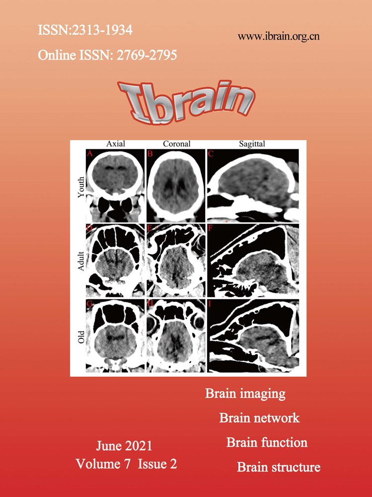
CT scans of different levels of brain were performed in juvenile group, adult group and old group. The order from top to bottom is juvenile group, adult group and old group. (A D G) Horizontal scan brain CT. (B E H) Coronal scan of brain CT. (C F I ) Sagittal scan of brain CT.

CT values of frontal lobe, parietal lobe, temporal lobe and occipital lobe in juvenile group, adult group and old group were compared. (A)The CT values of the right frontal lobe were compared among the three groups. (B) The CT values of the left frontal lobe were compared among the three groups. (C) The CT values of the right parietal lobe were compared among the three groups. (D)The CT values of the left parietal lobe among the three groups were compared with each other. (E) The CT values of the right temporal lobe were compared among the three groups. (F) The CT values of the left temporal lobe were compared among the three groups. (G) The CT values of the right occipital lobe were compared among the three groups. H. The CT values of the left occipital lobe were compared among the three groups. N=4.
Discussion
This study provides detailed descriptions and analysis of porcine with three age groups each brain region based on 64-slice Spiral CT. The close anatomical similarity between pigs and humans is an advantage. While we fully understand the CT value of each brain area in pigs of different ages, pigs are considered as an animal model for studying brain research like head injuries, mental illness and so on (Knoll RM et al., 2019; Roalf DR, et al., 2017). It is aware that different areas of the cerebral cortex have functional specific differences, each with different functions. The frontal lobe mainly controls movement and language functions. Sensory and language functions are managed by the parietal lobe. The temporal lobe is the auditory and language center and the occipital lobe is the visual center (Zavaglia M, et al., 2015). In University Hospital of Trondheim’s studies with 18-22 year-old young adults, better intelligence quotient (IQ) correlated with larger cortical surface area in frontal, temporal and occipital regions (Skranes J, et al., 2013), better executive function correlated with larger surface area in antero-medial frontal and temporal lobes (Ostgard HF, et al., 2016) and better visuo-motor performance correlated with larger cortical surface area in widespread regions of the brain (Sripada K, et al., 2015).
Therefore, the volume and surface area of each cortical area are closely related to its function. Our paper indicated that CT and its post-processing technology can clearly define the scans and various diameters of different brain levels in different age groups. The CT values of the right frontal lobe, temporal lobe, parietal lobe, occipital lobe, and left temporal lobe of the elderly group were significantly higher than those of the juvenile. What’s more, compared with the adult group, the elderly group has higher CT values in most brain areas. It explains that there are differences in CT values of brain regions of different ages (Yates TS, et al., 2021). The change of CT value can reflect the change of fluid in tissue, mainly the change of blood volume (Raslau FD, et al., 2019; Shi D, et al., 2020). Crucially, obtaining accurate baseline values, for the application of new innovative techniques in both experimental and clinical studies.
At present, CT technology is applied not only to the establishment of animal models, but also to the evaluation of animal surgery and disease diagnosis. In order to improve the clinical accuracy and feasibility, CT technology can scan the pig heart size (Lipiski M et al., 2020), accurately evaluate the ventricular volume parameters based on semi-automatic 3D threshold segmentation (Xu J, et al., 2019), evaluate the protection of novel femoral reaming technology on lung function and post-traumatic contusion (Halvachizadeh S, et al., 2021). Instruct delivery after acute myocardial infarction in pigs and evaluate its effect on myocardial infarction (Kim I, et al., 2019). Moreover, CT perfusion of animals can build a similar threshold for humans to improve the accuracy of postoperative imaging (Bressem KK, et al., 2019). CT dynamic quantitative iodine perfusion imaging can assess and monitor early myocardial ischemia (Scherer K, et al., 2019). It can be seen that the application of CT technology in animals has laid a certain foundation for the clinic, and the visualization of CT is conducive to pre-clinical research and planning. This is the distinct advantage of CT technology, which plays a dominant role in future clinical development.
To sum up, the application of imaging equipment to the study of experimental models of small pigs or the measurement of normal data has become a research hotspot. However, failed to compare with DSE pigs autopsy standard is the main deficiency. The study was conducted with healthy pigs, so the structure is clear in the computed tomography image. In abnormal structures, precise anatomical imaging of the brain may be more difficult. (Figure 4).

Study of CT in various parts of porcine model. CT: computed tomography.
Ethical statement
The animal study protocol was legally approved by the Animal Committee of Kunming Medical University (Approval No: kumu2021570). All experiments conformed to the Guide for the Care and Use of Laboratory Animals published by the US National Institutes of Health.
Acknowledgements
Not applicable.
Conflict of interest
There is no conflict of interest in this study.
Funding
The authors declare no financial support with regard to this work.
Transparency statement
The authors affirm that this manuscript is an honest, accurate, and transparent account of the study being reported; that no important aspects of the study have been omitted; and that any discrepancies from the study as planned (and, if relevant, registered) have been explained.
Authors' contribution
Lin Zhou, Rui-Ze Niu, Hong-Su Zhou and Zheng-Meng Wang contributed to the conception of the study; Feng-Lin Wang, Xin-Xin Yang and Issac Deng helped perform the analysis with constructive discussions; Zhao-Qiong Zhu, Xin-Fu Zhou, Liu-Lin Xiong revised and finalized it.