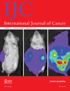
Fluorescence lifetime imaging microscopy of chemotherapy-induced apoptosis resistance in a syngenic mouse tumor model
M. Keese
Chirurgische Klinik, Universitätsklinikum Mannheim, 68167 Mannheim, Germany
European Molecular Biology Laboratory Heidelberg, 69117 Heidelberg, Germany
The first two authors contributed equally to this work.
Search for more papers by this authorV. Yagublu
Chirurgische Klinik, Universitätsklinikum Mannheim, 68167 Mannheim, Germany
European Molecular Biology Laboratory Heidelberg, 69117 Heidelberg, Germany
The first two authors contributed equally to this work.
Search for more papers by this authorK. Schwenke
Chirurgische Klinik, Universitätsklinikum Mannheim, 68167 Mannheim, Germany
Max-Planck-Institut für Molekulare Physiologie, Abteilung Systemische Zellbiologie, 44227 Dortmund, Germany
Search for more papers by this authorS. Post
Chirurgische Klinik, Universitätsklinikum Mannheim, 68167 Mannheim, Germany
Search for more papers by this authorCorresponding Author
P. Bastiaens
European Molecular Biology Laboratory Heidelberg, 69117 Heidelberg, Germany
Max-Planck-Institut für Molekulare Physiologie, Abteilung Systemische Zellbiologie, 44227 Dortmund, Germany
Tel: +49 (231) 133-2200, Fax: +49 (231) 133-2299
MPI für Molekulare Physiologie Otto-Hahn-Straße 11, D-44227 DortmundSearch for more papers by this authorM. Keese
Chirurgische Klinik, Universitätsklinikum Mannheim, 68167 Mannheim, Germany
European Molecular Biology Laboratory Heidelberg, 69117 Heidelberg, Germany
The first two authors contributed equally to this work.
Search for more papers by this authorV. Yagublu
Chirurgische Klinik, Universitätsklinikum Mannheim, 68167 Mannheim, Germany
European Molecular Biology Laboratory Heidelberg, 69117 Heidelberg, Germany
The first two authors contributed equally to this work.
Search for more papers by this authorK. Schwenke
Chirurgische Klinik, Universitätsklinikum Mannheim, 68167 Mannheim, Germany
Max-Planck-Institut für Molekulare Physiologie, Abteilung Systemische Zellbiologie, 44227 Dortmund, Germany
Search for more papers by this authorS. Post
Chirurgische Klinik, Universitätsklinikum Mannheim, 68167 Mannheim, Germany
Search for more papers by this authorCorresponding Author
P. Bastiaens
European Molecular Biology Laboratory Heidelberg, 69117 Heidelberg, Germany
Max-Planck-Institut für Molekulare Physiologie, Abteilung Systemische Zellbiologie, 44227 Dortmund, Germany
Tel: +49 (231) 133-2200, Fax: +49 (231) 133-2299
MPI für Molekulare Physiologie Otto-Hahn-Straße 11, D-44227 DortmundSearch for more papers by this authorAbstract
During cancer therapy with DNA-damaging drug-agents, the development of secondary resistance to apoptosis can be observed. In the search for novel therapeutic approaches that can be used in these cases, we monitored chemotherapy-induced apoptosis resistance in a syngenic mouse tumor model. For this, syngenic murine colorectal carcinoma cells, which stably expressed a FRET-based caspase-3 activity sensor, were introduced into animals to induce peritoneal carcinomatosis or disseminated hepatic metastases. This syngenic system allowed in vitro, in vivo and ex vivo analysis of chemotherapy induced apoptosis induction by optically monitoring the caspase-3 sensor state in the tumor cells. Tumor tissue analysis of 5-FU treated mice showed the selection of 5-FU-induced apoptosis resistant tumor cells. These and chemo-naive fluorescent tumor cells could be re-isolated from treated and untreated mice and propagated in cell culture. Re-exposure to 5-FU and second line treatment modalities in this ex-vivo setting showed that 5-FU induced apoptosis resistance could be alleviated by imatinib mesylate (Gleevec). We thus show that syngenic mouse systems that stably express a FRET-based caspase-3 sensor can be employed to analyse the therapeutic efficiency of apoptosis inducing chemotherapy.
References
- 1 Nita ME, Nagawa H, Tominaga O, Tsuno N, Fujii S, Sasaki S, Fu CG, Takenoue T, Tsuruo T, Muto T. 5-Fluorouracil induces apoptosis in human colon cancer cell lines with modulation of Bcl-2 family proteins. Br J Cancer 1998; 78: 986–92.
- 2 Tsuruo T, Naito M, Tomida A, Fujita N, Mashima T, Sakamoto H, Haga N. Molecular targeting therapy of cancer: drug resistance, apoptosis and survival signal. Cancer Sci 2003; 94: 15–21.
- 3 Longley DB, Johnston PG. Molecular mechanisms of drug resistance. J Pathol 2005; 205: 275–92.
- 4 Sugo N, Niimi N, Aratani Y, Takiguchi-Hayashi K, Koyama H. p53 Deficiency rescues neuronal apoptosis but not differentiation in DNA polymerase beta-deficient mice. Mol Cell Biol 2004; 24: 9470–7.
- 5 Sturm JW, Keese MA, Petruch B, Bonninghoff RG, Zhang H, Gretz N, Hafner M, Post S, McCuskey RS. Enhanced green fluorescent protein-transfection of murine colon carcinoma cells: key for early tumor detection and quantification. Clin Exp Metastasis 2003; 20: 395–405.
- 6 Samel S, Keese M, Lux A, Jesnowski R, Prosst R, Saller R, Hafner M, Sturm J, Post S, Lohr M. Peritoneal cancer treatment with CYP2B1 transfected, microencapsulated cells and ifosfamide. Cancer Gene Ther 2006; 13: 65–73.
- 7 Yang M, Jiang P, Sun FX, Hasegawa S, Baranov E, Chishima T, Shimada H, Moossa AR, Hoffman RM. A fluorescent orthotopic bone metastasis model of human prostate cancer. Cancer Res 1999; 59: 781–6.
- 8 Yang M, Baranov E, Jiang P, Sun FX, Li XM, Li L, Hasegawa S, Bouvet M, Al-Tuwaijri M, Chishima T, Shimada H, Moossa AR, et al. Whole-body optical imaging of green fluorescent protein-expressing tumors and metastases. Proc Natl Acad Sci U S A 2000; 97: 1206–11.
- 9 Ishikura H, Kondo K, Miyoshi T, Takahashi Y, Fujino H, Monden Y. Green fluorescent protein expression and visualization of mediastinal lymph node metastasis of human lung cancer cell line using orthotopic implantation. Anticancer Res 2004; 24: 719–23.
- 10 Heim R, Tsien RY. Engineering green fluorescent protein for improved brightness, longer wavelengths and fluorescence resonance energy transfer. Curr Biol 1996; 6: 178–82.
- 11 Funovics M, Weissleder R, Tung CH. Protease sensors for bioimaging. Anal Bioanal Chem 2003; 377: 956–63.
- 12 Welsh DK, Kay SA. Bioluminescence imaging in living organisms. Curr Opin Biotechnol 2005; 16: 73–8.
- 13 Rhee JM, Pirity MK, Lackan CS, Long JZ, Kondoh G, Takeda J, Hadjantonakis AK. In vivo imaging and differential localization of lipid-modified GFP-variant fusions in embryonic stem cells and mice. Genesis 2006; 44: 202–18.
- 14 Griesbeck O. Fluorescent proteins as sensors for cellular functions. Curr Opin Neurobiol 2004; 14: 636–41.
- 15 Chen Y, Mills JD, Periasamy A. Protein localization in living cells and tissues using FRET and FLIM. Differentiation 2003; 71: 528–41.
- 16 Bastiaens PI, Squire A. Fluorescence lifetime imaging microscopy: spatial resolution of biochemical processes in the cell. Trends Cell Biol 1999; 9: 48–52.
- 17 Mank M, Reiff DF, Heim N, Friedrich MW, Borst A, Griesbeck O. A FRET-based calcium biosensor with fast signal kinetics and high fluorescence change. Biophys J 2006; 90: 1790–6.
- 18 Evanko DS, Haydon PG. Elimination of environmental sensitivity in a cameleon FRET-based calcium sensor via replacement of the acceptor with Venus. Cell Calcium 2005; 37: 341–8.
- 19 Evans SK, Aiello DP, Green MR. Fluorescence resonance energy transfer as a method for dissecting in vivo mechanisms of transcriptional activation. Biochem Soc Symp 2006: 217–24.
- 20 Yudushkin IA, Schleifenbaum A, Kinkhabwala A, Neel BG, Schultz C, Bastiaens PI. Live-cell imaging of enzyme-substrate interaction reveals spatial regulation of PTP1B. Science 2007; 315: 115–9.
- 21 Tsourkas A, Weissleder R. Illuminating the dynamics of intracellular activity with ‘active’ molecular reporters. Mech Chem Biosyst 2004; 1: 133–45.
- 22 Jahnz M, Schwille P. Enzyme assays for confocal single molecule spectroscopy. Curr Pharm Biotechnol 2004; 5: 221–9.
- 23 Offterdinger M, Georget V, Girod A, Bastiaens PI. Imaging phosphorylation dynamics of the epidermal growth factor receptor. J Biol Chem 2004; 279: 36972–81.
- 24 Miyawaki A, Tsien RY. Monitoring protein conformations and interactions by fluorescence resonance energy transfer between mutants of green fluorescent protein. Methods Enzymol 2000; 327: 472–500.
- 25 Iino R, Murakami T, Iizuka S, Kato-Yamada Y, Suzuki T, Yoshida M. Real-time monitoring of conformational dynamics of the epsilon subunit in F1-ATPase. J Biol Chem 2005; 280: 40130–4.
- 26 Xu X, Gerard AL, Huang BC, Anderson DC, Payan DG, Luo Y. Detection of programmed cell death using fluorescence energy transfer. Nucleic Acids Res 1998; 26: 2034–5.
- 27 Stadelmann C, Lassmann H. Detection of apoptosis in tissue sections. Cell Tissue Res 2000; 301: 19–31.
- 28 Harpur AG, Wouters FS, Bastiaens PI. Imaging FRET between spectrally similar GFP molecules in single cells. Nat Biotechnol 2001; 19: 167–9.
- 29 Keese M, Offterdinger M, Tischer C, Girod A, Lommerse PH, Yagublu V, Magdeburg R, Bastiaens PI. Quantitative imaging of apoptosis commitment in colorectal tumor cells. Differentiation 2007; 75: 809–18.
- 30 Verveer PJ, Squire A, Bastiaens PI. Improved spatial discrimination of protein reaction states in cells by global analysis and deconvolution of fluorescence lifetime imaging microscopy data. J Microsc 2001; 202: 451–6.
- 31 Verveer PJ, Squire A, Bastiaens PI. Global analysis of fluorescence lifetime imaging microscopy data. Biophys J 2000; 78: 2127–37.
- 32 Pepperkok R, Scheel J, Horstmann H, Hauri HP, Griffiths G, Kreis TE. Beta-COP is essential for biosynthetic membrane transport from the endoplasmic reticulum to the Golgi complex in vivo. Cell 1993; 74: 71–82.
- 33 Ciccolini J, Peillard L, Evrard A, Cuq P, Aubert C, Pelegrin A, Formento P, Milano G, Catalin J. Enhanced antitumor activity of 5-fluorouracil in combination with 2′-deoxyinosine in human colorectal cell lines and human colon tumor xenografts. Clin Cancer Res 2000; 6: 1529–35.
- 34 Wakeling AE, Guy SP, Woodburn JR, Ashton SE, Curry BJ, Barker AJ, Gibson KH. ZD1839 (Iressa): an orally active inhibitor of epidermal growth factor signaling with potential for cancer therapy. Cancer Res 2002; 62: 5749–54.
- 35 Attoub S, Rivat C, Rodrigues S, Van Bocxlaer S, Bedin M, Bruyneel E, Louvet C, Kornprobst M, Andre T, Mareel M, Mester J, Gespach C. The c-kit tyrosine kinase inhibitor STI571 for colorectal cancer therapy. Cancer Res 2002; 62: 4879–83.
- 36 Halene S, Wang L, Cooper RM, Bockstoce DC, Robbins PB, Kohn DB. Improved expression in hematopoietic and lymphoid cells in mice after transplantation of bone marrow transduced with a modified retroviral vector. Blood 1999; 94: 3349–57.
- 37 Bellone G, Smirne C, Mauri FA, Tonel E, Carbone A, Buffolino A, Dughera L, Robecchi A, Pirisi M, Emanuelli G. Cytokine expression profile in human pancreatic carcinoma cells and in surgical specimens: implications for survival. Cancer Immunol Immunother 2006; 55: 684–98.
- 38 Bogenrieder T, Herlyn M. Axis of evil: molecular mechanisms of cancer metastasis. Oncogene 2003; 22: 6524–36.
- 39 Wouters BG, Koritzinsky M, Chiu RK, Theys J, Buijsen J, Lambin P. Modulation of cell death in the tumor microenvironment. Semin Radiat Oncol 2003; 13: 31–41.
- 40 Keese M, Magdeburg RJ, Herzog T, Hasenberg T, Offterdinger M, Pepperkok R, Sturm JW, Bastiaens PI. Imaging epidermal growth factor receptor phosphorylation in human colorectal cancer cells and human tissues. J Biol Chem 2005; 280: 27826–31.
- 41 Heymach JV, Nilsson M, Blumenschein G, Papadimitrakopoulou V, Herbst R. Epidermal growth factor receptor inhibitors in development for the treatment of non-small cell lung cancer. Clin Cancer Res 2006; 12: 4441s–5s.
- 42 Ellis AG, Doherty MM, Walker F, Weinstock J, Nerrie M, Vitali A, Murphy R, Johns TG, Scott AM, Levitzki A, McLachlan G, Webster LK, et al. Preclinical analysis of the analinoquinazoline AG1478, a specific small molecule inhibitor of EGF receptor tyrosine kinase. Biochem Pharmacol 2006; 71: 1422–34.
- 43 Cohen MH, Johnson JR, Pazdur R. U.S. Food and Drug Administration Drug Approval Summary: conversion of imatinib mesylate (STI571; Gleevec) tablets from accelerated approval to full approval. Clin Cancer Res 2005; 11: 12–9.
- 44 Hebbar M, Tournigand C, Lledo G, Mabro M, Andre T, Louvet C, Aparicio T, Flesch M, Varette C, de Gramont A. Phase II trial alternating FOLFOX-6 and FOLFIRI regimens in second-line therapy of patients with metastatic colorectal cancer (FIREFOX study). Cancer Invest 2006; 24: 154–9.
- 45 Venook A. Critical evaluation of current treatments in metastatic colorectal cancer. Oncologist 2005; 10: 250–61.
- 46 McLoughlin JM, Jensen EH, Malafa M. Resection of colorectal liver metastases: current perspectives. Cancer Control 2006; 13: 32–41.
- 47 Koppe MJ, Boerman OC, Oyen WJ, Bleichrodt RP. Peritoneal carcinomatosis of colorectal origin: incidence and current treatment strategies. Ann Surg 2006; 243: 212–22.
- 48 Hoffman R. Green fluorescent protein imaging of tumour growth, metastasis, and angiogenesis in mouse models. Lancet Oncol 2002; 3: 546–56.
- 49 Hengartner MO. The biochemistry of apoptosis. Nature 2000; 407: 770–6.
- 50 Degterev A, Boyce M, Yuan J. A decade of caspases. Oncogene 2003; 22: 8543–67.
- 51 Ashkenazi A, Dixit VM. Apoptosis control by death and decoy receptors. Curr Opin Cell Biol 1999; 11: 255–60.
- 52 Conlon I, Raff M. Size control in animal development. Cell 1999; 96: 235–44.
- 53 Padmanabhan S, Ravella S, Curiel T, Giles F. Current status of therapy for chronic myeloid leukemia: a review of drug development. Future Oncol 2008; 20: 359–77.
- 54 Karaman MW, Herrgard S, Treiber DK, Gallant P, Atteridge CE, Campbell BT, Chan KW, Ciceri P, Davis MI, Edeen PT, Faraoni R, Floyd M, et al. A quantitative analysis of kinase inhibitor selectivity. Nat Biotechnol 2008; 26: 127–32.
- 55 Masiello D, Mohi MG, McKnight NC, Smith B, Neel BG, Balk SP, Bubley GJ. Combining an mTOR antagonist and receptor tyrosine kinase inhibitors for the treatment of prostate cancer. Cancer Biol Ther 2007; 6: 195–201.
- 56 Judson I. Imatinib in advanced gastrointestinal stromal tumour: when is 800 mg the correct dose? Curr Opin Oncol 2008; 20: 433–7.
- 57
Samel S,
Keese M,
Kleczka M,
Lanig S,
Gretz N,
Hafner M,
Sturm J,
Post S
Supplementation and inhibition of NO-synthesis influences bacterial transit time during bacterial translocation in rats.
Shock
2003;
19:
378–82.
10.1097/00024382-200304000-00014 Google Scholar
- 58
Samel S,
Keese M,
Kleczka M,
Lanig S,
Gretz N,
Hafner M,
Sturm J,
Post S
Microscopy of bacterial translocation during small bowel obstruction and ischemia in vivo - a new animal model.
BMC Surg
2002;
13:
6.
10.1186/1471-2482-2-6 Google Scholar
- 59 Hoffman RM, Yang M Whole-body imaging with fluorescent proteins. Nature Protocols 2006; 1: 1429–38


