Whole-Exome Sequencing Identifies a Variant in TMEM132E Causing Autosomal-Recessive Nonsyndromic Hearing Loss DFNB99
Communicated by Michel Goossens
Contract grant sponsors: National Natural Science Foundation of China (81021001, 81300835, 81070905, 81072452); Research Project of Chinese Ministry of Education (113040A); Shandong Natural Science Foundation (ZR2012HQ015); Independent Innovation Foundation of Shandong University (2012TS112).
ABSTRACT
Autosomal-recessive nonsyndromic hearing loss (ARNSHL) features a high degree of genetic heterogeneity. Many genes responsible for ARNSHL have been identified or mapped. We previously mapped an ARNSHL locus at 17q12, herein designated DFNB99, in a consanguineous Chinese family. In this study, whole-exome sequencing revealed a homozygous missense mutation (c.1259G>A, p.Arg420Gln) in the gene-encoding transmembrane protein 132E (TMEM132E) as the causative variant. Immunofluorescence staining of the Organ of Corti showed Tmem132e highly expressed in murine inner hair cells. Furthermore, knockdown of the tmem132e ortholog in zebrafish affected the mechanotransduction of hair cells. Finally, wild-type human TMEM132E mRNA, but not the mRNA carrying the c.1259G>A mutation rescued the Tmem132e knockdown phenotype. We conclude that the variant in TMEM132E is the most likely cause of DFNB99.
Introduction
Hearing loss is one of the most common sensory disorders in humans. At least 50% of the cases are assumed to have a genetic basis, and more than two-thirds of the latter subset are classified as nonsyndromic hearing loss (NSHL) because of the absence of additional symptoms [Dror and Avraham, 2009; Hilgert et al., 2009]. Autosomal-recessive nonsyndromic hearing loss (ARNSHL) accounts for more than 80% of the NSHL cases [Hilgert et al., 2009]. To date, more than 81 loci for ARNSHL (DFNB) have been mapped, and 55 of the corresponding genes have been identified (http://hereditaryhearingloss.org/). Most of these genes have been found to play important roles in inner-ear development during embryogenesis.
In a previous study, we mapped an ARNSHL locus to an interval of 5.07 cM on chromosome 17q12 by genome-wide homozygosity mapping [Cheng et al., 2003]; we designated the locus as DFNB99. However, the search for the causative gene has been hampered due to the large size of the critical region.
Recently, a two-step approach combining linkage analysis and whole-exome sequencing has been proven to be highly successful for identifying genes involved in Mendelian diseases, including deafness [Rehman et al., 2010; Walsh et al., 2010; Delmaghani et al., 2012]. In the present study, we used exome sequencing to identify the mutation underlying DFNB99. We detected a missense mutation, c.1259G>A (rs139895222), p.Arg420Gln, in TMEM132E that cosegregates with deafness. Furthermore, we studied the function of tmem132e, the zebrafish ortholog of TMEM132E, and obtained critical evidence supporting that p.Arg420Gln is most likely the causative variant for DFNB99.
Materials and Methods
Research Subjects
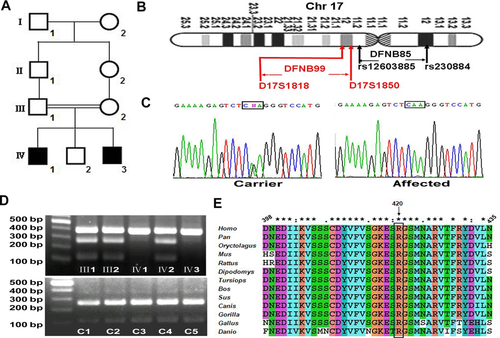
We identified two siblings with prelingual, bilateral, severe to profound sensorineural hearing loss in a Chinese family of consanguineous marriage (Fig. 1A). Romberg and tandem gait testing did not reveal any symptoms of vestibular dysfunction in either patient. Data for the probands in 95 unrelated Chinese families with ARNSHL were collected at the Institute of Otolaryngology, Chinese PLA General Hospital, Beijing. The patients in these families have severe to profound congenital and bilateral sensorineural hearing loss and mutations in GJB2, SLC26A4, and 12S rRNA genes were ruled out.
This study was approved by the medical ethics committees of Shandong University, China. Written informed consent was obtained from participating individuals. Blood samples were obtained from each participating individual, and genomic DNA was extracted by standard procedures.
Whole-Exome Sequencing
Whole-exome sequencing was performed on patient IV3 in this family (Beijing Genome Institute, Shenzhen, China). Exome capture involved use of the SureSelect Human All Exon Kit (Agilent, Santa Clara, CA) and sequencing involved the HiSeq2000 platform (Illumina, San Diego, CA).
Mutation Detection and PCR-RFLP Analysis
Sanger sequencing was used to detect the mutations in TMEM132E in this pedigree and in 95 unrelated probands with ARNSHL. Primers for amplification of exons and exon–intron boundaries of TMEM132E were designed with use of Primer Premier 5.0 (Premier Biosoft Interpairs, Palo Alto, CA). NC_000017.11 and NM_207313.1 were used as reference sequences. Primers and PCR product sizes are in Supp. Table S1. Amplification by PCR involved 40 ng genomic DNA with LA Taq DNA polymerase (Takara, Otsu, Shiga, Japan). PCR products were sequenced with use of the ABI PRISM 3730 DNA Analyzer (Applied Biosystems, Foster City, CA). Mutation nomenclature is based on the transcript NM_207313.1 and protein NP_997196.1. PCR-RFLP was used to determine the absence of the c.1259G>A variant in exon 7 of TMEM132E in 500 ethnically matched healthy controls. The c.1259G>A mutation destroyed one XhoI recognition site, which could be used to distinguish 1259G and 1259A alleles. The sequence around the variant was amplified and digested with XhoI (Takara, Otsu, Shiga, Japan) in accordance with the manufacturer's protocol, and restriction fragments were analyzed on 1.5% agarose gels.
Bioinformatics Analysis of Sequence Variants
Polymorphism Phenotyping v2 (PolyPhen-2) (http://genetics.bwh.harvard.edu/pph2) [Adzhubei et al., 2010] and Sorting Intolerant from Tolerant (SIFT) (http://sift.jcvi.org/) [ Ng and Henikoff, 2001] were used to predict the possible pathogenic effect of the p.Arg420Gln substitution in TMEM132E. Clustalw2 was used to generate alignments of the TMEM132E amino acid sequence among different species [Larkin et al., 2007].
TMEM132E Expression Profile
Quantitative RT-PCR
Adult mouse tissue including cochlea, heart, and lung tissue were dissected under a microscope. Total RNA was extracted with TRIZOL Reagent (Invitrogen, Carlsbad, CA) and cDNA was synthesized by using the RevertAid First Strand cDNA Synthesis Kit (Fermentas, Burlington, Ontario, Canada). The forward primer 5′-AGGTCACAGGGACTCGGGGC-3′ and reverse primer 5′-GCCTTCTACCGGCAAGGGGC-3′ were designed with NM_023438 used as the template. PCR sample solutions were prepared with use of Fast Start Universal SYBR Green Master, and real-time quantitative PCR involved use of LightCycler 480 II. Mouse glyceraldehyde-3-phosphate dehydrogenase was used as an internal control with the primers forward, 5′-AGGTCGGTGTGAACGGATTTG-3′ and reverse, 5′-TGTAGACCATGTAGTTGAGGTCA-3′. All reactions were performed in quadruplicate. Relative gene expression levels were determined by the delta Ct method.
Western blot analysis
Protein samples were isolated from adult mouse tissue by use of TRIZOL Reagent (Invitrogen, Carlsbad, CA) and processed on 10% SDS polyacrylamide gel and transferred to PVDF membrane (Millipore, Billerica, MA). Blots were probed with anti-TMEM132E antibody (ABGENT: AP11487b, 1:500 dilution) overnight at 4°C. After washing with TBST, membranes were treated with horseradish peroxidase-conjugated secondary antibody for 1 hr and visualized by use of ECL PLUS (GE Healthcare, Little Chalfont, Buckinghamshire, United Kingdom) as specified by the manufacturer. β-Actin was an internal control. The assay was performed in triplicate.
Immunofluorescence
Cochleas were dissected from P30 and P0 C57BL/6 mice and fixed with 4% paraformaldehyde overnight at 4°C. Cochleas from P30 mice were treated with 10% EDTA in phosphate-buffered saline (PBS) for 3 days. After being washed with PBS, samples were soaked in 30% sucrose, embedded in OCT compound (Tissue-Tek, Torrance, CA), and sectioned (7 μm) for immunofluorescence. Frozen sections were washed with PBS, blocked with 10% goat serum and 0.1% TritonX-100 in PBS, and incubated with anti-TMEM132E antibody (1:200 dilution) in a humidified chamber overnight at 4°C, washed and incubated with secondary antibody (Alexa Fluor 488 F(ab′)2 fragment of goat anti-rabbit IgG (H+L) antibody; Invitrogen) for 1 hr at 37°C, protected from light. Cell nuclei were stained with DAPI. Sections were washed, coverslipped, and photographed by laser-scanning confocal microscopy(LSM780; Zeiss, Jena, Oberkochen, Germany).
For whole-mount immunolabelling, cochlear sensory areas were microdissected and fixed in 4% paraformaldehyde in PBS overnight at 4°C. Tissues were washed twice in PBS and blocked in PBS containing 10% normal goat serum for 1 hr at room temperature, then incubated with anti-TMEM132E overnight at 4°C and processed as previously described. Tetramethyl rhodamine iso-thiocyanate (TRITC)-conjugated phalloidin (Sigma, St. Louis, MO) was used to label F-actin.
Experiments in Zebrafish
Zebrafish strains and breeding
Adult zebrafish of the Tubingen (Tu) strain were maintained at 28°C and bred according to a 14-hr on/10-hr off light cycle. Staging of zebrafish embryonic development involved standard morphological criteria.
Design and injection of morpholino oligonucleotides
Before designing specific morpholino oligonucleotides (MOs), the genomic sequence of the Tu strain was first confirmed to have only one orthologue of TMEM132E. Primers (Supp. Table S2) were designed by use of the existing database sequence to amplify the full-length cDNA of the wild-type zebrafish tmem132e gene. A 5′ fragment was amplified from zebrafish Tu RNA by RT-PCR with the ZF1and ZR1 primers, and a 3′ fragment was amplified with the ZF2 and ZR2 primers. Both fragments were cloned into the pMD18-T Vector, sequenced, and then compared with the genomic sequences in GenBank by a BLAST search.
This confirmed sequence was then used to design the antisense MOs, which were synthesized by GeneTools (Philomath, OR). A specific antisense MO, 5′-TTTTACCCTGTGAAAGAGACACAGT-3′, targeted the I2/E3 junction of zebrafish tmem132e and would result in the deletion of exon 3. A 5-nt mismatch control (MC) (5′-TTTaAgCCTcTGAAAcAcACACAGT-3′, mismatches in lower case) was used to titer an MO-specific nontoxic injection dose. Unless otherwise stated, 4 ng MO or MC was injected into 1- or 2-cell-stage embryos.
Total RNA was extracted from MO- or MC-injected larvae at 48 hr after fertilization. RT-PCR involved 5′-GACCCTCAACATCTATCTGCTC-3′ and 5′-ACTTTAAGGGGCAGTTTGGGCA-3′ primers to amplify a 674-bp fragment in MC-injected larvae and a 532-bp fragment corresponding to the truncated RNA present in morphant larvae.
Rescue experiments
The coding region of the wild-type TMEM132E was amplified from human brain RNA by RT-PCR (Invitrogen, Carlsbad, CA). Primers are in Supp. Table S2. A 5′ fragment was amplified with HF1 and HR1 primers, and a 3′ fragment was amplified with HF2 and HR2 primers. BamHI and XbaI sites were introduced into HF1 and HR2 before the start codon and after the stop codon of TMEM132E, respectively. Both fragments were cloned into the pMD18-T Vector (Takara, Otsu, Shiga, Japan). BamHI–AatII and AatII–XbaI sites were used to clone the fragments into the pCS2+ vector (Ambion, Austin, TX). To construct the full-length cDNA of mutant human TMEM132E, a 5′ fragment containing c.1259G>A was obtained by RT-PCR with HF1 and MR1 primers, a site-directed mutant primer set. The mutant coding region of human TMEM132E was also cloned into the pCS2+ vector. The Ambion's mMESSAGE mMACHINE Kit was used for the in vitro synthesis of the wild-type- and mutant-capped TMEM132E RNA. Approximately 100 pg of capped RNA was coinjected with MO or MC into each 1- or 2-cell-stage embryos.
Startle response
Each larvae was studied in a 35-mm cell culture dish. The vibration stimulus consisted of five light taps on the rim of the culture dish for 4 sec, which mimics the stimulus used in the behavioral assay to trigger the escape reflex. Movies were created with use of a high-speed camera (DP-72; Olympus, Shinjuku, Tokyo, Japan) at 1,000 frames/sec at 680 × 512 resolution. The test was repeated three times for each fish.
Recording of inner-ear microphonic potentials
Microphonic potentials in response to vibratory stimuli were measured from the otic vesicles of zebrafish larvae at 5 dpf. An individual larva was transferred to a recording chamber, visualized under an upright microscope with a water immersion objective (×40) and differential interference contrast optics (BX51WI; Olympus, Shinjuku, Tokyo, Japan), and immobilized in one drop of 1.5% low-melting agarose in standard extracellular solution (130 mM NaCl, 2 mM KCl, 2 mM CaCl2, 1 mM MgCl2, and 10 mM 4[2-hydroxyethyl[-1-piperazineethanesulfonic acid; 290 mOsm; pH 7.8). A vibratory stimulus was applied to the larval surface by use of a glass rod pulled to a tip 30 μm in diameter and driven by a calibrated piezoelectric actuator with sinusoidal stimuli of 2 μm peak-to-peak amplitude at 200 Hz. A glass recording electrode with resistance 5–6 M Ω when filled with extracellular solution was advanced, under visual control, through the swelling near the lateral semicircular canal and placed near the otolith of the posterior macula. Microphonic potentials were recorded with use of a MultiClamp 700B Microelectrode Amplifier (AXON; Molecular Devices, Sunnyvale, CA) in a current-clamp mode, low-pass filtered at 2 kHz, digitized at 50 kHz with a Digidata 1440A Digitizer (Molecular Devices, Sunnyvale, CA), and stored for analysis off-line with use of pClamp10.2 (Molecular Devices, Sunnyvale, CA).
Labeling of hair cells of neuromasts
To label hair cells of the inner ear, 0.001 μl of 200 μM 4-(4-[diethylamino]styryl) -N-methylpyridinium iodide, (4-Di-2-ASP; Sigma D3418) in 120-mM KCl was micropressure injected into one side of the otic cavities of the larvae after each larvae was anesthetized and deposited in the slot in the 1.0% agar plate prepared in a 60-mm glass dish. Then, images were taken of the larvae mounted on cover slips by laser-scanning confocal microscopy with a 63× objective lens (LSM780; Zeiss, Jena, Oberkochen, Germany).
To label lateral line neuromasts, larvae at 5 dpf anesthetized with 0.02% MESAB (3-aminobenzoic acid ethylester) were immersed in a solution of 200 μM 4-Di-2-ASP for 5 min at room temperature, and then washed three times in breeding fluorophore-free water. Images were taken of labeled fish under epifluorescence illumination with use of a charge-coupled-device camera with a 4× objective lens (DP-71; Olympus, Shinjuku, Tokyo, Japan).
Scanning electron microscopy
A total of 5-dpf MC- and MO-injected larvae were fixed in 2.5% glutaraldehyde overnight at 4°C, washed three times for 30 min each in 0.1 M PBS, then postfixed in 1% osmium tetroxide for 1 hr. Samples were again washed three times in PBS before being subjected to a graded series of alcohol (50%, 70%, 80%, 90%, and 100%). After three 5-min changes of 100% ethanol, samples were then transferred to isoamyl acetate for 30 min, dried, coated with palladium alloy, and mounted for viewing in the Hitachi S-570 SEM (Hitachi, Tokyo, Japan).
Results
DFNB99, a Candidate Disease Locus for ARNSHL, Does Not Overlap with DFNB85
The causative gene responsible for the ARNSHL family was previously mapped to an interval of 5.07 cM between D17S1850 and D17S1818 on chromosome 17q12 [Cheng et al., 2003]. This locus is adjacent to but does not overlap with DFNB85 located at 17p12-q11.2 (flanking single-nucleotide polymorphisms: rs12603885 and rs230884) [Shahin et al., 2010] (Fig. 1B). The interval includes 94 known protein-coding genes, and none of these genes has been previously reported to cause any forms of hearing loss in humans or animals. Thus, we designated this locus for ARNSHL as DFNB99, which was approved by the Human Genome Organization (HUGO) Gene Nomenclature Committee.
Identification of c.1259G>A Missense Mutation in TMEM132E
Whole-exome sequencing for patient IV3 in this family generated about 6.98 Gb of sequence data. Single-nucleotide variations were filtered in the dbSNP135, the 1000 Genomes Project, and HapMap8 databases with a 0.5% cutoff of minor allele frequency. Eight variants, in eight genes (TP53I13, CCDC55, ATAD5, TMEM132E, C17orf66, AATF, GPR179, and FBXO47) were detected in the homozygous region. TP53I13 and ATAD5 were found false positive and not confirmed by Sanger sequencing; the other five variants were predicted to be tolerated for protein function by use of PolyPhen-2 and SIFT software (Supp. Table S3). Only one missense mutation, c.1259G>A, in exon 7 of TMEM132E (GenBank: NM_207313) was confirmed by Sanger sequencing and was deemed meaningful on bioinformatics analysis (Fig. 1C). Sanger sequencing and PCR-RFLP assay confirmed complete cosegregation of the 1259A allele with hearing loss in the family. The patients (IV1 and IV3) were homozygotic for the 1259A allele, and their unaffected parents (III1 and III2) and brother (IV2) were heterozygotic for 1259A/G. Also, the 1259A allele was not found in 500 ethnically matched controls (Fig. 1D). This variant has already been submitted to dbSNP (http://www.ncbi.nlm.nih.gov/projects/SNP/snp_ref.cgi?rs=139895222).
Amino acid sequence alignment of TMEM132E in 13 species, including mouse, rat, chimpanzee, and zebrafish, showed that the arginine at position 420 is highly conserved (Fig. 1E). The substitution p.Arg420Gln was predicted to be likely detrimental to protein function by use of polyphen-2 (score of 1.00) and SIFT (score of 0.02).
We also screened the probands in 95 unrelated Chinese families with ARNSHL for TMEM132E mutations by sequencing all exons and exon–intron boundaries of TMEM132E (primers in Supp. Table S1). However, although five single-nucleotide variants were identified (Supp. Table S4), three are located in the 5′ UTR and two are synonymous coding variants. Thus, none seems to be pathogenic. TMEM132E mutations may be a rare cause of ARNSHL.
Tmem132e Is Highly Expressed in Hair Cells of Mouse Cochlea
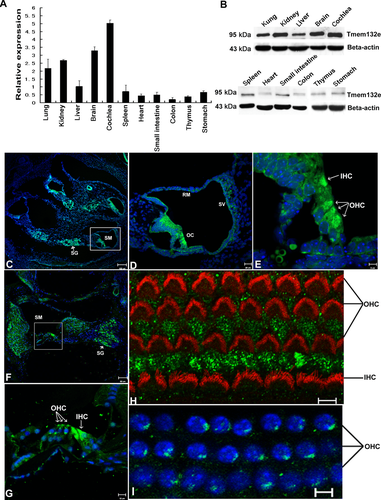
Tmem132e transcripts were present in all mouse tissues examined and expressed at the highest level in cochlea (Fig. 2A). In other organs, the expression ranged from very low (heart and colon) to high (brain and kidney). The protein levels in tissues agreed with mRNA findings (Fig. 2B). Immunofluorescence assay revealed prominent Tmem132e expression in the Organ of Corti, spiral ganglion, and stria vascularis in the inner ear section of P0 mice (Fig. 2C–E) and P30 mouse (Fig. 2F and G). In the Organ of Corti, Tmem132e immunostaining was more intense in the inner and outer hair cells than in pillar cells or supporting cells (Deiter's cells and Hensen's cells; Fig. 2E and G). Compared with the diffuse cytoplasmic distribution of Tmem132e in outer hair cells in the P0 mouse, in the P30 mouse, Tmem132e was mainly located at the apical and basal region of the outer hair cell body. Whole-mount immunostaining of the P30-mouse Organ of Corti was used to analyze any expression of Tmem132e at the bundle of hair cells. However, we did not find remarkable Tmem132e expression at the bundle of hair cells as labeled by TRITC-conjugated phalloidin (Fig. 2H) but found prominent Tmem132e expression at the basal region of the outer hair cells, which agreed with the findings in the P30-mouse cochlea (Fig. 2I).
Tmem132e Is Required for Proper Hair Cell Development in Zebrafish
The full-length cDNA of zebrafish tmem132e was obtained by RT-PCR and subcloned into the pMD18T vector. The open reading frame was 3,222 nt (JX995104), with the first two exons representing the clone XM_003198707 and the other seven exons corresponding to the 3′ portion of XM_687404. Alignments of the obtained sequence with the human TMEM132E showed 59% identity.
The antisense MOs, determined by comparing JX995104 with genomic DNA in GenBank by using BLAST, were designed to knock out exon 3 (Fig. 3A). We investigated the efficacy of the MOs by RT-PCR. Injection of tmem132e MO into 1- to 2-cell-stage embryos resulted in shortened transcripts of the expected size; injection of control MCs did not affect transcripts (Fig. 3B).
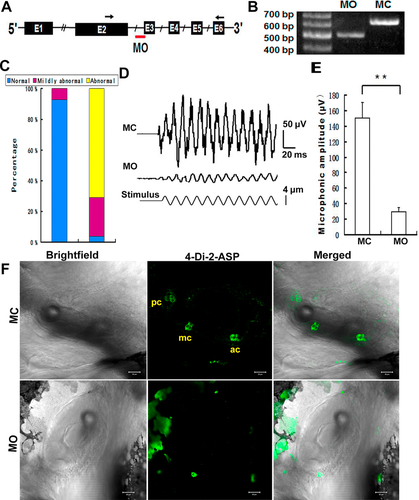
We found no difference in overall appearance of the otic vesicles and otoliths between MO and MC larvae. However, on assessing the startle response, reflecting hearing function, approximately 70% of MO-injected larvae showed a delayed response and abnormal swimming behavior (swimming in a circle as shown in Supp. Movie S1) (Fig. 3C). To further determine the hearing defect in MO larvae, we recorded the microphonic potentials evoked by vibration of the inner ear. MO larvae showed significantly reduced extracellular receptor potentials relative to controls with scramble morpholino injection (Fig. 3D and E). Microphonic potentials are an extracellular manifestation of the transduction currents carried by hair cells in the inner ear [Nicolson et al., 1998]. The defect of microphonic potential in the inner ear could result from nonfunctional or degenerating mechanoelectrical transduction components in hair cells.
We also determined the effect of tmem132e knockdown on the mechanotransductive activity of hair cells by staining with a cationic fluorophore, 4-Di-2-AS, which can permeate the mechanotransduction channels in hair cells and thus indicate the mechanotransduction function of hair cells [Gale et al., 2001]. In zebrafish, hair cells are distributed in the otic vesicle, and the lateral-line system mainly consists of the anterior lateral line (ALL) and the posterior lateral line (PLL). Hair cells of the cristae in the otic cavity of MC larvae were robustly labeled with 4-Di-2-AS, which was micropressure injected into the otic cavity, whereas hair cells in the inner ears of MO-injected larvae showed no uptake of the fluorophore (Fig. 3F). The findings were consistent with the recording of inner ear microphonic potentials, so these fish had impaired mechanoelectrical transduction in hair cells of the inner ear.
Larvae injected with MCs showed prominent fluorescent neuromasts of the lateral-line system (Fig. 4A), and MO morphants had few or no fluorescent neuromasts in ALLs or PLLs. The lateral branch of PLL (L-PLL) neuromasts, the major branch of the PLL, contains seven or eight neuromasts regularly spaced between the ear and the tip of the tail along each side of the zebrafish at 5 dpf [Metcalfe, 1985]. Because of this relatively simple spacing pattern, we could number the labeled L-PLL neuromasts of each injected larva to evaluate the effect of tmem132e MOs on hair cells. The injected fishes were classified into three categories based on total number of labeled L-PLLs on both sides: severely affected (zero to four labeled neuromasts), moderately affected (five to 10 labeled neuromasts), and normal (11–16 labeled neuromasts). We observed 97.6% larvae to be severely affected and 2.4% moderately affected in the tmem132e MO-injected group (n = 239), as compared with 0% and 9.4%, respectively, in the control MC-injected group (n = 186) (Fig. 4B).
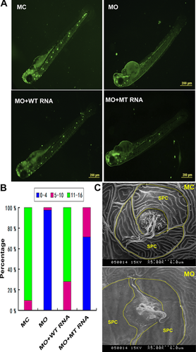
When two-cell embryos were coinjected with tmem132e MO and full-length human TMEM132E mRNA (n = 254) (Fig. 4A, [MO + WT RNA], and B) or human mRNA with c.1259G>A mutation (n = 244) (Fig. 4A, [MO+ MT RNA], and B), we observed 72% normal and 28% moderately affected larvae as compared with 0% normal and 2.8% moderately affected larvae with tmem132e MO and MT RNA coinjected. The frequency of larvae with affected hair cells are significantly different (P < 0.01). These results suggest that human TMEM132E mRNA but not the mRNA with c.1259G>A mutation can compensate for the loss of the endogenous zebrafish tmem132e.
By scanning electron microscopy, we examined the structure of neuromasts in MO-injected larvae and found that it significantly differed from that in control MC larvae (Fig. 4C). In control larvae, the perimeter of each neuromast was delineated by a pair of semilunar periderm cells surrounding the centrally located multiple hair bundles. In contrast, in neuromasts of MO-injected larvae, the hair bundles and semilunar periderm cells were disorganized. In severely affected larvae, no L-PLL neuromast was found because hair bundles were absent.
Discussion
In the present study, we characterized the DFNB99 locus that is adjacent to but does not overlap with DFNB85 and identified TMEM132E as the causative gene for this new form of prelingual, recessive deafness. TMEM132E belongs to a gene family consisting of TMEM132A, B, C, D, and E. TMEM132A is believed to contribute to cell survival by regulating the expression of certain ER stress-related genes in neuronal cells [Oh-hashi et al., 2010]. TMEM132D is expressed in neurons and has been identified as a novel candidate gene for panic disorder [Erhardt et al., 2011; Quast et al., 2012]. Recently, TMEM132E was found a susceptibility gene in panic disorder [Gregersen et al., 2014]. We here showed that Tmem132e is highly expressed in the brain. These data suggest that TMEM132E may play a role in neuronal function and relevant phenotypes.
Many DFNB genes are highly expressed in cochlear hair cells [Shabbir et al., 2006]. We found the expression of Tmem132e highest in the mouse cochlea. Furthermore, immunofluorescence analysis revealed Tmem132e prominently expressed in the apical and basal region of outer hair-cell cytoplasm but not in the hair bundle, in the Organ of Corti of mice. TMEM132E might play an important role in mammalian cochlear development and hearing.
The inner ear in zebrafish is anatomically and functionally similar to that in other vertebrates and is a well-established vertebrate model system for hair cells [Starr et al., 2004; Gleason et al., 2009; Han et al., 2011]. Moreover, zebrafish have an additional mechanosensory organ called the lateral line system, believed to have a common ancestral origin with that of the inner ear and consisting of stereotypically distributed patches of neuromasts made of hair cells surrounded by support cells [Fritzsch, 1988; Kornblum et al., 1990]. We observed that knocking down tmem132e expression in zebrafish caused a delayed startle response. Microphonic potentials are an extracellular manifestation of the transduction currents carried by hair cells in the inner ear [Nicolson et al., 1998]. The significant reduction in microphonic potential in the inner ear further illustrated nonfunctional or degenerating mechanoelectrical transduction components in hair cells. The reduced uptake of fluorophores in hair cells of both the otic cavity and lateral-line neuromasts provided additional evidence of defective mechanoelectrical transduction in Tmem132e-knockdown zebrafish.
Because the wild-type human TMEM132E mRNA could rescue the tmem132e-knockdown phenotype in zebrafish, the function of human TMEM132E may be highly similar to that of zebrafish and could compensate for the loss of endogenous zebrafish tmem132e. In addition, we used PSIPRED, Jpred3, and PredictProtein to predict the possible effect of the p.Arg420Gln variant on the protein structure but could predict no change (data not shown). Further studies are required to determine how c.1259G>A destroys the function of TMEM132E.
Conclusion
We provide evidence supporting the conclusion that the c.1259G > A variant in TMEM132E causes DFNB99. Although TMEM132E variants are probably not common in ARNSHL, our findings illustrate the importance of TMEM132E in the development and function of the auditory system. An appropriate mouse model as well as a thorough biochemical characterization of Tmem132e function is required to better understand the precise cause of deafness due to mutations in TMEM132E.
Acknowledgments
We thank the patients and their families for their participation. We thank Renzhi Zhan and Xuelian Ma (Department of Physiology, Shandong University) for the microphonic potentials recording. We thank Jiangang Gao (School of Life Science, Shandong University) for the analysis of whole-mount immunostaining in the Organ of Corti.
Disclosure statement: The authors declare no conflict of interest.


