Evaluation of the ultrasound findings of thyroid gland enlargement in Cape Coast Teaching Hospital, Ghana: A retrospective cross-sectional study
Abstract
Background and Aim
Goiter is a major source of morbidity in the world, especially in the developing world, where dietary iodine deficiency, a known cause of this condition, is endemic. The diagnosis is mostly by ultrasonography (USG) scan, which can give anatomical, pathological, and functional information for the management of goiter. This study aimed to determine the commonest ultrasound findings of goiter in Ghana.
Method
The records of all 213 patients with goiter diagnosed by USG scan over a 5-year period were retrieved. Data collected were sociodemographics, ultrasound features, thyroid nodules diameter, and Thyroid Imaging Reporting and Data System (TI-RADS) scores, which were analyzed using GNU PSPP, version 1.2.0-3. χ2 and two-tailed independent samples t-test were also employed, with p ≤ 0.05.
Results
A total of 213 patients with goiter diagnosed by USG scan were obtained over the study period. The mean age of the participants was 50.01 ± 17.27 years, with an age range of 16−92 years and females constituting the majority (82.16%). The commonest ultrasound features were well-defined solid nodules. The lesion sites for most patients were the whole thyroid (28.17%), both lobes (24.41%), and the right lobe (20.19%). The mean difference in sizes of cysts and solid nodules among genders was 0.26 (CI: −0.14 to 0.67, p = 0.20) and 0.12 (CI: −0.43 to 0.66, p = 0.67), respectively. The TI-RADS score featured TI-RADS 4 (36.62%), TI-RADS 1 (28.17%), TI-RADS 3 (25.82%), TI-RADS 5 (5.16%), and TI-RADS 2 (4.23%). Solid nodules (49.32%, p = 0.001) and cysts (35.71%, p = 0.003) were more common within 41−60 years and less frequent in those <21 years. A p ≤ 0.05 was considered significant in this study.
Conclusion
The predominant ultrasound features were well-defined solid nodules, simple cysts, and solid nodules with cystic changes, mostly located in the entire thyroid gland and least located in the isthmus only. Cysts and solid nodules were mostly seen in the 41−60 years age group.
Key points
What is known about this topic
-
Well-defined solid nodules, simple cysts, and solid nodules with cystic changes were some of the ultrasound features of an enlarged thyroid gland.
-
Lesion sites of an enlarged thyroid gland included the isthmus, left, and right lobes.
What this study adds
-
Most patients presenting with enlarged thyroid glands had lesions in the entire thyroid.
-
Thyroid Imaging Reporting and Data System (TI-RADS 4) was the most predominant TI-RADS score, and TI-RADS 2 was the least.
-
Few patients presented with lesions in the isthmus only, and the majority in the entire thyroid gland.
-
Cysts and solid nodules were mostly seen in those 41−60 years and least in those <21 years.
Clinical implications of this study
-
Knowing the locations, types, and sizes of thyroid lesions will aid clinicians in planning appropriate management for the patient.
-
Radiologists should adopt the TI-RADS lexicon in their reporting of thyroid imaging to enable proper communication amongst themselves and with the clinicians once they are familiar with the TI-RADS lexicon.
1 BACKGROUND
Thyroid gland enlargement also known as goiter, is a condition that occurs when each of the lateral lobes of the thyroid gland is larger than the person under examination's terminal phalanx of the thumb. In one of the guidelines of the World Health Organization in 2014, it was stated that the goiter prevalence goes up with the seriousness of iodine defect, and it is endemic in regions where the intake of iodine is less than 10 µg per day.1 The prevalence of thyroid gland enlargement worldwide has been reported to be 15.8% and as high as 28.3% in Africa and 4.7% in America.2
The causes of thyroid gland enlargement include iodine deficiency, follicular adenoma, Hashimoto thyroiditis, familial or sporadic multinodular goiter, thyroid cancer, colloid nodules, cysts, and many others. Common clinical presentations of this condition comprise cough, dysphagia, difficulty in breathing, and hoarseness of voice.3 Magnetic resonance imaging, computed tomography scan, plain radiography, radioisotope scintigraphy, fludeoxyglucose positron emission tomography (FDG-PET), and ultrasonography (USG) are the imaging modalities for the examination and diagnosis of this condition.4 However, high-resolution USG scan is the recommended first-line imaging modality due to its high sensitivity in imaging of the thyroid gland and does not involve the use of ionizing radiation.5
Thyroid gland ultrasound helps in the characterization of nodules and detection of other non-palpable but clinically significant nodules, that may require fine needle aspiration.3 Based on their sonographic appearances, thyroid USG scan may also be used to distinguish between malignant and benign thyroid masses.5 Since ultrasound has been reported as the most readily available imaging modality in Ghana, its use in the examination of the thyroid gland for differentiating cystic from solid masses, and its availability will result in the diagnosis of large numbers of thyroid pathologies.6
A reporting system for thyroid nodules on ultrasound has been proposed by the American College of Radiology (ACR) called Thyroid Imaging Reporting and Data System (TI-RADS). This was proposed to deal with the dilemma of how to report the nodules, which are overwhelmingly benign, to ensure consistency across practices. The ACR TI-RADS points, from 0 to 3 are assigned to the ultrasound features from various categories; composition, margin, shape, echogenic foci, and echogenicity, to obtain TI-RADS scores. These assigned points to the features are; cystic (0), mixed cystic and solid (1), solid nodule (2), anechoic (0), isoechoic (1), hypoechoic (2), very hypoechoic (3), wider than tall (0), taller than wide (3), well-defined (0), ill-defined (0), irregular (2), and macrocalcifications (1).7, 8 The scores and their descriptions are shown in Table 1.
| TI-RADS score | Points | Description | Risk of malignancy (%) |
|---|---|---|---|
| 1 | 0 | Benign | 0.3 |
| 2 | 2 | Not suspicious | 1.5 |
| 3 | 3 | Mildly suspicious | 4.8 |
| 4 | 4−6 | Moderately suspicious | 9.1 |
| 5 | ≥7 | Highly suspicious | 35 |
- Note: *ACR TI-RADS.7
- Abbreviation: TI-RAD, thyroid imaging reporting and data system.
The prevalence of thyroid gland enlargement has been reported to be high in Africa and some other continents. A recent study by Vanderpump et al., disclosed that people who are at particular risk, tend to be remote and live in upland areas in Latin America, South-East Asia, and Central Africa. The thyroid gland enlargement prevalence is known to be as high as 80% in areas of severe iodine deficiency.2, 9
-
To determine the commonest ultrasound features in Cape Coast, Ghana.
-
To ascertain predilection sites for thyroid lesions.
-
To determine any possible association between the ultrasound features and the sociodemographics.
2 METHODS
2.1 Study site, participants, and design
This retrospective cross-sectional study reviewed all the thyroid gland ultrasound scan reports, records, and sociodemographics of individuals who met the inclusion criteria (n = 213), who had thyroid gland ultrasound examinations in the radiology division of the Cape Coast Teaching Hospital (CCTH) from February 2016 to 2021. The CCTH, a 400-bed capacity facility, is the only tertiary public health facility in the south-central part of Ghana. It offers subspecialty services (including radiology services) and tertiary services to the populace of this part of the country and beyond. Currently, it is one of the leading medical research institutions in Ghana and the training center for the School of Medical Sciences of the University of Cape Coast and the Nursing and Midwifery Training College in Cape Coast. Cape Coast Metropolis (hosting CCTH) is one of the 17 districts of the Central Region of southern Ghana and a typical coastal community with lots of fish and seafood.
2.2 Data collection
The data obtained from the Lightwave Health Information System, which accommodates all the electronic health data for patients, included the age, sex, thyroid imaging features, and TI-RADS scores from the thyroid gland imaging reports as well as the diameter (in centimeters) of the solid nodules and cysts. Three radiologists with at least 8 years of experience in thyroid gland imaging and reporting did these reports. The ultrasound scan was carried out using the Mindray Diagnostic Digital Ultrasound System, with a 7.5 MHz linear probe, Model DCN3, 2014, manufactured by the Shenzhen Mindray Bio-Medical Electronics Company Limited. TI-RADS scores for all participants were acquired from their ultrasound reports by extracting the thyroid features from these reports and assigning their respective ACR TI-RADS points based on their constituent features. The ACR TI-RADS points assigned to the features seen were; cystic (0), solid with cystic changes (1), solid nodule (2), hypoechoic (2), isoechoic (1), anechoic (0), very hypoechoic (3), taller than wide (3), wider than tall (0), well-defined (0), ill-defined (0), irregular (2), and macrocalcifications (1).7, 8 The sum of all the points in a particular report gives the TI-RADS score for that particular patient, as described in (Table 1). All reports done within the study period were consecutively retrieved for analysis after the exclusion of those with incomplete information (n = 9). The age was categorized as; “<21 years,” “21−40 years,” “41−60 years,” “>60 years,” and sex as “male” and “female.”
-
Patients who underwent thyroid gland ultrasound examination within the study period with complete medical records.
-
Patients with comprehensive thyroid gland ultrasound reports.
-
Patients without complete medical records.
-
Patients without detailed thyroid gland ultrasound reports.
2.3 Statistical analysis
Data collected, including the TI-RADS scores and various ultrasound imaging features or lesions seen under each TI-RADS score grouping, were thoroughly assembled, screened, and coded for analysis using GNU PSPP (Category: Education, Science & Math), pspp version 1.2.0-3, developed by the Free Software Foundation, to derive the tables, frequencies, and percentages as well as performing cross-tabulations. The other results derived were presented in charts using LibreOffice Calc, developed by The Document Foundation (version 1:6.1.5-3+deb10u6). Two-tailed independent samples t-test was used to check for any differences in the mean diameter of solid nodules and cysts among the sexes. Equal variances were assumed for this test based on the results of Levene's test for equality of variances (p = 0.19). The χ2 test was also used to test for association among the variables (age, sex, solid nodules, and cysts). Statistical significance was also set at p ≤ 0.05, for this study.
3 RESULTS
Of the 213 participants included in this study, females constituted the majority (82.16%). The overall mean age was 50.01 (SD = 17.27) years spanning from 16 to 92 years. The males with goiter were generally older (57.16 ± 16.76 years) than the females (48.46 ± 17.04 years). Most of the patients were within the 41−60 years age group (45.54%), followed by those who were more than 60 years (27.23%) and those within 21−40 years (22.07%). Only 5.16% of them were below 21 years, as shown in (Table 2).
| Variable | Frequency/count | Percentage (%) |
|---|---|---|
| Sex | ||
| Males | 38 | 17.84% |
| Females | 175 | 82.16% |
| Age | Minimum (years) | Maximum (years) | Mean ± SD |
|---|---|---|---|
| Overall | 16 | 92 | 50.01 ± 17.27 years |
| Males | 19 | 89 | 57.16 ± 16.76 years |
| Females | 16 | 92 | 48.46 ± 17.04 years |
| Age categorization (years) | Gender | ||
|---|---|---|---|
| Males | Females | Overall (%) | |
| <21 | 1 | 10 | 11 (5.16) |
| 21−40 | 3 | 44 | 47 (22.07) |
| 41−60 | 20 | 77 | 97 (45.54) |
| >60 | 14 | 44 | 58 (27.23) |
| Total | 38 | 175 | 213 (100.00) |
- Abbreviation: SD, standard deviation.
The leading TI-RADS score in the study was TI-RADS 4 (36.62%, n = 78), followed by TI-RADS 1 (28.17%, n = 60) and TI-RADS 3 (25.82%, n = 55). The rarest TI-RADS scores were TI-RADS 2 (4.23%, n = 9) and TI-RADS 5 (5.16%, n = 11) as shown in Figure 1.

The mean size of cysts (diameter) for the females was 1.81 and 1.54 cm for the males, with a mean difference of 0.26 (CI: −0.14 to 0.67), but statistically, there was no difference between the mean sizes (p = 0.20). The average size of solid nodules (diameter) of females was 2.09 cm, and that of males was 1.97 cm with a mean difference of 0.12 (CI: −0.43 to 0.66). Statistically, there was also no difference between the mean sizes of solid nodules among the sexes (p = 0.67) (Table 3).
| Sex | Mean | Standard deviation | Mean difference | 95% confidence interval | t-score | p Value |
|---|---|---|---|---|---|---|
| Sizes of cysts (cm) | ||||||
| Females | 1.81 | 0.68 | 0.26 | −0.14 to 0.67 | 1.30 | 0.20 |
| Males | 1.54 | 0.54 | ||||
| Sizes of solid nodules (cm) | ||||||
| Females | 2.09 | 0.87 | 0.12 | −0.43 to 0.66 | 0.43 | 0.67 |
| Males | 1.97 | 0.74 | ||||
Both lobes, together with the isthmus, were the most affected area in the study, constituting 28.17% (n = 60), implying the majority of the patients presented with lesions in the entire thyroid (both lobes and isthmus), followed by both lobes only (24.41%, n = 52) and the right lobe only (20.19%, n = 43). The least affected areas were the isthmus only (10.33%, n = 22) and the left lobe only (16.90%, n = 36) (Figure 2).

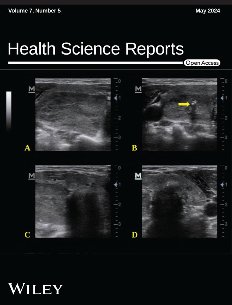
The most common ultrasound features were well-defined solid nodules (38.68%, n = 94) (Figures 3 and 4), simple cysts (25.93%, n = 63) (Figure 5), solid nodules with cystic changes (20.99%, n = 51), and calcifications (6.17%, n = 15) (Figure 4). The least observed feature was complex cysts or cystic with solid components (2.88%, n = 7) (Figure 3). The remaining results on the proportions of the ultrasound features are shown in Table 4.


| Ultrasound feature | Males | Females | Total (%) |
|---|---|---|---|
| Well-defined solid nodules | 11 | 83 | 94 (38.68) |
| Simple cysts | 13 | 50 | 63 (25.93) |
| Complex cysts | 0 | 7 | 7 (2.88) |
| Solid nodules with cystic changes | 14 | 37 | 51 (20.99) |
| Calcifications | 4 | 11 | 15 (6.17) |
| Ill-defined solid nodules | 4 | 9 | 13 (5.35) |
Solid nodules were more common for the 41−60 years age group (49.32%, n = 73), followed by those more than 60 years (31.08%, n = 46) and 21−40 years (16.89%, n = 25). The solid nodules were less frequent in those less than 21 years (2.70%, n = 4), with p = 0.001. The cysts were also more prevalent in the 41−60 years age category (35.71%, n = 25) and the 21−40 years age group (32.86%, n = 23). The rest are shown in Table 5.
| Variable | Age categorization (in years) | χ2 Value | p Value | |||
|---|---|---|---|---|---|---|
| <21 years | 21−40 years | 41−60 years | >60 years | |||
| Solid nodules | ||||||
| Yes | 4 (2.70%) | 25 (16.89%) | 73 (49.32%) | 46 (31.08%) | 15.74 | 0.001 |
| No | 7 (10.77%) | 22 (33.85%) | 24 (36.92%) | 12 (18.46%) | ||
| Cysts | ||||||
| Yes | 7 (10.00%) | 23 (32.86%) | 25 (35.71%) | 15 (21.43%) | 13.72 | 0.003 |
| No | 4 (2.80%) | 24 (16.74%) | 72 (50.35%) | 43 (30.07%) | ||
The distribution of solid nodules and cysts among males and females is shown in (Table 6). The result showed that solid nodules were more common in females (83.11%, n = 123) than males (16.89%, n = 25), though the result was not statistically significant (p-value = 0.57). The cysts were also more frequent in females (81.43%, n = 57) than in males (18.57%, n = 13), with p = 0.85.
| Variable | Sex | χ2 Value | p Value | |
|---|---|---|---|---|
| Males | Females | |||
| Solid nodules | ||||
| Yes | 25 (16.89%) | 123 (83.11%) | 0.30 | 0.57 |
| No | 13 (20.00%) | 52 (80.00%) | ||
| Cysts | ||||
| Yes | 13 (18.57%) | 57 (81.43%) | 0.04 | 0.85 |
| No | 25 (17.48%) | 118 (82.52%) | ||
4 DISCUSSION
Thyroid gland enlargement has affected many people in the world, ranging from mild to severe conditions, which has reduced the quality of life, especially in women.13, 14 Epidemiological data have revealed that pregnancy poses a significant challenge to the maternal thyroid gland. This is due to the increase in hormone requirements during gestation, resulting from increased thyroid-binding globulin, and peripheral thyroid hormone requirements, as well as the stimulatory effect of human chorionic gonadotropin on thyroid-stimulating hormone receptors.15-17 The direct actions of estrogen on the thyroid tissue also contribute to the development of thyroid gland enlargement, as reported by Li et al.18 In our study, we found that about 82.12% of the patients with thyroid gland enlargement were females. A similar result has been reported by Dauksiene et al., in Lithuania, who also found that thyroid gland enlargement is more prevalent in women than men, even though they reported a comparatively lower proportion (69.23% for women).19 This finding could also be due to the high consumption of cassava, which is a major staple in our setting among females, corroborated by other studies in Uganda, Nigeria, and Kenya.20-22 The consumption of cassava induces the development of thyroid gland enlargement, due to the presence of goitrogenic compounds like methimazole, resorcinol, potassium perchlorate, phloroglucinol, ethylene thiourea, 6-propyl-2-thiouracil, and pyrazole.22-24 This could further support the higher prevalence of thyroid gland enlargement in females.
In our study, we found that thyroid gland enlargement was more predominant in the 41−60 years age category, constituting 45.54%, which is in agreement with what was found by Saeed et al., in Saudi Arabia, who also reported a modal (43.93%) age group of 41−60 years.25 These findings are contrary to what was reported by Zekarias et al., who indicated that thyroid gland enlargement is more common in those who are less than 35 years old (67.7%).26 In assessing whether there is a difference in the sizes of solid nodules and cysts among the sexes, we found that the sizes of solid nodules and cysts in males were smaller than in females, but these differences in the sizes were not statistically significant, with p, 0.20 and 0.67 for males and females, respectively (Table 3). This is contrary to the findings of Germano et al., who found a significant difference in the sizes of thyroid nodules (solid and cystic) among the sexes.27
TI-RADS 4 was the most frequent TI-RADS score (36.62%) in our study, indicating that the majority of the patients had moderately suspicious thyroid lesions, followed by TI-RADS 1 (28.17%) and TI-RADS 3 (25.82%) (Figure 1). In a study by Borlea et al., in Romania, TI-RADS 4 also constituted the majority of 48.12%, but contrary to what we found, their second and third commonest TI-RADS scores were TI-RADS 5 (29.32%) and TI-RADS 2 (18.80%), respectively. The authors recorded no TI-RADS 3 in their study.28 In another study by Basha et al., their three most common TI-RADS scores were TI-RADS 2 (60.1%), TI-RADS 3 (18.0%), and TI-RADS 4 (12.7%).29 These disparities could be due to the differences in the demographics. We also found that the three most frequent ultrasound features of thyroid gland enlargement in our setting were solid nodules (44.03%), simple cysts (25.93%), and solid nodules with cystic changes (20.99%) (Table 4). Studies by Girardi et al., and Xi et al., reported solid nodules as the leading feature, followed by mixed nodules (solid with cystic changes) and calcifications, constituting 46.97%, 24.85%, and 20.91%, respectively, for Girardi et al., and 44.13%, 32.39%, and 23.47%, respectively for Xi et al.30, 31
The whole thyroid was most commonly affected (28.17%) in our study, followed by both lobes only (24.41%) and the right lobe only (20.19%) (Figure 2). Contrary to what we found, Saeed et al., reported that most patients presented with lesions in the right lobe only (41.60%), followed by both lobes only (37.00%) and the left lobe only (19.10%). The isthmus was the least affected area of the thyroid gland (1.20%) in their study, as also seen in ours (10.33%).25 Other studies have also reported the right lobe as the most affected area of the thyroid and the isthmus as the least.32, 33 Even though the isthmus is the least affected site of the thyroid gland in the studies discussed above, it nonetheless could be an extra risk factor to examine for an accurate surgical approach in undetermined thyroid lesions or cancer at fine-needle aspiration.34
For the age distribution of solid nodules and cysts, we found that solid nodules and cysts were more predominant in those 41–60 years, constituting 49.32% and 35.71%, respectively, and less common in those less than 21 years, which also constituted 2.70% and 10.00%, respectively. Similar findings have been reported by Lin et al., who also found solid nodules and cysts to be more common in the 41–60 years age category.35 Our study also revealed that solid nodules and cysts are more prevalent in females than males, even though these results were not statistically significant, with p = 0.57 and p = 0.85, respectively (Table 6). This could be due to the high incidence of thyroid diseases in females, as also seen in our study (Table 2).36 In Japan, similar findings have also been corroborated by Uchino et al. In their study, cysts and solid nodules constituted the majority of 70.83% and 71.43%, respectively in females.37
5 LIMITATION
The relatively small sample size used in this retrospective study was a limitation, hence the findings for this study cannot be generalized. Future multicenter prospective studies with larger sample sizes may be required. Histopathology findings and the thyroid function test were not included in the study since the focus of this was mainly on ultrasonographic features.
6 CONCLUSION
Well-defined solid nodules, simple cysts, and solid nodules with cystic changes were the most common ultrasound features of enlarged thyroid gland in our study. The diameters of solid nodules and cysts were higher for females than for males, but these findings were not statistically significant. TI-RADS 4 was the most predominant TI-RADS score, and TI-RADS 2 was the least. This indicates that the majority of the patients had moderately suspicious thyroid lesions, which is an issue of public health concern. Few patients presented with lesions in the isthmus only, and the majority in the entire thyroid gland. Cysts and solid nodules were mostly seen in those aged 41−60 years and least in those aged <21 years.
AUTHOR CONTRIBUTIONS
Emmanuel Kobina Mesi Edzie: Conceptualization; methodology; investigation; writing—original draft; writing—review and editing; validation; formal analysis; project administration. Klenam Dzefi-Tettey: Conceptualization; methodology; investigation; validation; writing—original draft; writing—review and editing; formal analysis. Edmund Kwakye Brakohiapa: Conceptualization; methodology; validation; writing—original draft; writing—review and editing; formal analysis; supervision. Obed Nimo: Writing—original draft; writing—review and editing; methodology; investigation; formal analysis; supervision; validation. Peter Appiah-Thompson: Writing—original draft; writing—review and editing; methodology; validation; formal analysis; supervision; conceptualization. Michael Kofi Amedi: Writing—original draft; writing—review and editing; methodology; investigation; validation; conceptualization. Ansumana Bockarie: Writing—original draft; writing—review and editing; validation; conceptualization; investigation. Frank Quarshie: Writing—review and editing; writing—original draft; methodology; investigation; validation; data curation; formal analysis; software. Emmanuel Onimole: Methodology; writing—original draft; writing—review and editing; visualization; formal analysis; resources. Emmanuel Akorli: Writing—original draft; writing—review and editing; investigation; methodology; formal analysis; validation. Richard Anthony: Writing—original draft; writing—review and editing; methodology; investigation; formal analysis; supervision. Richard Ato Edzie: Writing—original draft; writing—review and editing; methodology; investigation; data curation; formal analysis. Nana Ama Amankwa: Writing—original draft; writing—review and editing; investigation; methodology; validation; formal analysis. Aaron Amartey: Writing—original draft; writing—review and editing; methodology; validation; supervision; formal analysis; software. Bernard Osei: Writing—original draft; writing—review and editing; investigation; methodology; validation; formal analysis; data curation. Bright Oppong: Writing—original draft; writing—review and editing; validation; formal analysis; methodology; investigation; supervision. Abdul R. Asemah: Writing—original draft; writing—review and editing; methodology; investigation; validation; formal analysis; data curation; project administration. Philip Narteh Gorleku: Writing—original draft; writing—review and editing; conceptualization; methodology; investigation; supervision; validation.
ACKNOWLEDGMENTS
We are thankful to the management and staff of the records and Ultrasound scan units of Cape Coast Teaching Hospital (CCTH) for their support in making this study successful. All the authors supported this research.
CONFLICT OF INTEREST STATEMENT
The authors declare no conflict of interest.
ETHICS STATEMENT
The committee for ethical review, Cape Coast Teaching Hospital, gave approval for this research to be carried out, with clearance number CCTHERC/EC/2020/096. Anonymity and confidentiality were ensured throughout the study. This study conformed to the 1975 Declaration of Helsinki. Informed consent was not obtained for this large number of retrospective patients as we could not have contacted all the patients because patients might have changed contacts, dead at the time of data collection, or been long discharged. These were made known to the CCTHERC, and no concerns were raised.
TRANSPARENCY STATEMENT
The lead author, Emmanuel Kobina Mesi Edzie, affirms that this manuscript is an honest, accurate, and transparent account of the study being reported, that no important aspects of the study have been omitted, and that any discrepancies from the study as planned (and if relevant, registered) have been explained.
Open Research
DATA AVAILABILITY STATEMENT
The data that support the findings of this study are available from Cape Coast Teaching Hospital (CCTH). Restrictions apply to the availability of these data, to protect patients' confidentiality. Data are available from the authors with the permission of CCTH through the email address: [email protected]. The corresponding author (Emmanuel Kobina Mesi Edzie) had full access to all of the data in this study and takes complete responsibility for the integrity of the data and the accuracy of the data analysis.