Hepatology Highlights
IMAGINE There Is No Needle
Muzammil Muhammad Khan,* Maria Alejandra Luna-Cuadros,** and Daryl T.Y. Lau1
Optimal assessment of advanced fibrosis in NAFLD remains a challenge. In a prospective NAFLD registry, Cassinotto et al. selected 577 consecutive patients who had liver biopsy, Fibrosis-4 index (FIB-4), vibration-controlled transient elastography (VCTE), and two-dimensional shear-wave elastography with SuperSonic Imagine (2D-SWE-SSI). Using liver biopsy as the gold standard, multistep strategies were compared to identify advanced fibrosis (F ≥ 3). In a two-step approach, FIB-4 followed by VCTE or 2D-SWE-SSI had comparable accuracy, but 15%-25% would still require a liver biopsy to determine advanced fibrosis. For patients with a gray-zone elastography score (8-10 kPa), a three-step strategy with FIB-4/2D-SWE-SSI + VCTE or FIB-4/VCTE + 2D-SWE-SSI would further reduce the need for biopsy to 7.3%. Combined noninvasive modalities can provide accurate assessment of advanced fibrosis, but these strategies need to be validated in diverse patient populations and clinical settings. (Hepatology 2020;73:2196-2205).
HNF4α Halts Disease Progression in NASH
Tejasav S. Sehrawat,2 Ojasav Sehrawat,3 and Robert E. Schwartz4
Xu et al. describe the important role of liver-enriched transcription factor hepatocyte nuclear factor 4 alpha (HNF4α) in preventing disease progression from NAFLD to NASH. They demonstrate a protective role of HNF4α in a high-fat/cholesterol/fructose murine model of NASH using gain- and loss-of-function studies. Lower levels of inflammatory (e.g., monocyte chemoattractant protein 1, Tnfα) and fibrotic markers (e.g., collagen, type I, alpha 1; hydroxyproline) were noted in mice overexpressing HNF4α delivered by adeno-associated virus serotype 8/albumin injections. Additionally, they show lower hepatic triglyceride levels and lipid droplet accumulation in these mice. In contrast, opposite results were observed in Hnfα–/– mice. HNF4α overexpression failed to exert similar protective effects in p53Hep–/– mice. Furthermore, they establish that this protective effect is exerted through an HNF4α-mediated coordinated modulation of lipolytic, p53, and bile acid signaling in hepatocytes. Taken together, these data suggest a potential therapeutic role of targeting HNF4α in NAFLD and NASH. (Hepatology 2020;73:2251-2265).
Foreseen With Fourteen
Anthony J. Choi*** and Robert S. Brown, Jr.4
Although our understanding of HCC tumor heterogeneity continues to advance, the current clinical staging systems do not take it into account. Morris et al. set out to quantify interpatient biological heterogeneity by analyzing 317 proteins in 767 HCC patients and 200 controls. Using 14 proteins, the investigators developed the “Hepatoscore-14,” which categorized the degree of aberration in biomarker signature as low, medium, or high. When applied prospectively to patients, the low, medium, and high Hepatoscore groups had significantly different overall survival rates. Furthermore, when applied within already existing prognostic classifications (e.g., metastasis, performance status) and staging systems (e.g., Child-Turcotte-Pugh score, Barcelona Clinic Liver Cancer stage), significant survival differences were again demonstrated. These findings suggest that accounting for tumor heterogeneity will improve prognostication and may provide insight into research eligibility and treatment modalities that ultimately improve outcomes for HCC. (Hepatology 2020;73:2278-2292).
A Lock-Smith 4 Macrophages in HCC
Gopanandan Parthasarathy2 and Robert E. Schwartz4
Tan et al. investigated the role of lysyl oxidase-like 4 (LOXL4) in HCC with a focus on its effect on macrophages. In a choline-deficient, amino-acid–defined diet plus carbon tetrachloride mouse model of HCC, mRNA levels of LOXL4 were increased in hepatic parenchymal cells, but protein levels colocalized in macrophages. In LOXL4 knockout mice, tumor growth and infiltration of tumor-associated macrophages (TAMs) were reduced. Conversely, injection of recombinant LOXL4 (rLOXL4) increased protein levels within TAMs and increased tumor growth. Specifically, rLOXL4 potently accelerated maturation of bone-marrow–derived rather than resident macrophages, and macrophages treated with rLOXL4 maintained Ly6C as a surface marker and were immunosuppressive on T cells in vitro. This was attributable to up-regulation of programmed death ligand 1 (PD-L1) by rLOXL4, which was dose dependent and specific to macrophages. Finally, in 90 patients with HCC, LOXL4 and PD-L1 expression on macrophages was correlated with each other and inversely with overall survival. Taken together, LOXL4 represents an interesting therapeutic target for HCC treatment. (Hepatology 2020;73:2326-2341).
I Mast React!
Enis Kostallari2 and Robert E. Schwartz4
Cholestatic liver injury is characterized by ductular reaction and accompanied by an increased number of mast cells (MCs) surrounding damaged bile ducts. In this study, Kyritsi et al. demonstrate that introduction of MCs into wild-type or MC-deficient KitW-sh mice induced lobular damage, necrosis, ductular reaction, and inflammation. MC administration to mice also promoted angiogenesis, HSC activation, and liver fibrosis. To further evaluate the effect of MCs in the liver, the investigators used an Mdr2 knockout mice (Mdr2–/–) HDC–/– double-knockout mouse model, which shows reduced liver damage, inflammation, and biliary senescence as compared to Mdr2–/– mice. Injection of MCs to these double-knockout mice increased liver damage, inflammation, senescence, and liver fibrosis in a TGF-β1 signaling manner. However, the effect of MCs on the liver was attenuated when MCs lacked TGF-β1 signaling. As a conclusion, MC-derived TGF-β1 may be a target in chronic cholestatic liver disease. (Hepatology 2020;73:2397-2410).
Activation Proliferation Liverosis Network
Nidhi Jalan-Sakrikar2 and Robert E. Schwartz4
Apelin (APLN) ligand modulates several biological pathways, including organ fibrosis, through its receptor, APJ. Serum APLN levels are elevated in patients with chronic liver diseases along with an increased hepatic expression of APJ. Chen et al. evaluated whether the APLN-APJ axis plays a role in ductular reaction and hepatic fibrosis during cholestasis. Liver sections of patients with primary sclerosing cholangitis showed increased immunoreactivity for APLN and APJ compared to healthy controls. Mouse models of cholestatic liver disease also displayed increased APLN levels in serum and in isolated cholangiocytes. This was in conjunction with increased intrahepatic bile duct mass. Antagonizing the APLN-APJ axis, either pharmacologically or genetically, ameliorated experimentally induced cholestasis and fibrosis in mice by reducing cholangiocyte proliferation. Mechanistically, APLN through APJ stimulated the formation of reactive oxygen species (ROS) that triggered extracellular signal-regulated kinase signaling to induce cholangiocyte as well as HSC activation and proliferation. Thus, the APLN-APJ axis represents a novel therapeutic target for cholestatic liver diseases. (Hepatology 2020;73:2411-2454).
Drugs Don’t Work in Patients Who Don’t Take Them
David M. Salerno5 and Robert S. Brown, Jr.4
Poor adherence, prescription access issues, and medication misuse can increase morbidity and hospital admissions in patients with decompensated cirrhosis. Thomson et al. reviewed the Optum Clinformatics Data Mart for outpatient prescription claims between 2010 and 2015 for >12,000 patients with decompensated cirrhosis. Alcohol-associated liver disease was the most prevalent etiology (38%), ascites was the most common complication (87%), and patients were taking a median of 3.6 medications. First-fill rates for medications to treat ascites, HE, and varices were low at 56%, 63%, and 60%, respectively. Less than 20% of patients had enough medication to cover >50% of the follow-up days. During a median follow-up of 2.3 years, potentially inappropriate medication use was common, with >50% of patients filling a prescription for opiates and 10% for nonsteroidal anti-inflammatory drugs. A multidisciplinary approach and integration of pharmacy records with medical records would help address gaps between intended and actual use. (Hepatology 2020;73:2429-2440).
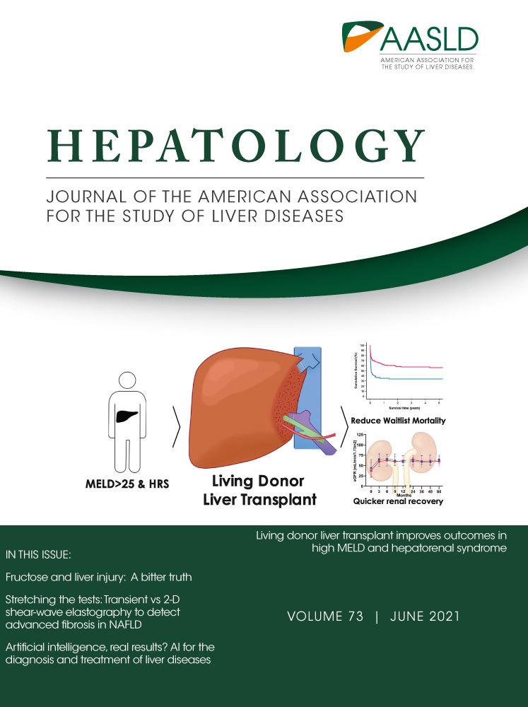
Living Donor Transplants Live Well!
Chaitanya Allamneni**** and Ravi S. Vora6
Traditionally, Model for End-Stage Liver Disease (MELD) >25 and need for renal replacement therapy have been viewed as contraindications to living donor liver transplant (LDLT) because of high risk of mortality and graft loss in this group. Wong et al. analyzed patients from 2008 to 2017 on an intention-to-treat (ITT) basis comparing ITT-LDLT to ITT-DDLT (deceased donor liver transplantation) outcomes. Within the ITT-LDLT group, risk of death was lower, transplant rate was higher, and 5-year overall survival was greater for MELD >25 and hepatorenal syndrome patients. There was a significant survival advantage within the first month after listing for ITT-LDLT. Perioperative outcomes and 5-year survival were similar between LDLT and DDLT groups. Overall ITT-LDLT reduced waitlist mortality and allowed earlier access to transplant, suggesting that the application of LDLT should not be restricted by an arbitrary MELD cutoff. (Hepatology 2020;73:2441-2454).
…and the Golden cytoGlobe for Limiting Fibrosis Goes to…
Jessica L. Maiers2 and Robert E. Schwartz4
ROS are a major contributor to HSC activation and liver fibrosis. Fibrotic livers display increased ROS levels as well as reduced oxidative scavenging by antioxidant systems. Previous work had shown that overexpression of the ROS scavenger, cytoglobin, was sufficient to limit HSC activation in response to ROS-induced damage, but the therapeutic potential of cytoglobin was unclear. The article by Dat et al. showed that overexpression of cytoglobin attenuated fibrogenesis in mouse models of cholestatic liver disease or in response to diet-induced steatohepatitis. This group then engineered a his-tagged cytoglobin and studied whether cytoglobin could be uptaken by HSCs and limit both HSC activation and fibrogenesis. Clathrin-mediated uptake of His-cytoglobin reduced expression of canonical HSC activation genes in either culture- or ROS-activated HSCs in vitro, and this effect was mediated, at least in part, by interferon γ. In vivo studies further showed that His-cytoglobin could safely and effectively protect mice from either thioacetamide- or a DDC-induced liver fibrosis. In summary, delivery of the ROS scavenger, cytoglobin, is a potential antifibrotic strategy for patients with chronic liver disease. (Hepatology 2020;73:2527-2545).