The Role of the Dorsolateral Prefrontal Cortex in Ego Dissolution and Emotional Arousal During the Psychedelic State
ABSTRACT
Lysergic acid diethylamide (LSD) is a classic serotonergic psychedelic that induces a profoundly altered conscious state. In conjunction with psychological support, it is currently being explored as a treatment for generalized anxiety disorder and depression. The dorsolateral prefrontal cortex (DLPFC) is a brain region that is known to be involved in mood regulation and disorders; hypofunction in the left DLPFC is associated with depression. This study investigated the role of the DLPFC in the psycho-emotional effects of LSD with functional magnetic resonance imaging (fMRI) and magnetoencephalography (MEG) data of healthy human participants during the acute LSD experience. In the fMRI data, we measured the correlation between changes in resting-state functional connectivity (RSFC) of the DLPFC and post-scan subjective ratings of positive mood, emotional arousal, and ego dissolution. We found significant, positive correlations between ego dissolution and functional connectivity between the left & right DLPFC, thalamus, and a higher-order visual area, the fusiform face area (FFA). Additionally, emotional arousal was significantly associated with increased connectivity between the right DLPFC, intraparietal sulcus (IPS), and the salience network (SN). A confirmational “reverse” analysis, in which the outputs of the original RSFC analysis were used as input seeds, substantiated the role of the right DLPFC and the aforementioned regions in both ego dissolution and emotional arousal. Subsequently, we measured the effects of LSD on directed functional connectivity in MEG data that was source-localized to the input and output regions of both the original and reverse analyses. The Granger causality (GC) analysis revealed that LSD increased information flow between two nodes of the ‘ego dissolution network’, the thalamus and the DLPFC, in the theta band, substantiating the hypothesis that disruptions in thalamic gating underlie the experience of ego dissolution. Overall, this multimodal study elucidates a role for the DLPFC in LSD-induced states of consciousness and sheds more light on the brain basis of ego dissolution.
1 Introduction
Psychedelics have been utilized as therapeutic tools across the world for millennia (Carod-Artal, Carod-Artal 2015). The revitalization of psychedelic research in the last decade has led to many new clinical trials, which have demonstrated that LSD and other psychedelic compounds may be promising treatments for psychiatric disorders. In conjunction with therapy, psilocybin is showing promise for treating end-of-life distress (Griffiths et al. 2016), smoking cessation (Johnson et al. 2017), major depressive disorder (MDD) (Carhart-Harris et al. 2021), and alcohol use disorder (Bogenschutz et al. 2022). Additionally, LSD has shown impressive results in clinical trials for generalized anxiety disorder (GAD) and MDD (Beutler et al. 2024; Holze et al. 2022).
Beyond psychedelics, there are other established interventions that may treat psychiatric conditions through potentially related mechanisms. Accelerated intermittent Theta Burst Stimulation (iTBS), a version of repeated Transcranial Magnetic Stimulation (rTMS), versus sham has shown an ~90% response rate in limited clinical trials for the treatment of MDD (Cole et al. 2022). This intervention targets the left dorsolateral prefrontal cortex (lDLPFC) in a high-frequency, excitatory manner (Vida et al. 2023). A randomized sham-controlled study of rTMS excitation over the rDLPFC was found to effectively reduce symptoms of mania in conjunction with medication (Praharaj et al. 2009), and is now a third-line treatment for mania in Canada (Yatham et al. 2018). Moreover, low-frequency, inhibitory TMS over the right DLPFC (rDLPFC) is also effective for treating depression (Chen et al. 2013). These opposing effects of excitation and inhibition in the left and right DLPFC suggest a hemispheric lateralization of function within the DLPFC and its relevance to mood regulation and mood disorder. A recent meta-analysis showed that the left DLPFC plays a role in executive function while the right DLPFC has a stronger role in other aspects of cognitive processing and emotional responses (Lin and Feng 2024).
Under LSD, ego dissolution strongly alters mood while also (acutely) resembling the mania-like symptoms of acute, first-episode psychosis (Preller and Vollenweider 2018). Ego dissolution can either be euphoric or anxiety-inducing and is characterized by experiences of interconnectedness, the blurring of the boundary between self and other, and a sense that ordinarily insignificant things have become profoundly meaningful (Friesen 2022). Meanwhile, emotional arousal underpins the experience of mania and psychosis and is also a fundamental aspect of the psychedelic experience. Clinical populations with bipolar I disorder and schizophrenia typically experience difficulties with emotional regulation (Johnson et al. 2016), which can contribute to the experience of psychosis (Haralanova et al. 2012). In the psychedelic state, emotional arousal can manifest as euphoria, anxiety, or a loss of self-control, which can be examined through questionnaires such as the Altered States of Consciousness (ASC) scale (Dittrich 1998).
In this study, we investigate whether the left and the right DLPFC play different roles in mediating the subjective experience of LSD. This study is a new analysis of data collected by Carhart-Harris, Kaelen, et al. (2016), Carhart-Harris, Muthukumaraswamy, et al. (2016). In a randomized, within-subjects study of healthy volunteers, participants were administered LSD and placebo in two separate dosing sessions and asked to complete a post-scan Visual Analogue Scale (VAS) including measures of positive mood, ego dissolution, and emotional arousal. Subjective ratings were correlated with resting-state functional connectivity (RSFC) of the left, right, and combined DLPFC, as captured with functional magnetic resonance imaging (fMRI). We predicted that changes in functional connectivity of both the left and right DLPFC induced by LSD would correlate with reported changes in ego dissolution, since psychedelics evoke a spectrum of effects that encompasses emotional arousal, including shifts in mood and mania-like symptoms. Furthermore, we expected changes in lDLPFC seed-based FC under LSD would be associated with subject-reported changes in positive mood, based on the observation of changes in depressive symptoms resulting from TMS over the lDLPFC. Lastly, we expected alterations in rDLPFC under LSD to correlate with reported changes in emotional arousal, based on observed changes in arousal and mania-like symptoms after excitation of the rDLPFC in TMS.
2 Methods
2.1 Participants
fMRI Blood Oxygen Level Dependent (BOLD) and MEG data were collected from 20 participants, of which 15 (four females; mean age, 30.5 ± 8.0 years) were deemed suitable for analysis. One participant withdrew from the study due to excessive anxiety, while four others exhibited excessive head motion. For full inclusion criteria, please refer to Carhart-Harris, Kaelen, et al. (2016), Carhart-Harris, Muthukumaraswamy, et al. (2016). Ethical approval was granted by the National Research Ethics Service committee London–West London. The research complied with the revised declaration of Helsinki (2000), the International Committee on Harmonization Good Clinical Practice guidelines, and the National Health Service Research Governance Framework.
2.2 Study Design
Participants underwent two scanning sessions, one with placebo (PLCB) and one with LSD, separated by at least fourteen days. Participants were administered either PLCB or 75 μg of LSD intravenously via a 10 mL solution infused over two minutes. After a 60-min acclimatization period, subjects were led to an MRI scanner for Arterial Spin Labeling (ASL) and fMRI and then to magnetoencephalography (MEG) 165 min after drug administration. As stated above, the ASL data was not analyzed in this study. Both the fMRI and MEG recordings that were analyzed were eyes-closed resting-state scans. Eyes-open resting-state, eyes-closed music-listening, and video-watching recordings were also acquired but were not included in this analysis due to a primary focus on eyes-closed rest (Carhart-Harris, Kaelen, et al. 2016; Carhart-Harris, Muthukumaraswamy, et al. 2016).
2.3 Subjective Reports
Directly after each scan, participants were administered a Visual Analogue Scale (VAS), which asks a series of questions about their subjective experience. The six domains of questions were the Intensity of the Experience, Simple Imagery, Complex Imagery, Positive Mood, Ego dissolution, and Emotional Arousal. The average (± S.D.) changes from PLCB to LSD were 10.4 ± 4.8 for Emotional Arousal, 6.7 ± 6.0 for Positive Mood, and 5.7 ± 7.2 for Ego Dissolution. At the end of each scan day, participants were also given the 11-dimensional Altered States of Consciousness (ASC) questionnaire, which asked participants to retrospectively rate their subjective experience at the peak of the compound's effects.
2.4 fMRI
2.4.1 fMRI Acquisition
MRI data was captured using a 3 T GE HDx system. Two BOLD-weighted fMRI data sets were acquired via a gradient echo planar imaging sequence. The pulse sequence consisted of TR/TE = 2000/35 ms, field-of-view = 220 mm, 64 × 64 acquisition matrix, parallel acceleration factor = 2, and 90° flip angle. Thirty-five oblique axial slices were captured in an interleaved manner, each being 3.4 mm thick with zero slice gap (3.4 mm isotropic voxels). Each of the two BOLD scans had a precise duration of 7:20 min. An additional 7:20 min run was conducted between these two scans, but it was excluded from this study because of its added music component.
2.4.2 fMRI Preprocessing
Preprocessing of the fMRI data was performed previously by Carhart-Harris, Kaelen, et al. (2016), Carhart-Harris, Muthukumaraswamy, et al. (2016) using a combination of four distinct yet complementary softwares: FMRIB Software Library (FSL) (Smith et al. 2004), AFNI (Cox 1996), Freesurfer (Dale et al. 1999), and Advanced Normalization Tools (ANTS) (Avants et al. 2011). Participants were excluded if more than 15% of their volumes were scrubbed, with a scrubbing threshold of frame-wise head displacement (FWHD) > 0.5 mm. Preprocessing stages included (1) removal of the first three volumes; (2) de-spiking; (3) slice time correction; (4) motion correction; (5) brain extraction; (6) rigid body registration to anatomical scans; (7) non-linear registration to 2 mm MNI brain; (8) and scrubbing. Scrubbed volumes were replaced with the mean of the surrounding volumes.
Further pre-processing steps included (1) spatial smoothing; (2) band-pass filtering; (3) linear and quadratic detrending; (4) regression of nine nuisance regressors. Six nuisance regressors were motion-related, and three were anatomically related. The three anatomical nuisance regressors calculated were ventricles (FreeSurfer, eroded in 2 mm space), draining veins (FSL's CSF minus FreeSurfer's Ventricles, eroded in 1 mm space) and local white matter (WM) (FSL's WM minus FreeSurfer's subcortical gray matter (GM) structures, eroded in 2 mm space). Regarding WM regression, AFNI's 3dLocalstat was used to calculate the mean local WM time-series for each voxel, using a 25 mm radius sphere centered on each voxel (Jo et al. 2010).
2.4.3 fMRI Resting State Network Analysis
2.4.3.1 Seed Location
The seed-based analysis was performed with the use of FMRIB Software Library (FSL). Previous research of the DLPFC has utilized Brodmann Area 9/46, which does not account for recent developments in the understanding of the heterogeneity of this brain region (Cieslik et al. 2013; Jung et al. 2022). Thus, our study derived novel seed regions from rTMS studies that targeted the lDLPFC at (−42, 44, 30) in the Montreal Neurological Institute (MNI) coordinate system (Weigand et al. 2018). While this is an effective TMS stimulation point, it is not conducive for resting-state analysis because it is on the edge of the brain and is therefore prone to generating artifacts. To solve this problem, the lDLPFC seed was shifted by six units in each axis to (−36, 38, 24), thereby ensuring that an 8 mm sphere centered around it would not overlap with the edge of the brain. The rDLPFC seed was centered at the mirror location (36, 38, 24) in the right hemisphere.
2.4.3.2 Functional Connectivity Analysis
Resting-state seed-to-voxel connectivity was measured between each brain voxel and the lDLPFC, rDLPFC, and combined DLPFC seeds. In the analyses of just the left or right DLPFC, the opposing seed's activity was regressed out in the general linear model (GLM). We first analyzed the difference in connectivity between the PLCB and LSD conditions (Delta analysis), then examined correlations between the connectivity of each seed and the VAS ratings of Ego Dissolution, Emotional Arousal, and Positive Mood (Covariate analysis). All analyses were computed using FSL-Randomise (5000 permutations per test and contrast) (Winkler et al. 2014). Variance smoothing was employed at 6 mm for the between conditions t-test. Clusters were considered significant if pFWE < 0.05, corrected for multiple comparisons using threshold-free cluster enhancement within trial (Smith and Nichols 2009).
2.4.3.3 Confirmational Analysis
The seeds in this study are novel; in particular, the individual and combined DLPFC seeds have traditionally not been used for resting-state FC analyses. Therefore, to ensure the robustness of our results, we performed a confirmational analysis in which we leveraged the absolute valued and binarized output clusters from the above analysis as the input seeds for new Delta and Covariate analyses. Our hypothesis was that the confirmational analysis would yield clusters containing the original DLPFC seeds. This would confirm that the subjective experiences of ego dissolution, emotional arousal, and positive mood are specific to the FC of the DLPFC.
2.5 MEG
2.5.1 MEG Acquisition
Participants were recorded with a CTF 275-gradiometer MEG, though four of the sensors were turned off because of excessive sensor noise. Each scan lasted approximately seven minutes. MEG data was acquired at 600 Hz. There were two scans associated with the eyes-closed resting-state condition; we randomly selected one per subject. Simultaneous electrocorticography (ECG), vertical and horizontal electrooculography (EOG), and electromyography (EMG) recordings were acquired.
2.5.2 MEG Preprocessing and Source Reconstruction
Preprocessing of the MEG data in this study was similar, but not identical, to the pipeline described in Carhart-Harris, Kaelen, et al. (2016), Carhart-Harris, Muthukumaraswamy, et al. (2016). All preprocessing was performed in FieldTrip (Oostenveld et al. 2011). Data was high-pass filtered at 1 Hz with a 2nd-order Butterworth filter, downsampled initially to 400 Hz, and segmented into 2-s epochs. Line noise at 50 and 100 Hz was removed with a Discrete Fourier Transform filter. Outlier epochs and channels were manually deleted by visual inspection. An automatic algorithm was used to remove muscle artifacts from right-hemisphere temporal channels at the edge of the MEG dewar, which are most likely to be contaminated by such artifacts (Muthukumaraswamy et al. 2015). In particular, such channels with high activity (z > 6) and frequency between 105 and 145 Hz were suppressed. Independent component analysis (ICA) using the logistic infomax algorithm was applied to detect ECG and EOG artifacts (Bell and Sejnowski 1995). Components were manually removed by visual inspection. Finally, data was downsampled again to 200 Hz to reduce the size of the data. The key differences between our preprocessing pipeline and that of Carhart-Harris, Kaelen, et al. (2016), Carhart-Harris, Muthukumaraswamy, et al. (2016) are the lack of automatic ICA artifact removal and the downsampling to 200 Hz. (Note that the results still hold with the original preprocessing).
Source reconstruction followed a procedure similar to the one described in (Mediano et al. 2024). The fMRI analysis yielded four networks: sets of regions that correlated with ratings of ego dissolution and emotional arousal, in two different “directions” (“forward,” or original, and “reverse,” or confirmational). For each network, a template consisting of the centroids of the constituent regions was inversely warped to each subject's native-space anatomical MRI. A head model for each participant was constructed using the single-shell method. Based on the corresponding leadfield model, a linearly constrained minimum variance (LCMV) beamformer was applied to the inversely warped template, with the regularization parameter set to 5% of the average of the diagonal elements of the sensor covariance matrix (Van Veen et al. 1997).
2.5.3 Granger Causality (GC) Analysis
Directed functional connectivity was estimated between the constituent regions in each network with GC. Broadly speaking, GC describes the extent to which the past activity of brain region x predicts the future of brain region y above and beyond the past activity of y (Granger 1963, 1969).
Here, GC estimation was based on linear autoregressive (AR) modelling of the data (Barnett and Seth 2014; Geweke 1984), and computed from the AR model parameters by a state-space method (Barnett and Seth 2015; Solo 2016) [An AR model may be readily transformed into an equivalent innovations-form state-space model (Hannan and Deistler 2012)] which devolves to the solution of a Discrete-time Algebraic Riccati Equation (DARE). Model orders (number of lags) for the AR models were selected using the Hannan-Quin information criterion (Hannan and Quinn 1979) and model parameters obtained via Ordinary Least Squares (OLS). Pairwise-conditional GCs between sources (i.e., GCs between pairs of sources conditioned on the remaining sources in the network) were computed in the time domain, and in the frequency domain at a spectral resolution of 1024; frequency-domain GCs were then integrated across the frequency ranges 0–4, 4–8, 8–13, 13–30, 30–48, and 48–100 Hz to obtain band-limited GC (Barnett and Seth 2011) in the delta, theta, alpha, beta, low-gamma, and high-gamma bands, respectively. As a sanity check, we verified that band-limited Granger causality values summed to the corresponding time-domain GC. All of these analyses were conducted with the Multivariate Granger Causality toolbox, version 2.0 (MVGC-2) (Barnett and Seth 2014).
Time domain and band-limited GCs were computed for each of the four networks in both experimental conditions (PLCB and LSD). In the time domain, within-condition statistical significance against a null hypothesis of vanishing GC was evaluated using an F-test (Barnett and Seth 2011). For band-limited GCs, statistical inference was evaluated by permutation testing (500 epoch-wise permutations), since an analytical sampling distribution for conditional band-limited GC remains unknown. An asymptotic sampling distribution in the unconditional case has recently been obtained by Gutknecht and Barnett (2023). Subject-level p-values were aggregated at the group level by Fisher's method. Between conditions, Wilcoxon signed-rank tests were used to compare time-domain and band-limited GCs [delta and high gamma were excluded from this analysis due to the segmentation of the data into 2-s epochs, which is the length of one slow delta cycle, and the presence of artifacts in high gamma MEG data (Muthukumaraswamy 2013)]. In all significance tests, the Benjamini-Hochberg False Discovery Rate (FDR) was applied to correct for multiple comparisons; in the band-limited case, a second level of FDR correction was applied to adjust for multiple comparisons across the four frequency bands. The total number of multiple comparisons in the band-limited case was nregions × (nregions—1) × 4, where nregions is the number of brain regions in the network of interest.
3 Results
3.1 Delta Analysis (fMRI)
In the “Delta” analysis, we first measured the effects of LSD on seed-to-voxel resting-state functional connectivity (RSFC) between the combined, bilateral DLPFC seed (Figure 1) and the rest of the brain, irrespective of the correlation with subjective ratings. This analysis revealed that LSD increased connectivity between the seed and the two major hubs of the Default Mode Network (DMN), the medial prefrontal cortex and the posterior cingulate cortex (Figure 2). It also revealed a reduction in RSFC between this seed and cortical regions including the left angular gyrus, right supramarginal gyrus, left precuneus, and a region in the left DLPFC. As the DLPFC is part of the frontoparietal network (FPN), this result substantiates the hypothesis that psychedelics increase between-network connectivity between the FPN and the DMN (Carhart-Harris, Muthukumaraswamy, et al. 2016; Girn et al. 2023; Müller et al. 2018; Roseman et al. 2014).

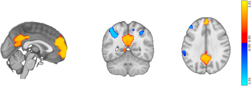
We also measured the RSFC of the left and right DLPFC on their own. LSD significantly reduced the RSFC between the lDLPFC and the rest of the brain, especially in the sensorimotor cortex (Figure S1). (Note that global signal regression was not performed on the data.) LSD did not have a significant effect on the RSFC of the rDLPFC.
3.2 Covariate Analysis (fMRI)
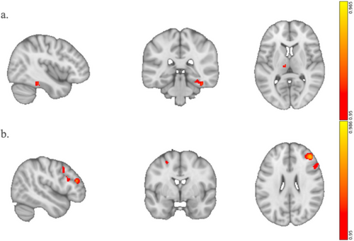
We first examined correlations between the connectivity of the combined DLPFC (both left and right hemisphere) and subjective ratings of ego dissolution, emotional arousal, and positive mood. We found significant correlations between ego dissolution and two clusters with peaks at the left thalamus and the right fusiform gyrus (rFG) (Figure 3a). Given the novel nature of our original seeds, we sought to test the robustness of this correlation. Therefore, we conducted a confirmational analysis in which the thalamus and rFG were the seed regions. If the analysis returned a cluster within the DLPFC, it would verify the specificity of the DLPFC in ego dissolution. Indeed, this confirmational analysis revealed two main clusters, which contained not only the rDLPFC but also the right inferior frontal gyrus (rIFG) (Figure 3b). Scatter plots of the correlation between ego dissolution and RSFC are illustrated in Figure S3; the correlation is higher in the confirmational analysis, demonstrating the specificity of the rDLPFC cluster in mediating ego dissolution.
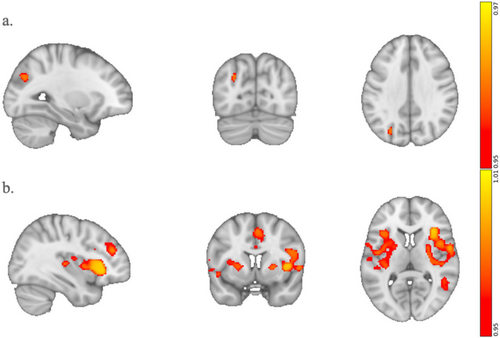
Emotional arousal significantly correlated with connectivity between the rDLPFC and the IPS (Figure 4a). The confirmational analysis reproduced a portion of the rDLPFC while also revealing new regions, namely the right and left anterior insula (rAI, lAI), dorsal anterior cingulate cortex (dACC), and middle temporal gyrus (MTG), that correlated with emotional arousal (Figure 4b). The rAI, lAI, and dACC are main nodes of the Salience Network. As shown in the scatter plots of Figure S4, the correlation between emotional arousal and RSFC is once again larger in the confirmational analysis, indicating that the rDLPFC cluster is more specific to regulating emotional arousal than the original rDLPFC seed.
We did not find any significant correlations between positive mood and the connectivity of the left, right, or combined DLPFC.
We also performed exploratory correlations between the three seeds and the 11-dimensional ASC taken at the end of each scanning day. There were two significant correlations before FDR. Reduced RSFC between the lDLPFC seed and the left Lingual Gyrus was correlated with anxiety (Figure S2a). Secondly, increased RSFC between the rDLPFC and the left Putamen correlated with Changed Meaning of Percepts (Figure S2b).
3.3 GC Analysis (MEG)
Within each network, we measured Granger Causality (GC), an estimate of directed functional connectivity, between each pair of constituent regions, conditioned on all other regions in the network. We computed both time-domain (broadband) and frequency-domain GC in each network.
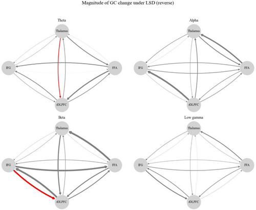
Within each condition (PLCB and LSD), all pairwise-conditional time-domain GC values were significant for all four of the networks (p < 0.002 in all cases). All frequency-domain GC values were significant in all four of the frequency bands—theta, alpha, beta, and low gamma—in all of the networks. Between PLCB and LSD, there were no significant differences in time-domain GC for any pair of regions in any of the networks. However, frequency-domain GC did exhibit significant differences between conditions (Figure 5). In particular, LSD significantly increased theta-band GC from the thalamus to the rDLPFC (p = 0.0366) and beta-band GC from the IFG to the rDLPFC in the ego reverse network (p = 0.0366). There were no significant between-condition differences in frequency-domain GC for the ego forward, emotional forward, or emotional reverse network. Our results suggest that LSD specifically alters GC in a portion of the rDLPFC, i.e., the one in the ego reverse network, rather than the entire rDLPFC seed that was used in the ego forward network.
4 Discussion
In this study, we examined the effect of LSD on the functional connectivity of the left, right, and combined DLPFC, as measured with fMRI in healthy human participants. Then, we explored associations between subjective ratings of the LSD experience and the FC of each DLPFC seed. In the fMRI analysis, we find that LSD overall increases FC between the combined DLPFC and the DMN. Furthermore, the covariate of ego dissolution is positively correlated with increased FC between the combined DLPFC and rFG and thalamus, as well as between rFG and thalamus and the rDLPFC and rIFG. On the other hand, emotional arousal is positively correlated with increased FC between the rDLPFC and IPS, as well as between the IPS and the rDLPFC and nodes within the SN. Our complementary analysis of GC in the MEG dataset revealed that LSD significantly increases bottom-up connectivity from the left thalamus to the rDLPFC in the theta band, as well as beta band connectivity from the rIFG to the rDLPFC.
4.1 Lateralization of Function in the DLPFC
Our study was motivated by the evidence that TMS therapy and psychedelic-assisted therapy report similar therapeutic outcomes via two different treatment modalities. If both methodologies can reduce subjective ratings of depression, they may do so through the engagement of similar mechanisms. TMS for depression has evolved from a multitude of targets to a specific coordinate in the lDLPFC (Cash et al. 2021). We utilized a generalized coordinate and its mirror in the right hemisphere as the seeds for our RSFC analyses (Weigand et al. 2018). This data could then be correlated with subjective reports of the acute LSD experience, namely VAS ratings of ego dissolution, emotional arousal, and positive mood. Through our forward and reverse analyses, we were able to confirm the importance of the rDLPFC in the subjective experiences of emotional arousal and ego dissolution, two intertwined but separable experiences.
These results contribute to a relatively new understanding of the lateralization of function between the left and right DLPFC. As stated in the Introduction, this lateralization is exhibited in the different therapeutic effects associated with stimulating the left versus the right DLPFC with TMS. In particular, while stimulating the left DLPFC can reduce the severity of depression symptoms, stimulating the right DLPFC can inhibit mania. Our findings regarding the neural correlates of ego dissolution and emotional arousal are consistent with the lateralization of function in the DLPFC as revealed by TMS. We also found that the rDLPFC is particularly implicated in bottom-up shifts in network balance.
We propose that the function of primarily the right, and not the left, DLPFC shapes the experience of emotional arousal under psychedelics, which explains our finding that emotional arousal significantly correlates with the RSFC of only the rDLPFC. We contend that emotional arousal on LSD corresponds to a state phenomenologically similar to mania, particularly with regard to the variety of emotional valence. Emotional arousal captures both positive and negative changes in emotion on psychedelics; it is an unvalenced measure of subjective experience. This generalizable phenomenon is best understood as an umbrella term that can encompass multiple dimensions, which can be associated with positive or negative feelings. Secondly, one could construe increases in emotional arousal under LSD as consistent with an increase in perceived ‘salience’ (Friesen 2022; Wießner et al. 2023), thus invoking the “aberrant salience” model of mania (Sass and Pienkos 2013) and psychosis more generally (Kapur 2003).
On the other hand, we propose that ego dissolution recruits the functions of both the left and right DLPFC, hence ego dissolution is correlated with the RSFC of both hemispheres. Indeed, ego dissolution can be highly euphoric yet also resemble a state resembling manic, first-episode psychosis, where ‘self’ abnormalities are known to be common (Moe 2016; Sass and Pienkos 2013). Ego dissolution predicts increases in positive emotional reactivity to psychedelic use (Orłowski et al. 2022) and can also predict improved well-being. Note also that a “mystical” loss of boundaries between the self and the other can also characterize manic psychosis. People experiencing this state have described a “breach in the barriers of individuality” leading to a sense of spiritual communion with the universe (Custance 1952; Landis 1964; Parnas et al. 2005; Sass and Pienkos 2013). In some cases, this state can trigger beliefs of, for instance, telepathy, or other such so-called “narcissistic delusions” such as “delusions of reference”. The phenomenology of this state is very similar to that of ego dissolution on psychedelics, which can also sometimes give rise to claimed telepathic-like experiences (Johnstad 2020).
4.2 The DLPFC and Emotional Arousal
Our fMRI RSFC forward analysis found significant correlations between emotional arousal and connectivity between the rDLPFC and left IPS. The IPS is thought to be involved in regulating emotional arousal during threats. For instance, an fMRI and MEG study found global brain connectivity increases in the left IPS when analyzing threat detection (Balderston et al. 2017). Low frequency rTMS over either left or right IPS during threat of shock has been shown to reduce arousal (Balderston et al. 2020). These findings have led to a theory of connectivity in which the IPS mediates hyperarousal in anxiety disorders through DLPFC regulation (Brown et al. 2023).
The reverse analysis, in which the IPS cluster was used as the seed, returned a cluster in the rDLPFC. While this cluster was smaller than the original rDLPFC seed, it nevertheless confirmed that the rDLPFC is specifically involved in mediating emotional arousal on LSD. The outputs of the reverse analysis also contain clusters in the bilateral AI, dACC, and MTG. The lAI, rAI, and dACC are well-known hubs of the Salience Network (SN), which processes the emotional significance or salience of information. Previous work has shown that the SN acts as an intermediary between the central executive network (CEN) and the DMN. Some recent work also indicated a potential over-involvement of the SN in depression (Lynch et al. 2024). Hubs in these three networks show differences in information flow and dominance of signaling contingent on the task (Menon and Uddin 2010; Molnar-Szakacs and Uddin 2022). Aberrations in network transitions are theorized to be at the root of psychological illness (Menon 2011). Furthermore, as explained in Section 4.1, experiences of aberrant salience are a key aspect of emotional arousal on LSD; emotional sensitivity to the environment is heightened because everything appears to be extremely significant. Future work on this network may take into account the DMN for GC analysis or examine individual dyads within the network for other subjective categories like anxiety.
Our MEG analysis of these forward and reverse networks displayed statistically significant connections between all pairs of regions, across all frequency bands combined, under both placebo and LSD, which would suggest that regions form a genuine network in the brain. However, there were no statistically significant changes from placebo to LSD in emotional arousal in the frequency or time domain. Other spectral patterns, such as cross-freuqency coordination, may explain changes in emotional arousal on LSD (Clarke-Williams et al. 2024; Vinck et al. 2023).
4.3 The DLPFC and Ego Dissolution
The experience of ego dissolution, or “the loss of a sense of oneself,” is an essential aspect of the classical psychedelic experience (Millière 2017). Furthermore, there is a relationship between ego dissolution and statistically significant reductions in depression scores within adults, even in the long term (Carhart-Harris, Kaelen, et al. 2016; Roseman et al. 2018; Weiss et al. 2024). Hence, reliable biomarkers and mechanisms of the psychedelic experience of ego dissolution are worth further investigation. By identifying the network of regions that mediate the experience, we may be able to stimulate them with TMS, either independently of or in conjunction with a psychedelic treatment, to augment ego dissolution and thereby improve or better predict the therapeutic outcomes (Copa et al. 2024).
The psychedelic experience of ego dissolution has been correlated with brain activity in a wealth of previous research. A previous RSFC study, which was conducted on the same LSD data examined in this paper, revealed that global connectivity changes in the left and right angular gyrus and left and right insula correlated with ego dissolution (Tagliazucchi et al. 2016). Ego dissolution has mainly been attributed to connectivity changes within the DMN (Carhart-Harris, Muthukumaraswamy, et al. 2016; Lebedev et al. 2015) but also reduced interhemispheric connectivity within the salience network (Lebedev et al. 2015). This ‘disintegration’ of the high-level networks has been conceived as a ‘relaxation’ of top-down inhibitory control, which, according to the so-called ‘RElaxed Beliefs Under pSychedelics’ (REBUS) model, demonstrates a ‘flattening’ of the functional hierarchy in the brain and corresponding ‘relaxed beliefs’ or assumptions about the self and environment. In a variety of mental illnesses, including depression, beliefs or assumptions can become (pathologically) over-weighted; thus, their relaxation under (and potentially after) psychedelic experience may explain their potential therapeutic utility (Moe 2016; Sass and Pienkos 2013). Previous work has made significant strides toward establishing changes in functional cortical hierarchy under psychedelics (Girn et al. 2022; Luppi et al. 2021; Shinozuka et al. 2025).
This study correlated ego dissolution with a network of brain regions, rather than a single region or the whole brain. We began by selecting a novel seed that comprised the combined left and right DLPFC. We identified a significant correlation between ego dissolution and changes in RSFC between the combined DLPFC and the thalamus & FFA. A confirmational, or reverse analysis, in which the FFA & thalamus became the input seeds, yielded two main outputs: a smaller rDLPFC cluster and the rIFG.
These analyses, which were performed in fMRI, capture undirected connectivity. We then measured directed connectivity in an MEG dataset that was acquired on the same set of participants. In particular, we evaluated GC on the source-reconstructed timeseries of the regions in the forward and reverse networks. While many methods exist for capturing directed connectivity, GC has strong advantages in identifying time-delayed interactions, is robust to noisy data, and is flexible in working with the frequency domain. While we did not directly correlate GC with the ego dissolution VAS scales, this analysis nevertheless revealed the effect of LSD on the direction of information flow between the identified clusters across different frequency bands: theta, alpha, beta, and low gamma. While the effects of LSD on directed FC across the whole brain have been previously documented (Barnett et al. 2020), our analysis reveals changes in information flow specific to areas correlated with ego dissolution. Our findings are inconsistent with previous literature, which found that LSD exclusively decreased GC (Barnett et al. 2020). However, our estimates of GC were conditional on all other regions in the network, whereas previous analyses were either unconditional or conditional on timeseries that were averaged across multiple regions.
The reverse analysis revealed two statistically significant increases in GC: from the thalamus to the rDLPFC in the theta band and from the IFG to the rDLPFC in the beta band. The centroid of the thalamus seed is specifically located within the medial dorsal (MD) thalamus, which is structurally and functionally connected to the DLPFC. In fact, the primary source of input to the parvocellular MD is the DLPFC (Byne et al. 2009; Mitchell and Chakraborty 2013; Pergola et al. 2015). Increased bottom-up information flow from the thalamus to higher-order regions of the brain, such as the prefrontal cortex, has been proposed as a critical component of the neural mechanisms underlying the psychedelic experience (Avram et al. 2021; Onofrj et al. 2023; Vollenweider and Geyer 2001). The thalamus plays a role in gating sensory information to the cortex, screening out irrelevant stimuli (McCormick and Bal 1994). Aberrant connectivity from the thalamus to the cortex can lead to a “sensory overload,” inundating the cortex with excessive interoceptive and exteroceptive information (Vollenweider and Geyer 2001). Altered connectivity between the thalamus and the DLPFC is a robust biomarker of schizophrenia and psychosis, conditions in which patients assign too much meaning to irrelevant stimuli (Anticevic et al. 2014; Pergola et al. 2018; Steullet 2020; Welsh et al. 2010; Woodward et al. 2012). Similarly, some studies have shown that psychedelics exclusively increase functional connectivity between the thalamus and many regions of cortex (Bedford et al. 2023; Müller et al. 2017; Tagliazucchi et al. 2016), though other studies have found some decreases in thalamocortical connectivity (Avram et al. 2022; Gaddis et al. 2022; Preller et al. 2018, 2020). Psilocybin specifically alters activity in a region of the thalamus that overlaps most with the MD nucleus, which is consistent with our findings (Gaddis et al. 2022). A recent theory proposes that theta-band spiking in the thalamus shifts thalamocortical coupling to a dysrhythmic state that underlies altered states of consciousness, which is also aligned with our observations of elevated theta-band thalamocortical connectivity on LSD (Onofrj et al. 2023).
The DLPFC is generally involved in determining whether information is pertinent enough to enter and stay in working memory (Altamura et al. 2010; Friedman and Robbins 2022; Petrides 2000; Rassi et al. 2023; Rosero Pahi et al. 2020). Crucially, the ability of the PFC to maintain and assign relevance to neural representations is mediated by the MD thalamus (Bolkan et al. 2017; Marton et al. 2018; Mitchell and Chakraborty 2013; Parnaudeau et al. 2015; Rikhye et al. 2018; Schmitt et al. 2017) Rodent studies have demonstrated that MD neurons stabilize context-relevant representations in the PFC, thereby enabling them to be maintained within working memory, while suppressing context-irrelevant representations (Rikhye et al. 2018).
Thus, enhanced directed connectivity from the MD thalamus may cause information from typically deemed irrelevant contexts to interfere with representations in the PFC, leading to a “hyper-flexible” state in which the brain is less constrained by the demands of the present cognitive context. We speculate that, during self-reflection, heightened connectivity between the MD thalamus and the cortex may amplify neural representations that do not ordinarily pertain to the self. These amplifications could induce instability across this network including thalamic dependent internal sensory perception within the rIFG (Dobrushina et al. 2021) and distort functionality in any one of the other regions in the ego dissolution network, which have all been correlated with self-identification (Herwig et al. 2012; Kaplan et al. 2008; Ma and Han 2012). This broadening of relevance could explain the experience of the “loss of the sense of self” found in ego dissolution; on psychedelics the neural mechanisms of the self “over-represent” or assign too much relevance to external stimuli, yielding a sense of vast interconnectedness with the environment.
Furthermore, increased beta-band connectivity from the rIFG to the rDLPFC, as we observed, could be involved in integrating, or attempting to integrate, experiences of ego dissolution. In particular, the rIFG and the rDLPFC are both part of a network that integrates disconfirmatory evidence, i.e., present stimuli that violate a prior belief (Ehlis et al. 2024; Lavigne et al. 2015). In other words, when prior held beliefs about the self are challenged by conflicting evidence, the rIFG and the rDLPFC work together to integrate this information and thereby maintain or update the belief. This could be consistent not only with the rIFG's established role in integrating internal representations (Adolfi et al. 2017; Dobrushina et al. 2021), but also REBUS' hypothesis that psychedelics revise prior beliefs by altering top-down inhibition from higher-order regions like the rIFG and the rDLPFC.
The observed effects of LSD on directed connectivity are similar to the neural signatures of schizophrenia, which relate to a longstanding, but contentious, view that psychedelics are “psychotomimetics” or models of psychosis (Nichols and Walter 2021; Umbricht et al. 2003; Vollenweider et al. 1998). Schizophrenia and mania are associated with a number of structural deficits in the DLPFC, including cortical thinning, reduced gray matter volume, and lower fractional anisotropy of outgoing white matter tracts (Abé et al. 2023; Ellison-Wright and Bullmore 2009; van Haren et al. 2011). Furthermore, as stated above, aberrations in connectivity between the MD thalamus and the PFC are commonly observed in patients with schizophrenia, perhaps explaining observations that task-irrelevant representations interfere more with cognition in these populations (Wagner et al. 2013).
While some of the changes in brain activity and phenomenology on LSD are also found in individuals with schizophrenia, it is important to note that schizophrenia is a chronic condition, whereas the LSD trip is a transient experience. Also, disruptions to self-awareness in schizophrenia differ significantly from those of LSD. For instance, patients over-attribute agency to themselves and overestimate their causal power over the world, whereas people who take LSD tend not to do so (Hur et al. 2014). Furthermore, individuals affected by schizophrenia often exhibit long-standing emotional withdrawal and cognitive blunting, which are markedly different from the emotional arousal and cognitive flexibility experienced on LSD (Doss et al. 2021; Martínez et al. 2021; Oorschot et al. 2013).
In summary, we propose that amplified connectivity from the thalamus to the DLPFC may cause representations of the outside world to interfere with self-related cognition, thereby giving rise to the experience of ego dissolution. The same neural mechanisms may underpin delusional beliefs about the self in patients afflicted with schizophrenia and psychosis.
4.4 Limitations
This data has a small sample size; data from only 15 participants was included in the analysis. The convergent and significant results we found in both the fMRI and MEG analyses indicate that the observed effects are real. There is also a potential that this data is confounded by the physiological effects of LSD, namely its vasoconstrictive effects. However, while vasoconstriction alters the fMRI signal, it does not affect MEG activity in the same way (Özbay et al. 2019; Wilson et al. 2016). The significant increases in directed information flow in the MEG analysis suggest that vasoconstriction does not underlie the observed changes in RSFC.
The study design may not have given sufficient time for the effects of LSD to wash out; there was only a 14-day separation between the LSD and placebo sessions, and LSD can affect brain activity for up to a month afterwards (Barrett et al. 2020; McCulloch et al. 2022). However, the order of LSD and placebo was counterbalanced across participants. If the changes in RSFC depended on whether participants received LSD first or second, then we likely would not have obtained significant results. If a longer washout period had been given, we suspect that we would have seen even stronger changes in RSFC.
Because the data is resting-state, it is difficult to relate different patterns of brain activity to specific psychological behaviors or processes that are modulated via LSD. To provide stronger evidence, tasks must be deployed to examine changes in self-awareness in response to working memory demands, while acknowledging the risk of generalized deficit confounds in this regard.
We did not correlate the directed connectivity of the MEG data to ratings of ego dissolution because these ratings were not available for all of the included participants. However, because the undirected connectivity between the same regions did correlate with ego dissolution in the fMRI dataset, we are confident that the directed connectivity plays some role in shaping the experience of ego dissolution. That being said, whole-brain MEG analyses may have uncovered a broader set of brain regions that correlate with ego dissolution and emotional arousal.
Finally, accurate source reconstruction of MEG data to subcortical regions like the thalamus is generally challenging, especially in resting-state data (Krishnaswamy et al. 2017). fMRI analysis of subcortical activity may be more reliable, so dynamic causal modeling on the fMRI data, which can be used to estimate directed connectivity, may be worth exploring to assess the robustness of the MEG GC findings.
5 Conclusion
This study set out to show that changes in functional connectivity with the dorsolateral prefrontal cortex correspond to the subjective effects of the psychedelic state. In the fMRI analysis, ratings of emotional arousal correlated with connectivity between the salience network & IPS, and the right, but not left, DLPFC, implying a lateralization of function in the DLPFC. On the other hand, ego dissolution was associated with increased connectivity between the combined left & right DLPFC and clusters in the thalamus & the fusiform gyrus. A GC analysis of these networks elucidated the directionality of information flow between the constituent regions of each network. LSD elevated theta-band GC from the thalamus to the rDLPFC, which exemplifies an increase in bottom-up information flow and the flattening of the hierarchy of the brain on psychedelics.
Our study of both undirected and directed connectivity of the DLPFC on LSD has unveiled new methods and opportunities for research in psychedelic science. Firstly, distinguishing the lDLPFC and rDLPFC could improve understanding of not only psychedelics but also psychiatric disorders. In particular, analyzing the two hemispheres separately may reveal the lateralization of the DLPFC's function, whereas measuring their activity together could elucidate the extent to which the two hemispheres function in unison. Secondly, studies informed by the structural heterogeneity of the DLPFC and its receptor makeup could further elucidate this region's role in the psychedelic experience and psychiatric illnesses. Thirdly, this is one of the first studies to analyze the effects of psychedelics with both fMRI and MEG data. Future studies on psychedelics could leverage this multimodal approach, which takes advantage of the high spatial resolution of fMRI and high temporal resolution of MEG (Hall et al. 2014). Finally, the confirmational analysis played an essential role in our understanding of these networks, enabling us to ensure the validity of our novel seeds and substantiating the lateralization of function in the DLPFC.
Our findings provide novel insights into the neural mechanisms of ego dissolution and other altered states of consciousness on psychedelics, which may be similar to the neural pathways that are affected in psychosis and schizophrenia. Future research could determine whether the same neural mechanisms play a role in the therapeutic effects of psychedelics for disorders such as depression. Perhaps the reason ego dissolution is correlated with a reduction in depression is that, for a moment in time, the self-perpetuating definition of ‘who we believe we are’ stops long enough for us to envision broader possibilities and recreate ourselves.
Author Contributions
C.R.C. conceptualized the study, conducted all fMRI analyses, created Figures 1-4, and wrote the manuscript. K.S. preprocessed and source-reconstructed the MEG data, conducted most of the Granger causality analyses on the MEG data, and wrote the manuscript together with C.R.C. R.T. advised on analyses, created Figure 5, and edited the manuscript. L.R., S.M., D.J.N., and R.C.-H. conceptualized the fMRI and MEG experiments and collected the data. L.B. provided essential guidance on the Granger causality analysis, including help with writing scripts and performing the within-condition significance tests. M.K. and O.D. served as master's thesis advisors for C.R.C. and oversaw the fMRI analyses.
Acknowledgments
Clayton R. Coleman would like to express sincere gratitude to his thesis advisors Matt Howard and Ottavia Dipasquale and the staff at King's College London IoPPN—especially Fernando Zelaya, Owen O'Daly, and Eamonn Walsh—for their invaluable support. Special thanks to Mitul Mehta for his thesis review and for suggesting the conformational analysis. Clayton R. Coleman is funded by the Usona Institute. Kenneth Shinozuka is funded by the Clarendon Fund, a Department of Psychiatry studentship, the John Henry Jones Scholarship from Balliol College, the Research Council U.K., and the Usona Institute. Robert Tromm is funded by the Usona Institute. Robin Carhart-Harris is funded by a Ralph Metzner endowment. Lionel Barnett is supported by European Research Council Advanced Investigator Grant CONSCIOUS; grant number 101019254.
Conflicts of Interest
The authors declare no conflicts of interest.
Open Research
Data Availability Statement
LSD data was collected by the Centre for Psychedelic Research at Imperial College London (Carhart-Harris et al. 2020). The full dataset includes arterial spin labeling (ASL), functional magnetic resonance imaging (fMRI), and magnetoencephalography (MEG). We did not examine the ASL data in this analysis. A brief description of data collection and preprocessing procedures is given below, but the full methodology can be found in the primary paper (Carhart-Harris, Kaelen, et al. 2016; Carhart-Harris, Muthukumaraswamy, et al. 2016).
Code is available upon request by contacting the lead author at [email protected]. Processed fMRI data is open-source and available at https://openneuro.org/datasets/ds003059/versions/1.0.0. MEG data may be available upon request by contacting Leor Roseman at [email protected].


