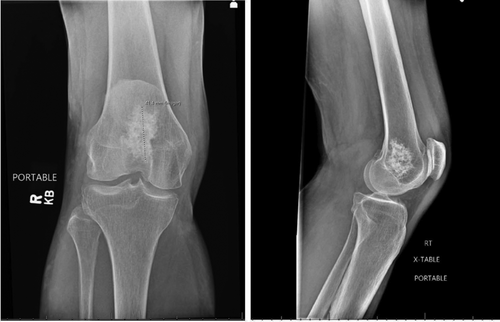
A 70-year-old female with knee pain after a fall
Funding and support: By JACEP Open policy, all authors are required to disclose any and all commercial, financial, and other relationships in any way related to the subject of this article as per ICMJE conflict of interest guidelines (see www.icmje.org). The authors have stated that no such relationships exist.
1 CASE PRESENTATION
A 70-year-old female presented to the emergency department with right knee pain. She suffered a mechanical fall while walking on a trail outdoors and landed on her knee. The patient was able to continue ambulating, but developed worsening knee pain and swelling overnight. She denied any other injury from the fall.
Physical examination revealed a small abrasion on the anterior aspect of her knee. Her knee was diffusely tender to palpation, and she had decreased range of motion secondary to discomfort. There was no ligament laxity noted on examination. The x-rays were negative for acute fractures but showed another finding as seen in Figure 1.

2 DIAGNOSIS: CHONDROID TUMOR
The x-ray showed an incidental finding of a 4.1 cm patchy sclerotic lesion in the distal femur, indicating a chondroid lesion. Chondroid tumors represent a family of tumors that develop from the chondroid matrix.1 The most common types are osteochondroma, enchondroma, and chondroblastoma.1, 2 Chondroid tumors encompass a spectrum of benign and malignant neoplasms that exhibit varying degrees of cartilaginous differentiation. They are among the most common incidental benign bone lesions found on imaging, and do not typically cause symptoms.1, 2 Patients with chondroid tumors may have increased susceptibility to fractures or breaks.3
On x-ray, chondroid tumors have the appearance of “popcorn” or sclerotic calcifications.4 If the lesion is noted to be concerning on x-ray, magnetic resonance imaging can be obtained to further differentiate the lesion type.5 If advanced imaging is indicative of malignant lesions, the patient should be referred to oncology for potential biopsy, surgical resection, and radiation.6
ACKNOWLEDGMENTS
A special thank you to our patient. No prior presentations or financial support.


