Right upper quadrant abdominal pain with fever
1 CASE PRESENTATION
A 52-year-old woman with a medical history of poorly controlled diabetes mellitus, ischemic heart disease, end-stage renal disease, and congestive heart failure presented to the emergency department with a chief complaint of fever and chills off and on for 1 week. Tenderness in the right upper quadrant was detected. Laboratory workup revealed leukocytosis with left shift and hyperglycemia.
2 DIAGNOSIS
2.1 Emphysematous liver abscess
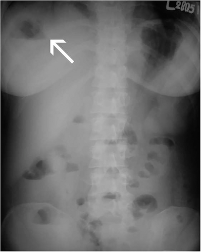
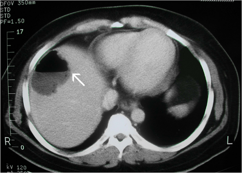
An abdominal plain film, which showed a fine air-fluid level (arrow) in the liver at an upright position (Figure 1) enabled us to diagnose the air-containing liver abscess at a glance. Computed tomography (CT) with contrast demonstrated a large abscess with gas formation at the right liver (Figure 2). Klebsiella pneumoniae developed in the blood and pus cultures. After CT-guided drainage and intravenous antibiotics for 6 weeks, the patient recovered uneventfully. Gas-producing liver abscess is rare, but it may have a fulminating course in patients with diabetes mellitus. Critical diagnosis and adequate management decrease the mortality.1 In this case, the standing plain film assisted in the immediate diagnosis.


