Man with episodic abdominal pain and jaundice
1 PATIENT PRESENTATION
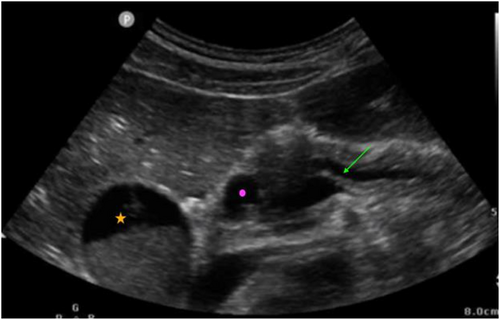
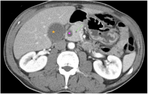
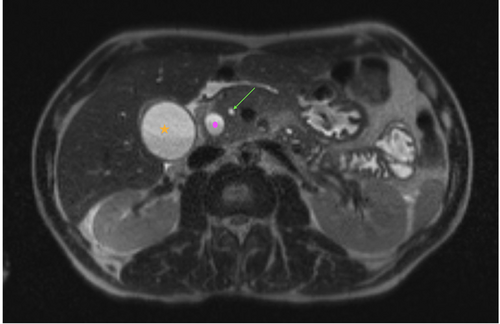
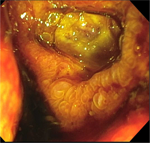
A 51-year-old man without a past medical history presented to our emergency department with 2 months of intermittent epigastric pain radiating to the back, tan stools, fatigue, and weight loss. He denied current alcohol use. He appeared diffusely jaundiced. His abdomen was soft with mild epigastric tenderness and a negative Murphy sign. Serum analysis showed total bilirubin 13.7, aspartate transaminase 207, alanine transaminase 463, alkaline phosphatase 634, lipase > 4000, elevated carcinoembryonic antigen/carbohydrate antigen 19-9, and negative hepatitis panel. Imaging included point-of-care (POC) ultrasound (Figure 1, Video 1), computed tomography (CT; Figure 2), magnetic resonance imaging (MRI; Figure 3), and endoscopic retrograde cholangiopancreatograph (ERCP; Figure 4).
2 DIAGNOSIS
2.1 Double duct sign
Our patient had obstructive jaundice. POC ultrasound revealed a distended gallbladder with sludge without signs of acute cholecystitis. The dilated common bile duct >10 mm (normal < 6 mm) and dilated pancreatic duct >5 mm (normal < 3 mm) were consistent with the rare double duct sign. Dilatation without a clear etiology was confirmed on MRI and CT. ERCP revealed a duodenal ulcer with biopsy positive for invasive, poorly differentiated adenocarcinoma. Finally, a pancreaticoduodenectomy revealed a tumor of the pancreatic head.
Etiologies of the double duct sign include neoplasms of the pancreatic head and ampulla of Vater and less commonly choledocholithiasis and chronic pancreatitis.1 Overall prevalence of associated malignancy varies from 58% to 100%.2 However, the presence of jaundice is strongly associated with malignancy whereas absence with benign conditions.3-5 Isodense culprit neoplasms are often not visualized on advanced imaging. As initial presentation of pancreatic cancer is insidious, the ominous presence of this sign should raise concerns for malignancy.
ACKNOWLEDGMENTS
The authors would like to thank Daniela Galan Alonso, MD, Department of Radiology, Jackson Memorial Hospital.


